

The DOI matches its Crossref record; 70 of the 71 references it cites were found in a registry or an index.
Phenotypic profile of alternative activation marker CD163 is different in Alzheimer’s and Parkinson’s disease
Open accessAbstract
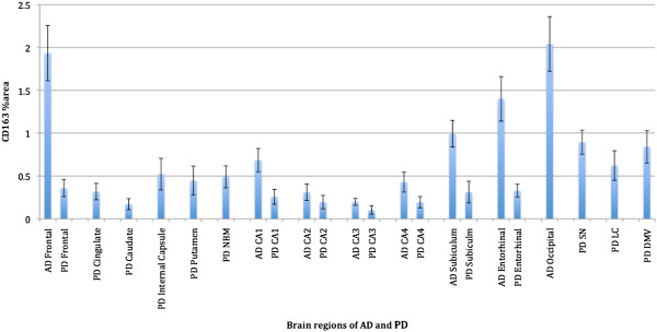
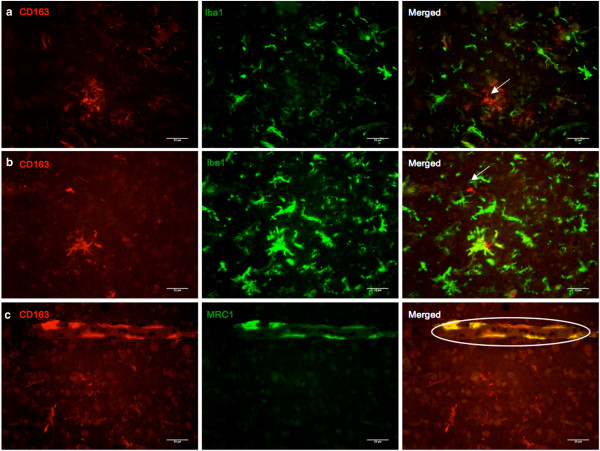
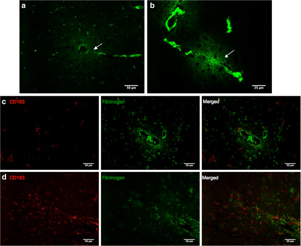
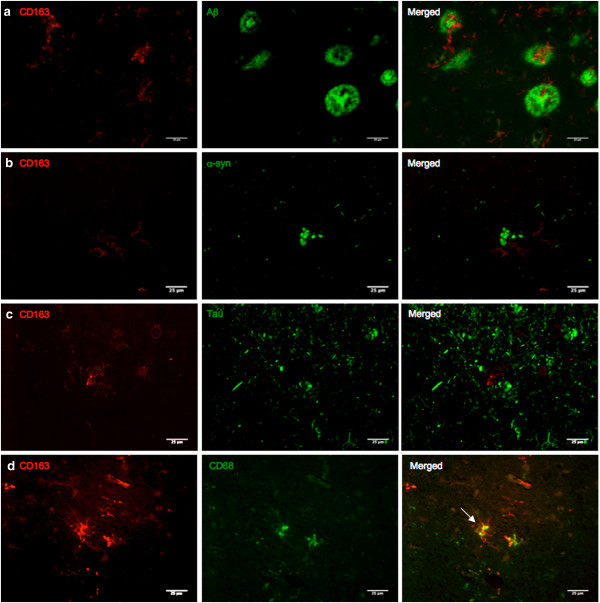
BACKGROUND: Microglial activation is a pathological feature common to both Alzheimer's and Parkinson's diseases (AD and PD). The classical activation involves release of pro-inflammatory cytokines and reactive oxygen species. This is necessary for maintenance of tissue homeostasis and host defense, but can cause bystander damage when the activation is sustained and uncontrolled. In recent years the heterogeneous nature of microglial activation states in neurodegenerative diseases has become clear and the focus has shifted to alternative activation states that promote tissue maintenance and repair. We studied the distribution of CD163, a membrane-bound scavenger receptor found on perivascular macrophages. CD163 has an immunoregulatory function, and has been found in the parenchyma in other inflammatory diseases e.g. HIV-encephalitis and multiple sclerosis. In this study, we used immunohistochemistry to compare CD163 immunoreactivity in 31 AD cases, 27 PD cases, and 16 control cases. Associations of microglia with pathological hallmarks of AD and PD were investigated using double immunofluorescence. RESULTS: Parenchymal microglia were found to be immunoreactive for CD163 in all of the AD cases, and to a lesser extent in PD cases. There was prominent staining of CD163 immunoreactive microglia in the frontal and occipital cortices of AD cases, and in the brainstem of PD cases. Many of them were associated with Aß plaques in both diseases, and double staining with CD68 demonstrates their phagocytic capability. Leakage of fibrinogen was observed around compromised blood vessels, raising the possibility these microglia might have originated from the periphery. CONCLUSIONS: Increase in microglia's CD163 immunoreactivity was more significant in AD than PD, and association of CD163 immunoreactive microglia with Aβ plaques indicate microglia's attraction towards extracellular protein pathology, i.e. extracellular aggregates of Aβ as compared to intracellular Lewy Bodies in PD. Double staining with CD163 and CD68 might point towards their natural inclination to phagocytose plaques. Fibrinogen leakage and compromise of the blood brain barrier raise the possibility that these are not resident microglia, but systemic macrophages infiltrating the brain.
Overview
The claims and statistics of this paper haven’t been checked yet.
Run the check — free, no account needed. It reads the open-access text and checks the sources it cites.
Integrity signals
Registry
Cites
Statistics
Figures
Verification
Claims the paper makes
The claims and statistics of this paper haven’t been checked yet.
Run the check — free, no account needed. It reads the open-access text and checks the sources it cites.
Claims other papers make about this one
Each is checked against this paper’s own text: does it say what the citing paper says it does?
The source backs part of the claim, not all of it — or backs it for a different population, condition or comparison.
In the citing paper
“CD163, a hub gene of MG-M4, is known to be involved in the breakdown of the blood-brain barrier.53,54”
From hdWGCNA identifies co-expression networks in high-dimensional transcriptomics data (2023) · cites this paper as [54]
Experimental — accuracy not yet measuredThe source backs part of the claim, not all of it — or backs it for a different population, condition or comparison.
In the citing paper
“CD163 is a hemoglobin-haptoglobulin scavenger receptor that is considered to be expressed exclusively on perivascular macrophages in the brain under normal conditions, as well as peripheral cells of monocyte lineage [38, 56].”
From Loss of homeostatic microglial phenotype in CSF1R-related Leukoencephalopathy (2020) · cites this paper as [38]
Experimental — accuracy not yet measuredThe cited source says what the claim attributes to it.
In the citing paper
“Like Dal Bianco et al.20 Pey et al.120 also found higher levels of CD163 staining in the hippocampus, as well as the frontal and occipital cortices.”
From Markers of microglia in post-mortem brain samples from patients with Alzheimer’s disease: a systematic review (2018) · cites this paper as [120]
The check wasn’t confident either way, so it gives no verdict — never disputed.
In the citing paper
“CD163, a hub gene of MG-M4, is known to be involved in the breakdown of the blood-brain barrier.53,54”
From hdWGCNA identifies co-expression networks in high-dimensional transcriptomics data (2023) · cites this paper as [54]
Experimental — accuracy not yet measuredIn the citing paper
“Increased expression of proteins associated with non-microglia myeloid cells, such as CD163, has been observed in the brains of PD patients.”
From Microglia in neurodegenerative diseases: mechanism and potential therapeutic targets (2023) · cites this paper as [227]
Self-check
Percentages, sums and reported differences re-computed from the paper’s own numbers.
The self-check needs the paper’s full text.
Citations
Cites
Alternative activation of macrophages
Gordon, S. (2003) · Nature Reviews Immunology · doi:10.1038/nri978 · Cited by 6,189
Diagnosis and management of dementia with Lewy bodies: Third report of the DLB consortium
McKeith, I. G., Dickson, D. W., Lowe, J., Emre, M., O'Brien, J. T., Feldman, H., Cummings, J., Duda, J. E., Lippa, C., Perry, E. K., et al. (2005) · Neurology · doi:10.1212/01.wnl.0000187889.17253.b1 · Cited by 5,069
Kettenmann, H., Hanisch, U.-K., Noda, M., & Verkhratsky, A. (2011) · Physiological Reviews · doi:10.1152/physrev.00011.2010 · Cited by 3,547
Microglia in neurodegenerative disease
Perry, V. H., Nicoll, J. A. R., & Holmes, C. (2010) · Nature Reviews Neurology · doi:10.1038/nrneurol.2010.17 · Cited by 1,543
Holmes, C., Boche, D., Wilkinson, D., Yadegarfar, G., Hopkins, V., Bayer, A., Jones, R. W., Bullock, R., Love, S., Neal, J. W., et al. (2008) · The Lancet · doi:10.1016/s0140-6736(08)61075-2 · Cited by 1,416 · cited 1×
“Much interest has been shown in Aβ and its removal from the brain via natural methods or vaccination [58]; CD163 immunoreactive microglia clusters around Aβ plaques support the idea that they might serve a similar purpose.”
BackgroundMicroglial Dysfunction and Defective β-Amyloid Clearance Pathways in Aging Alzheimer's Disease Mice
Hickman, S. E., Allison, E. K., & El Khoury, J. (2008) · The Journal of Neuroscience · doi:10.1523/jneurosci.0616-08.2008 · Cited by 1,329 · cited 2×
“via downregulation of Aβ receptors [22] and neuronal degeneration, but TNF-α is protective due to its pre-conditioning effect on metabolic excitotoxicity, Aβ toxicity and ischaemia [23].”
BackgroundSimard, A. R., Soulet, D., Gowing, G., Julien, J.-P., & Rivest, S. (2006) · Neuron · doi:10.1016/j.neuron.2006.01.022 · Cited by 1,265
Tansey, M. G., & Goldberg, M. S. (2010) · Neurobiology of Disease · doi:10.1016/j.nbd.2009.11.004 · Cited by 1,045
Macrophage activation switching: an asset for the resolution of inflammation
Porcheray, F., Viaud, S., Rimaniol, A.-C., Léone, C., Samah, B., Dereuddre-Bosquet, N., Dormont, D., & Gras, G. (2005) · Clinical and Experimental Immunology · doi:10.1111/j.1365-2249.2005.02934.x · Cited by 840
Buechler, C., Ritter, M., Orsó, E., Langmann, T., Klucken, J., & Schmitz, G. (2000) · Journal of Leukocyte Biology · doi:10.1002/jlb.67.1.97 · Cited by 704
Cited by
Sorted by citations
Items 101–125 of 125
Neuroinflammation and Immune Dysfunction in the Mechanisms of Development of Parkinson’s Disease
Idova, G. V., Лазаревна, А. Е., & Zhanaeva, S. Y. (2023) · Neuroscience and Behavioral Physiology · doi:10.1007/s11055-023-01549-8 · Cited by 2
Identification of Key Pathways and Genes in Dementia via Integrated Bioinformatics Analysis
Vastrad, B. M., & Vastrad, C. M. (2025) · SN Comprehensive Clinical Medicine · doi:10.1007/s42399-025-01876-6 · Cited by 2
Immunotherapies for Movement Disorders: Parkinson’s Disease and Amyotrophic Lateral Sclerosis
Schutt, C. R., Gendelman, H. E., & Mosley, R. L. (2016) · Springer eBooks · doi:10.1007/978-3-319-44022-4_46 · Cited by 2
Vastrad, B. M., Pattanashetti, S., & Vastrad, C. M. (2023) · bioRxiv (Cold Spring Harbor Laboratory) · doi:10.1101/2023.12.05.570305 · Cited by 2
Cell-based immunotherapy for neurodegenerative disease: A promising avenue
Huerta, T. J., Urbina-Muñoz, V., Urra-Alvarez, V., Villablanca, C., Gómez‐Pérez, L. S., Saavedra, B., Contreras, T., & Vidal, R. L. (2025) · Neural Regeneration Research · doi:10.4103/nrr.nrr-d-25-00816 · Cited by 1
NEUROINFLAMMATION AND IMMUNE DYSFUNCTION IN THE PATHOGENESIS OF PARKINSON’S DISEASE
Idova, G. V., Лазаревна, А. Е., & Zhanaeva, S. Y. (2023) · Журнал высшей нервной деятельности им И П Павлова · doi:10.31857/s0044467723040056 · Cited by 1
Immune response during idiopathic Parkinson's disease: From humans to animal models
Nissen, S. K., Ferreira, S. A., Klæstrup, I. H., & Romero‐Ramos, M. (2021) · International review of movement disorders · doi:10.1016/bs.irmvd.2021.08.006 · Cited by 1
Pathologic α-Synuclein Species Activate LRRK2 in Pro-Inflammatory Monocyte and Macrophage ResponsesPreprint
Xu, E., Boddu, R., Abdelmotilib, H., Kelly, K., Sokratian, A., Harms, A. S., Schonhoff, A. M., Bryant, N., Harmsen, I. E., Schlossmacher, M. G., et al. (2020) · bioRxiv (Cold Spring Harbor Laboratory) · doi:10.1101/2020.05.04.077065 · Cited by 1
Nagayach, A., & Patro, I. K. (2015) · University
Defining Microglial Phenotypes in Alzheimer’s Disease
Walker, D. G., & Lue, L. (2018) · InTech eBooks · doi:10.5772/intechopen.75511
Sonninen, T.-M., Plotnikova, L. M., Laham-Karam, N., Goldsteins, G., Koıstınaho, J., & Lehtonen, Š. (2021) · Elsevier eBooks · doi:10.1016/b978-0-323-85764-2.00009-0
Kozloski, G. A. (2019) · Materials and Methods · doi:10.13070/mm.en.9.2758
Kozloski, G. A. (2019) · Materials and Methods · doi:10.13070/mm.en.9.2844
Seyvald, Y. E., & Калиничев, А. Г. (2023) · Scientific Bulletin of the Omsk State Medical University · doi:10.61634/2782-3024-2023-12-69-81
Qi, P., Huang, M., & Zhu, H. (2025) · Combinatorial Chemistry & High Throughput Screening · doi:10.2174/0113862073322931241030104813
Vastrad, B. M., & Vastrad, C. M. (2025) · Genome Instability & Disease · doi:10.1007/s42764-025-00147-x
Young, J. J., Park, H.-J., Kim, M., Par‐Young, J., Bartlett, H. H., Kim, H. S., Ünlü, S., Osmani, L., Shin, M. S., Bucala, R., et al. (2023) · Figshare · doi:10.6084/m9.figshare.24716931
Muñoz‐Castro, C., Mejias‐Ortega, M., Sánchez‐Mejías, E., Navarro, V., Trujillo‐Estrada, L., Jiménez, S., León, J. A. G., Fernandez-Valenzuela, J. J., Sanchez-Mico, M. V., Romero‐Molina, C., et al. (2024) · Figshare · doi:10.6084/m9.figshare.26571967
Swanson, M. E. V., Scotter, E. L., Smyth, L. C. D., Murray, H. C., Ryan, B., Turner, C., Faull, R. L. M., Dragunow, M., & Curtis, M. A. (2020) · Figshare · doi:10.6084/m9.figshare.13120600
Young, J. J., Park, H.-J., Kim, M., Par‐Young, J., Bartlett, H. H., Kim, H. S., Ünlü, S., Osmani, L., Shin, M. S., Bucala, R., et al. (2024) · Figshare · doi:10.6084/m9.figshare.24716955
Kerslake, L. D., Noonan, C., Davies, D., Kim, D., Eshou, V. A., Goldsbury, C. S., & Cullen, K. M. (2026) · Neurobiology of Aging · doi:10.1016/j.neurobiolaging.2026.03.004
Understanding the role of perivascular macrophages in Parkinson's disease pathophysiology
Fuentealba, J. R. (2017) · theses.fr (ABES) · doi:10.70675/b2e8f905z4f72z497eza017zc5b9d7fe53a1
Djuretić, J., Ivanović, J., Jezdić, K., Todorović, V., Bufan, B., Santrač, A., & Aranđelović, J. (2026) · npj Aging · doi:10.1038/s41514-026-00364-y
Yang, L.-J., Deng, N., Zhu, M., Shi, X., Yang, J., & Nie, H. (2026) · Frontiers in Cell and Developmental Biology · doi:10.3389/fcell.2026.1907664
Takeda, Y., Kuramoto, Y., Tsuji, S., Doe, N., Yamahara, K., & Yoshimura, S. (2026) · Brain Research · doi:10.1016/j.brainres.2026.150617
Metadata
- Authors
- Pey, P., Pearce, R. K., Kalaitzakis, M. E., Griffin, W. S. T., & Gentleman, S. M.
- Affiliations
- Imperial College London (GB)
- University of Arkansas for Medical Sciences (US)
- Corresponding author
- Pey, Peixuan
- Gentleman, Steve M
- Venue
- Acta Neuropathologica Communications
- Published
- 2014-02-14
- Volume
- 2
- Issue
- 1
- Pages
- 21-21
- ISSN
- 2051-5960 (ISSN-L)
- PMC
- PMC3940003
- OpenAlex
- W2147311470
- Cited by
- 128
- References
- 71
- Citations by year
- 2026: 7 · 2025: 12 · 2024: 10 · 2023: 18 · 2022: 14 · 2021: 11 · 2020: 16 · 2019: 11 · 2018: 8 · 2017: 9 · 2016: 7 · 2015: 5
- FWCI
- 3.64
- Type
- journal article
- Language
- en
- Open access
- gold
- doi.org
- Licence
- Locations
- Acta Neuropathologica Communications (publishedVersion, cc-by)
- Europe PMC (PubMed Central) (publishedVersion, cc-by)
- PubMed Central (publishedVersion, cc-by)
- PubMed (publishedVersion)
- Funders
- Parkinson's UK (G0909, J-1402)
- Imperial College London
- National Institutes of Health (AG12411)
- National Institute on Aging (AG 12411, P01AG012411)
- Topics
- Life Sciences › Neuroscience › Neurology › Neuroinflammation and Neurodegeneration Mechanisms
- Life Sciences › Neuroscience › Cellular and Molecular Neuroscience › Alzheimer's disease research and treatments
- Life Sciences › Neuroscience › Neurology › Neurological Disease Mechanisms and Treatments
- Keywords
- CD163, Alzheimer's disease, Parkinson's disease, amyloid plaques, Lewy bodies, CD68, blood-brain barrier, perivascular macrophages, phagocytosis, neuroinflammation
- MeSH terms
- CD68 Molecule, CD163 Antigen, Aged, Aged, 80 and over, Alzheimer Disease, Brain, Calcium-Binding Proteins, Cell Count, DNA-Binding Proteins, Female, Fibrinogen, Humans, Macrophages, Male, Membrane Glycoproteins, Microfilament Proteins, Middle Aged, Neurologic Examination, Parkinson Disease, Receptors, Cell Surface, Receptors, Immunologic, Antigens, Differentiation, Myelomonocytic, Antigens, CD, Amyloid beta-Peptides, Microglia, Plaque, Amyloid
About these checks
Registry, retraction, Cites and statistics results are computed, not judged, with one exception: a printed reference that no index lists is read once by a model before it counts as a citation error. Claims are extracted by a model and kept only when their quote appears verbatim in the text; each cited source is then checked, with a model’s help, for whether it says what the claim relies on. Those verdicts can be wrong — read the claim’s quote and the cited source before relying on one.
Statistics and arithmetic are recomputed, not judged.
Confidence bands are shown only for fields calibrated on our annotated test set. None is calibrated yet, so no band is shown.
What counts as a claim ›How we measure confidence ›How we check this ›
Are you an author, or do you see a problem with this page? Request a review
