

Shared expression of phenotypic markers in systemic sclerosis indicates a convergence of pericytes and fibroblasts to a myofibroblast lineage in fibrosis
Open accessThe DOI matches its Crossref record; 49 of the 49 references it cites were found in a registry or an index.
Abstract
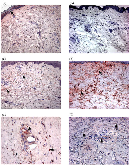
The mechanisms by which microvascular damage leads to dermal fibrosis in diffuse cutaneous systemic sclerosis (dcSSc) are unclear. We hypothesized that microvascular pericytes constitute a cellular link between microvascular damage and fibrosis by transdifferentiating into myofibroblasts. We used a combination of immunohistochemistry and double immunofluorescence labelling of frozen skin biopsies taken from normal and dcSSc patients to determine whether a phenotypic link between pericytes and myofibroblasts exists in dcSSc. Using α-smooth muscle actin, the ED-A splice variant of fibronectin (ED-A FN) and Thy-1 to identify myofibroblasts, we demonstrated the presence of myofibroblasts in fibrotic dcSSc skin. Myofibroblasts were totally absent from control skin, atrophic stage dcSSc skin and non-lesional skin. Using double immunofluorescence labelling, both myofibroblasts and pericytes were shown to express ED-A FN and Thy-1 in dcSSc skin but not in control skin. Proliferating cell nuclear antigen was also expressed by myofibroblasts and pericytes in dcSSc skin while being absent in control skin. These observations suggest that the presence of myofibroblasts may represent a transitional phase during the fibrotic stages of dcSSc and that Thy-1 +ve pericytes participate in the fibrogenic development of dcSSc by synthesizing ED-A FN, which may be associated with a proliferation and transition of pericytes and fibroblasts to myofibroblasts, thus linking microvascular damage and fibrosis.
Overview
The claims and statistics of this paper haven’t been checked yet.
Free: it reads the open-access text and checks the first 5 claims that cite a source we can check. Create a free account to check every claim.
Integrity signals
Registry
Cites
Statistics
Figures



Verification
Claims the paper makes
The claims and statistics of this paper haven’t been checked yet.
Free: it reads the open-access text and checks the first 5 claims that cite a source we can check. Create a free account to check every claim.
Claims other papers make about this one
Each is checked against this paper’s own text: does it say what the citing paper says it does?
The source backs part of the claim, not all of it — or backs it for a different population, condition or comparison.
In the citing paper
“Pericytes already express αSMA, and can become myofibroblasts if they leave their cellular niche and start to express proteins such as collagen type I and FN1-EDA.”
From Unraveling SSc Pathophysiology; The Myofibroblast (2018) · cites this paper as [27]
Experimental — accuracy not yet measuredIn the citing paper
“However, of note, in late stage fibrotic atrophic SSc skin these cells can no longer be detected (27).”
From Unraveling SSc Pathophysiology; The Myofibroblast (2018) · cites this paper as [27]
In the citing paper
“That this process occurs in SSc is suggested by a study that shows that pericytes in SSc skin, but not in healthy skin, express FN1-EDA and other myofibroblast markers (27).”
From Unraveling SSc Pathophysiology; The Myofibroblast (2018) · cites this paper as [27]
Self-check
Percentages, sums and reported differences re-computed from the paper’s own numbers.
The self-check needs the paper’s full text.
Citations
Cites
Myofibroblasts and mechano-regulation of connective tissue remodelling
Tomasek, J. J., Gabbiani, G., Hinz, B., Chaponnier, C., & Brown, R. A. (2002) · Nature Reviews Molecular Cell Biology · doi:10.1038/nrm809 · Cited by 4,290 · cited 1×
“Originally described in wound tissue, the primary role of myofibroblasts is contraction of early granulation tissue [6].”
BackgroundScleroderma (systemic sclerosis): classification, subsets and pathogenesis
(2001) · Classic Papers in Rheumatology · doi:10.3109/9780203214237-95 · Cited by 3,950
Self-citationThe myofibroblast in wound healing and fibrocontractive diseases
Gabbiani, G. (2003) · The Journal of Pathology · doi:10.1002/path.1427 · Cited by 1,630 · cited 1×
“Like pericytes, myofibroblasts express alpha smooth muscle actin (αSMA) and are strongly associated with fibrotic tissue [5].”
BackgroundDesmoulière, A., Redard, M., Darby, I. A., & Gabbiani, G. (1995) · Europe PMC (PubMed Central) · Cited by 1,448
PCNA: structure, functions and interactions
Kelman, Z. (1997) · Oncogene · doi:10.1038/sj.onc.1200886 · Cited by 929 · cited 1×
“Proliferating cells were labelled with a rabbit polyclonal antibody against proliferating cell nuclear antigen (PCNA) (Abcam, UK) [34].”
MethodSerini, G., Bochaton-Piallat, M.-L., Ropraz, P., Geinoz, A., Borsi, L., Zardi, L., & Gabbiani, G. (1998) · The Journal of Cell Biology · doi:10.1083/jcb.142.3.873 · Cited by 847 · cited 1×
“Thus, the de novo synthesis of ED-A FN appears to be a pre-requisite of α-SMA expression and myofibroblast differentiation [ 10 ].”
BackgroundBone marrow–derived progenitor cells in pulmonary fibrosis
Hashimoto, N., Jin, H., Liu, T., Chensue, S. W., & Phan, S. H. (2004) · Journal of Clinical Investigation · doi:10.1172/jci18847 · Cited by 720 · cited 1×
“This corroborates two recent studies of murine lung fibrosis in which collagen-synthesizing cells were found to be distinct from α-SMA+ve myofibroblasts [ 42 ,43] and a previous analysis of dcSSc skin, in which the presence of myofibroblasts did not correlate with α1(I) procollagen mRNA [14].”
ResultHinz, B., Mastrangelo, D., Iselin, C. E., Chaponnier, C., & Gabbiani, G. (2001) · The American Journal of Pathology · doi:10.1016/s0002-9440(10)61776-2 · Cited by 640 · cited 1×
“As ED-A FN is a pre-requisite for myofibroblast formation [9], the expression of ED-A FN by pericytes is likely to be of significance in the differentiation of perivascular fibroblasts and pericytes into myofibroblasts, and may represent a key step in linking microvascular damage and fibrosis.”
BackgroundBone marrow–derived progenitor cells in pulmonary fibrosis
Hashimoto, N., Jin, H., Liu, T., et al. (2004) · Journal of Clinical Investigation · doi:10.1172/jci200418847 · Cited by 621
Nailfold videocapillaroscopy assessment of microvascular damage in systemic sclerosis.
Houtman, N. M. (2000) · PubMed · Cited by 601
Research works printed but not linked: 1
- Jelaska A, Korn JH: Role of apoptosis and transforming growth factor beta1 in fibroblast selection and activation in systemic sclerosis. Arthritis Rheum. 2000, 43: 2230-2239. 10.1002/1529-0131(200010)43:10<2230::AID-ANR10>3.0.CO;2-8. · doi:10.1002/1529-0131(200010)43:10<2230::aid-anr10>3.0.co;2-8
Cited by
Items 101–200 of 240
Matrix remodeling in systemic sclerosis
Leask, A. (2015) · Seminars in Immunopathology · doi:10.1007/s00281-015-0508-2 · Cited by 42
Kavian, N., Servettaz, A., Marut, W., Nicco, C., Chéreau, C., Weill, B. J., & Batteux, F. (2011) · Arthritis & Rheumatism · doi:10.1002/art.34354 · Cited by 41
Phenotype characterization of pericytes during tissue repair following low-level laser therapy
Medrado, A. R. A. P., Costa, T. F., Prado, T., Reis, S. R. D. A., & Andrade, Z. A. (2010) · Photodermatology Photoimmunology & Photomedicine · doi:10.1111/j.1600-0781.2010.00521.x · Cited by 35
“However, besides immunohistochemistry, ultrastructural features are decisive for pericytes identification (8, 14).”
BackgroundSvegliati, S., Amico, D., Spadoni, T., Fischetti, C., Finke, D., Moroncini, G., Paolini, C., Tonnini, C., Grieco, A., Rovinelli, M., et al. (2017) · Frontiers in Immunology · doi:10.3389/fimmu.2017.00075 · Cited by 43
“High levels of PDGF and PDGF receptor β (PDGFR β) have been found in skin lesions from patients with scleroderma (17, 18) and may contribute to the differentiation of perivascular pericytes into vascular smooth muscle cells, fibroblasts, and myofibroblasts (19).”
BackgroundMacro- and microvascular disease in systemic sclerosis
Kavian, N., & Batteux, F. (2015) · Vascular Pharmacology · doi:10.1016/j.vph.2015.05.015 · Cited by 40
p53 and Myofibroblast Apoptosis in Organ Fibrosis
McElhinney, K., Irnaten, M., & O'Brien, C. J. (2023) · International Journal of Molecular Sciences · doi:10.3390/ijms24076737 · Cited by 39
TRPV4 ION Channel Is Associated with Scleroderma
Goswami, R., Cohen, J., Sharma, S., Zhang, D. X., Lafyatis, R., Bhawan, J., & Rahaman, S. O. (2016) · Journal of Investigative Dermatology · doi:10.1016/j.jid.2016.10.045 · Cited by 35
Romano, E., Rosa, I., Fioretto, B. S., Matucci‐Cerinic, M., & Manetti, M. (2021) · Life · doi:10.3390/life11070610 · Cited by 39
“In particular, in the affected skin of SSc patients with the diffuse cut neous subset pericytes were proved to express type I collagen and the ED-A fibronectin splice variant, an isoform that is de novo expressed during wound healing and fibrotic changes [89], while in mouse pulmonary pericytes stimulation with TGF-β1…”
BackgroundAn update on the pathogenic role of IL-6 in rheumatic diseases
Kaneko, Y., & Takeuchi, T. (2021) · Cytokine · doi:10.1016/j.cyto.2021.155645 · Cited by 34
Stromal cells differentially regulate neutrophil and lymphocyte recruitment through the endothelium
McGettrick, H. M., Buckley, C. D., Filer, A. D., Rainger, G. E., & Nash, G. B. (2010) · Immunology · doi:10.1111/j.1365-2567.2010.03307.x · Cited by 39
Therapeutic targets in systemic sclerosis
Denton, C. P. (2007) · Arthritis Research & Therapy · doi:10.1186/ar2190 · Cited by 40
“Its importance in organbased fibrosis is unclear, but in the skin recent studies have clearly shown that platelet-derived growth factor signalling is critical to pericyte activation and that this sequence occurs as SSc progresses [15].”
BackgroundSelf-citationClinical correlation of brachial artery flow-mediated dilation in patients with systemic sclerosis
Takahashi, T., Asano, Y., Amiya, E., Hatano, M., Tamaki, Z., Takata, M., Ozeki, A. N., Watanabe, A., Kawarasaki, S., Taniguchi, T., et al. (2013) · Modern Rheumatology · doi:10.3109/14397595.2013.854064 · Cited by 32
Immortalized CNS pericytes are quiescent smooth muscle actin-negative and pluripotent
Dore‐Duffy, P., Mehedi, A., Wang, X., Bradley, M., Trotter, R., & Gow, A. (2011) · Microvascular Research · doi:10.1016/j.mvr.2011.04.003 · Cited by 31
“Purified pericytes also demonstrate high myogenic potential in culture and in vivo (Rajkumar et al., 2005; Crisan et al., 2007; Dellavalle et al., 2007; Kadoya et al., 2008).”
Fibrogenesis in nonalcoholic steatohepatitis
Minicis, S. D., & Svegliati‐Baroni, G. (2011) · Expert Review of Gastroenterology & Hepatology · doi:10.1586/egh.11.28 · Cited by 36
Wilson, S. E. (2022) · Cells · doi:10.3390/cells11020309 · Cited by 33
“These include dermal fibroblasts [51], keratinocytes via EMT [52,53], adipocytes [54], as well as pericytes [55] and fibrocytes [56] that migrate from the dermal blood vessels.”
BackgroundSystemic Sclerosis and the Scleroderma-Spectrum Disorders
Varga, J., & Denton, C. P. (2009) · Elsevier eBooks · doi:10.1016/b978-1-4160-3285-4.10077-4 · Cited by 31
Self-citationMolecular and Cellular Basis of Hypertrophic Scarring
Kwan, P. O., Desmoulière, A., & Tredget, E. E. (2018) · Elsevier eBooks · doi:10.1016/b978-0-323-47661-4.00045-9 · Cited by 31
Fibroblast abnormalities in the pathogenesis of systemic sclerosis
Usategui, A., Rey, M. J. D., & Pablos, J. L. (2011) · Expert Review of Clinical Immunology · doi:10.1586/eci.11.39 · Cited by 27
Cellular and molecular mechanisms in the pathophysiology of systemic sclerosis
Hua-Huy, T., & Dinh‐Xuan, A. T. (2015) · Pathologie Biologie · doi:10.1016/j.patbio.2015.03.003 · Cited by 26
“Today, it has been recognized that myofibroblasts come from different precursors, including pericytes in the vessel walls, adventitial fibroblasts, bone marrow-derived fibrocytes, epithelial and endothelial cells [64].”
BackgroundRosato, E., Cianci, R., Barbano, B., Menghi, G., Gigante, A., ROSSI, C., Zardi, E. M., Amoroso, A., Pisarri, S., & Salsano, F. (2009) · Acta Pharmacologica Sinica · doi:10.1038/aps.2009.128 · Cited by 26
Etich, J., Bergmeier, V., Pitzler, L., & Brachvogel, B. (2016) · Connective Tissue Research · doi:10.1080/03008207.2016.1210606 · Cited by 28
Extreme Diversity of the Human Vascular Mesenchymal Cell Landscape
Bruijn, L. E., van den Akker, B. E. W. M., Rhijn, C. M. J., Hamming, J. F., & Lindeman, J. H. N. (2020) · Journal of the American Heart Association · doi:10.1161/jaha.120.017094 · Cited by 27
Giulio Gabbiani and the discovery of myofibroblasts
Ribatti, D., & Tamma, R. (2019) · Inflammation Research · doi:10.1007/s00011-018-01211-x · Cited by 26
Nakamura, M., & TOKURA, Y. (2010) · British Journal of Dermatology · doi:10.1111/j.1365-2133.2010.10021.x · Cited by 25
The role of myofibroblasts in the progression of oral submucous fibrosis: A systematic review
Wadhwan, V., Venkatesh, A., Reddy, V., & Malik, S. S. (2019) · Journal of Oral and Maxillofacial Pathology · doi:10.4103/jomfp.jomfp_238_18 · Cited by 24
The benefits and prospects of interleukin-6 inhibitor on systemic sclerosis
Shima, Y. (2018) · Modern Rheumatology · doi:10.1080/14397595.2018.1559909 · Cited by 24
“Interestingly, pathological examination revealed that myofibroblasts disappeared from the tissue of atrophic-stage SSc, while they were detected in the dermis of progressive-stage SSc [48].”
BackgroundMicrovascular targets for anti-fibrotic therapeutics.
Pu, K.-M., Sava, P., & Gonzalez, A. L. (2013) · PubMed Central · Cited by 27
Vadasz, Z., Balbir‐Gurman, A., Meroni, P. L., Farge, D., Levi, Y., Ingegnoli, F., Braun‐Moscovici, Y., Rosner, I., Slobodin, G., Rozenbaum, M., et al. (2019) · Lara D. Veeken · doi:10.1093/rheumatology/kez035 · Cited by 23
Li, F.-F., Chen, B.-J., Li, W., Li, L., Zha, M., Zhou, S., Bachem, M. G., & Sun, Z.-L. (2015) · Journal of Diabetes Research · doi:10.1155/2016/6924593 · Cited by 24
“Evidence for the involvement of PSCs in islet fibrosis is mainly based on staining of α-SMA and/or GFAP for marker detection [25, 48, 49].”
BackgroundA System Out of Breath: How Hypoxia Possibly Contributes to the Pathogenesis of Systemic Sclerosis
van Hal, T. W., van Bon, L. G., & Radstake, T. R. D. J. (2011) · International Journal of Rheumatology · doi:10.1155/2011/824972 · Cited by 24
“These cells also show expression of the cell-surface glycoprotein Thy1 [57], which is essential in the differentiation of fibroblasts to myofibroblasts [58].”
BackgroundPharmacotherapy of systemic sclerosis
Postlethwaite, A. E., Harris, L. J., Raza, S. H., Kodura, S., & Akhigbe, T. (2010) · Expert Opinion on Pharmacotherapy · doi:10.1517/14656561003592177 · Cited by 25
“2 Myofibroblasts in systemic sclerosis Myofibroblasts that are characterized by expression of a smooth muscle actin, ability to contract granulation tissue in vivo and collagen gels in vitro, and expression of the EDA splice variant of fibronectin are found in biopsies of lesional skin of patients with dcSSc in increa…”
BackgroundCells from the skin of patients with systemic sclerosis secrete chitinase 3-like protein 1
Ho, Y. Y., Baron, M. G., Recklies, A. D., Roughley, P. J., & Mort, J. S. (2014) · BBA Clinical · doi:10.1016/j.bbacli.2013.12.001 · Cited by 22
“These cell types include bonemarrow-derivedmesenchymal stem cells [41], pericytes [42], endothelial progenitor cells [43], and circulating progenitor cells, such as fibrocytes and monocytes [41, 44, 45].”
BackgroundCorallo, C., Cheleschi, S., Cutolo, M., Soldano, S., Fioravanti, A., Volpi, N., Franci, D., Nuti, R., & Giordano, N. G. (2019) · Arthritis Research & Therapy · doi:10.1186/s13075-019-1931-x · Cited by 26
Getting Out of a Sticky Situation: Targeting the Myofibroblast in Scleroderma
Leask, A. (2012) · The Open Rheumatology Journal · doi:10.2174/1874312901206010163 · Cited by 25
“These results are particularly intriguing as, as described above, myofibroblasts in fibrosis (including in scleroderma) have been postulated to originate from microvascular pericytes; these sets of observations are supportive of this interesting possibility [6, 23, 24] (please see below).”
BackgroundLeask, A. (2021) · Wound Repair and Regeneration · doi:10.1111/wrr.12889 · Cited by 23
“of the skin of scleroderma patients, but not in neighboring, clinicallyunaffected tissue.(25) Lesional and nonlesional SSc retain this pheno-”
BackgroundThe Role of Pro-fibrotic Myofibroblasts in Systemic Sclerosis: From Origin to Therapeutic Targeting
Romano, E., Rosa, I., Fioretto, B. S., Matucci‐Cerinic, M., & Manetti, M. (2021) · Current Molecular Medicine · doi:10.2174/0929867328666210325102749 · Cited by 26
Experimental systems to study the origin of the myofibroblast in peritoneal fibrosis
Padwal, M., & Margetts, P. J. (2016) · Kidney Research and Clinical Practice · doi:10.1016/j.krcp.2016.07.003 · Cited by 22
“This was also supported in models of fibrosis such as scleroderma [25] and liver and renal fibrosis [12,26].”
MethodTargeting the jagged/notch pathway: a new treatment for fibrosis?
Leask, A. (2010) · Journal of Cell Communication and Signaling · doi:10.1007/s12079-010-0101-3 · Cited by 21
“[Although the importance of pericytes in this process is also understood ( Rajkumar et al. 1999, 2005; Shiwen et al. 2009; Liu et al. 2010; Humphreys et al. 2010; Zeisberg and Duffield 2010)].”
Engineering Advanced In Vitro Models of Systemic Sclerosis for Drug Discovery and Development
Pieri, A. D., Korman, B. D., Jüngel, A., & Wuertz‐Kozak, K. (2021) · Advanced Biology · doi:10.1002/adbi.202000168 · Cited by 30
Leflunomide prevents ROS-induced systemic fibrosis in mice
Morin, F., Kavian, N., Chouzenoux, S., Cerles, O., Nicco, C., Chéreau, C., & Batteux, F. (2017) · Free Radical Biology and Medicine · doi:10.1016/j.freeradbiomed.2017.03.035 · Cited by 20
Oleate Induces a Myofibroblast-Like Phenotype in Mesangial Cells
Mishra, R., & Simonson, M. S. (2007) · Arteriosclerosis Thrombosis and Vascular Biology · doi:10.1161/atvbaha.107.157339 · Cited by 25
Emerging targets for the treatment of scleroderma
Leask, A. (2012) · Expert Opinion on Emerging Drugs · doi:10.1517/14728214.2012.678833 · Cited by 20
“The molecular mechanism underlying the persistence of the myofibroblast has been extensively studied, and appears to be a dysregulated ‘fibroproliferative’ TGF-b/ endothelin-1/CCN2 signaling pathway [13-17].”
BackgroundYuasa, T., Juniantito, V., Ichikawa, C., Yano, R., Izawa, T., Kuwamura, M., & Yamate, J. (2012) · Experimental and Toxicologic Pathology · doi:10.1016/j.etp.2012.07.005 · Cited by 17
Immunophenotypical Characterization of Macrophages in Rat Bleomycin-Induced Scleroderma
Juniantito, V., Izawa, T., Yuasa, T., Ichikawa, C., Yano, R., Kuwamura, M., & Yamate, J. (2012) · Veterinary Pathology · doi:10.1177/0300985812450718 · Cited by 18
“In skin with fibrotic lesions, spindle-shaped cells reacting to aSMA are regarded as myofibroblasts.(33) In control skin, no myofibroblasts were found in the dermis, although vascular smooth muscles and arrector pilli muscles reacted to a-SMA.”
BackgroundResultInfluentialSingle-Cell Analysis of ADSC Interactions with Fibroblasts and Endothelial Cells in Scleroderma Skin
Frommer, M. L., Langridge, B., Awad, L., Jasionowska, S., Denton, C. P., Abraham, D., Abu‐Hanna, J., & Butler, P. E. M. (2023) · Cells · doi:10.3390/cells12131784 · Cited by 17
Self-citationServaas, N. H., Hiddingh, S., Chouri, E., Wichers, C. G. K., Affandi, A. J., Ottria, A., Bekker, C. P. J., Cossu, M., Silva‐Cardoso, S. C., van der Kroef, M., et al. (2022) · Arthritis & Rheumatology · doi:10.1002/art.42319 · Cited by 18
“The resulting mix of inflammatory molecules induces the differentiation of resident epithelium, endothelium, monocytes and fibroblasts into myofibroblast that deposit excessive amounts of extracellular matrix, eventually leading to permanent tissue scarring[6], [7].”
BackgroundTuleta, I., & Frangogiannis, N. G. (2025) · American Journal of Physiology-Cell Physiology · doi:10.1152/ajpcell.00403.2025 · Cited by 20
Calponin Expression in Renal Tubulointerstitial Fibrosis Induced in Rats by Cisplatin
Yuasa, T., Yano, R., Izawa, T., Kuwamura, M., & Yamate, J. (2014) · Journal of Toxicologic Pathology · doi:10.1293/tox.2013-0048 · Cited by 15
Janson, D., Saintigny, G., Zeypveld, J., Mahé, C., & Ghalbzouri, A. E. (2014) · European Journal of Dermatology · doi:10.1684/ejd.2014.2312 · Cited by 16
“Staining with alpha smooth muscle actin (α-SMA), a marker of myofibroblasts and smooth muscle tissue, is present in several skin appendages, such as blood vessels and smooth muscle cells, but not in fibroblasts (10, 11).”
BackgroundJuniantito, V., Izawa, T., Yuasa, T., Ichikawa, C., Tanaka, M., Kuwamura, M., & Yamate, J. (2012) · Experimental and Toxicologic Pathology · doi:10.1016/j.etp.2012.05.002 · Cited by 15
Molecular and Cellular Pathways as Treatment Targets for Biologic Therapies in Systemic Sclerosis
Dimitroulas, T., Daoussis, D., Garyfallos, A., Sfikakis, P. P., & Kitas, G. D. (2015) · Current Medicinal Chemistry · doi:10.2174/0929867322666150209161224 · Cited by 15
Hashimoto, A., Karim, M., Kuramochi, M., Izawa, T., Kuwamura, M., & Yamate, J. (2020) · Toxicologic pathology (Print) · doi:10.1177/0192623319893310 · Cited by 12
Rigau, A. R., Liang, M., Devakumar, V., Neelagar, R., Matei, A., Györfi, A., Bergmann, C., Filla, T., Fedorchenko, V., Schett, G. A., et al. (2024) · Annals of the Rheumatic Diseases · doi:10.1136/ard-2024-226336 · Cited by 20
“32 33 We further analysed alterations of the distribution of subpopulations between the upper and lower dermis in patients with SSc compared with healthy individuals (figure 4C and D).”
The Role of Mesenchymal Stromal Cells in Tissue Repair and Fibrosis
Akasaka, Y. (2022) · Advances in Wound Care · doi:10.1089/wound.2021.0037 · Cited by 17
“140 The role of pericytes as myofibroblast precursors has been indicated by studies of fibrogenesis in several organs.”
Krishna, C. V., Singh, J., Thangavel, C., & Rattan, S. C. (2016) · American Journal of Physiology-Gastrointestinal and Liver Physiology · doi:10.1152/ajpgi.00445.2015 · Cited by 13
Leask, A. (2016) · Methods in molecular biology · doi:10.1007/978-1-4939-6430-7_34 · Cited by 13
Pharmacological Treatment of Fibrosis: a Systematic Review of Clinical Trials
Siani, A. (2020) · SN Comprehensive Clinical Medicine · doi:10.1007/s42399-020-00292-2 · Cited by 14
“Typical examples of mesenchymal precursors of myofibroblasts include fibrocytes, pericytes, SM cells, as well as adipose and bone marrow-derived stromal/ stem cells [19, 20].”
BackgroundMechanisms of Vascular Disease
Manetti, M., & Kahaleh, B. (2016) · Springer eBooks · doi:10.1007/978-3-319-31407-5_16 · Cited by 12
Talotta, R., Atzeni, F., Ditto, M. C., Gerardi, M. C., Batticciotto, A., Bongiovanni, S. F., & Sarzi‐Puttini, P. (2017) · Journal of Scleroderma and Related Disorders · doi:10.5301/jsrd.5000254 · Cited by 11
Zanin-Silva, D. C., Santana-Gonçalves, M., Vasconcelos, M., Lima-Júnior, J. R., Dias, J. B. E., Moraes, D. A., Covas, D. T., Malmegrim, K. C. R., Ramalho, L. N. Z., & Oliveira, M. C. (2022) · Arthritis Research & Therapy · doi:10.1186/s13075-022-02779-w · Cited by 13
“In wound healing processes and systemic sclerosis, myofibroblasts express α-SMA temporarily during their differentiation, and staining decreases to undetectable levels as fibrosis progresses [28, 29].”
BackgroundHutchenreuther, J., & Leask, A. (2016) · Cell and Tissue Research · doi:10.1007/s00441-016-2419-5 · Cited by 13
“Staining of clinical samples from patients with diffuse cutaneous systemic sclerosis (dcSSc) showed that both myofibroblasts and pericytes in their dermis expressed α-SMA and the EDA domain containing the isoform of fibronectin responsible for myofibroblast activation in fibrosis (Rajkumar et al. 2005).”
Mechanisms of Vascular Disease
Kahaleh, B., & Mulligan‐Kehoe, M. J. (2011) · Springer eBooks · doi:10.1007/978-1-4419-5774-0_19 · Cited by 12
Yuasa, T., Izawa, T., Kuwamura, M., & Yamate, J. (2010) · Journal of Toxicologic Pathology · doi:10.1293/tox.23.1 · Cited by 11
Extracellular matrix bioengineering and systems biology approaches in liver disease
Nieto, N., & Lütolf, M. P. (2011) · Systems and Synthetic Biology · doi:10.1007/s11693-011-9085-4 · Cited by 12
“cells (De Minicis et al. 2007; Tuchweber et al. 1996; Knittel et al. 1999; Hinz 2007; Rajkumar et al. 2005; Desmouliere et al. 2003); or bone marrow-derived myofibroblasts (Forbes”
BackgroundNew Light is Shed on the Enigmatic Origin of the Lung Myofibroblast
Rosas, I. O., Kottman, R. M., & Sime, P. J. (2013) · American Journal of Respiratory and Critical Care Medicine · doi:10.1164/rccm.201308-1494ed · Cited by 12
“To date, pericytes have been implicated in the pathogenesis of renal (11), dermal (12), and spinal cord (13) fibrosis, but their role in lung disease, especially scarring, has yet to be defined.”
BackgroundSingle-cell RNA sequencing reveals dysregulated fibroblast subclusters in prurigo nodularisPreprint
Patel, J. R., Joel, M. Z., Lee, K. K., Kambala, A., Cornman, H. L., Oladipo, O. O., Taylor, M. T., Deng, J., Parthasarathy, V., Cravero, K., et al. (2023) · bioRxiv (Cold Spring Harbor Laboratory) · doi:10.1101/2023.01.29.526050 · Cited by 13
A Positive Loop Formed by SOX11 and Periostin Upregulates TGF-β Signals Leading to Skin Fibrosis
Nanri, Y., Nunomura, S., Honda, Y., Takedomi, H., Yamaguchi, Y., & Izuhara, K. (2022) · Journal of Investigative Dermatology · doi:10.1016/j.jid.2022.12.008 · Cited by 10
Immunogenetics of Systemic Sclerosis
Gumkowska-Sroka, O., Kotyla, K., & Kotyla, P. J. (2024) · Genes · doi:10.3390/genes15050586 · Cited by 10
“Furthermore, IL-6 plays a pivotal role in fostering fibrotic alterations, directly facilitating the differentiation of fibroblasts into myofibroblasts [134].”
BackgroundSchneider, G. S., Bubel, M., Pohlemann, T., & Oberringer, M. (2015) · Molecular and Cellular Biochemistry · doi:10.1007/s11010-015-2451-x · Cited by 11
“Fibroblasts, MF and pericytes, especially MFP, may thus contribute to tissue fibrosis [9, 38].”
BackgroundHeterogeneous muscle gene expression patterns in patients with massive rotator cuff tears
Gibbons, M. C., Fisch, K. M., Pichika, R., Cheng, T., Engler, A. J., Schenk, S., Lane, J. G., Singh, A., & Ward, S. R. (2018) · PLoS ONE · doi:10.1371/journal.pone.0190439 · Cited by 11
The Role of Autophagy and Apoptosis in Affected Skin and Lungs in Patients with Systemic Sclerosis
Spasovski, V., Andjelković, M., Parezanovic, M., Komazec, J., Ugrin, M., Klaassen, K., & Stojiljković, M. (2023) · International Journal of Molecular Sciences · doi:10.3390/ijms241311212 · Cited by 8
“One of the sources of myofiroblasts are pericytes, specialized mesenchymal cells, associated with the walls of small blood vessels and smooth muscle cells of blood vessels [31,32].”
BackgroundRenaud, L., Wilson, C. L., Lafyatis, R., Schnapp, L. M., & Feghali‐Bostwick, C. A. (2024) · iScience · doi:10.1016/j.isci.2024.110010 · Cited by 9
“18,19 Additionally, peri-cytes have the ability to differentiate into different lineages and are often considered as mesenchymal stem cells of crucial importance in SSc. 20 Interestingly, SSc pericytes, fibroblasts, and myofibroblasts all express A disintegrin and metalloproteinase domain 12 ( ADAM12…”
Molecular and cellular basis of hypertrophic scarring
Kwan, P. O., Desmoulière, A., & Tredget, E. E. (2012) · Elsevier eBooks · doi:10.1016/b978-1-4377-2786-9.00045-x · Cited by 11
Ultrastructural and functional characterization of myofibroblasts in lung diseases
Karvonen, H. M. (2014) · Cited by 7
Arioka, M., Matsunaga, H., Ishikane, S., Ito, K., Hashimoto, K., Wang, Y., Igawa, K., & Takahashi‐Yanaga, F. (2025) · Life Sciences · doi:10.1016/j.lfs.2025.123914 · Cited by 7
Cellular Transdifferentiation: A Crucial Mechanism of Fibrosis in Systemic Sclerosis
Jiménez, S. A., & Piera-Velázquez, S. (2023) · Current Rheumatology Reviews · doi:10.2174/0115733971261932231025045400 · Cited by 7
Didier, K., Robbins, A., Antonicelli, F. D., Pham, B. N., Giusti, D., & Servettaz, A. (2019) · La Revue de Médecine Interne · doi:10.1016/j.revmed.2019.05.016 · Cited by 8
Etiology and Pathogenesis of Scleroderma
Varga, J. (2013) · Elsevier eBooks · doi:10.1016/b978-1-4377-1738-9.00083-9 · Cited by 8
Etiology and Pathogenesis of Scleroderma
Varga, J. (2016) · Elsevier eBooks · doi:10.1016/b978-0-323-31696-5.00083-8 · Cited by 9
Can, G., Sarıoğlu, S., Bırlık, M., Kenar, G., Soysal, Ö., Solmaz, D., Gerdan, V., Önen, F., Akkoç, N., & Akar, S. (2021) · TURKISH JOURNAL OF MEDICAL SCIENCES · doi:10.3906/sag-2012-25 · Cited by 8
“Among these cells, only endothelial cells are mesenchymal cells subject to apoptosis, however pericytes and smooth muscle cells rapidly proliferate [6].”
BackgroundYamamoto, A., Saito, T., Hosoya, T., Kawahata, K., Asano, Y., Sato, S., Mizoguchi, F., Yasuda, S., & Kohsaka, H. (2021) · Arthritis & Rheumatology · doi:10.1002/art.42042 · Cited by 7
“In addition, increased numbers of proliferating fibroblasts in the fibrotic skin lesions of patients with SSc have been reported, especially in those with early SSc (9).”
BackgroundTam, A. Y. Y., Khan, K., Xu, S., Bergin, M., Huang, L., Arroyo-Colon, E., Cheng, D., Verderio, E. A. M., Ong, V. H., Denton, C. P., et al. (2025) · Arthritis & Rheumatology · doi:10.1002/art.43104 · Cited by 7
Self-citationPathophysiology of Fibrosis in Systemic Sclerosis
Trojanowska, M., & Varga, J. (2016) · Springer eBooks · doi:10.1007/978-3-319-31407-5_18 · Cited by 6
The Expression and Function of ABC Transporters at the Blood-Brain Barrier
Zhang, W., Bamji‐Mirza, M., Chang, N., Haqqani, A. S., & Stanimirovic, D. B. (2015) · CRC Press eBooks · doi:10.1201/b18606-8 · Cited by 5
Role of programmed cell death 4 in myofibroblast differentiation in oral submucous fibrosis
Desai, K. M., Kale, A. D., Angadi, P. V., Datar, U. V., Belaldavar, C., & Arany, P. R. (2021) · Journal of Oral and Maxillofacial Pathology · doi:10.4103/jomfp.jomfp_86_21 · Cited by 5
“[25] While the precise origin of the myofibroblasts remains unclear, phenotypic similarities have suggested that they arise from peri‐vascular (pericyte) and stromal fibroblasts.[26] A key signaling pathway, TGF‐β1, has been well established as a primary inducer of the αSMA differentiated myofibroblast phenotype.”
BackgroundBarosi, G., & Gale, R. P. (2011) · Leukemia Research · doi:10.1016/j.leukres.2011.01.013 · Cited by 5
Khanna, D., Kramer, F., Höfler, J., Ghadessi, M., Sandner, P., Allanore, Y., Denton, C. P., Kuwana, M., Matucci‐Cerinic, M., Pope, J. E., et al. (2024) · Rheumatology · doi:10.1093/rheumatology/keae150 · Cited by 7
Self-citationYin, M., Sun, L., Wu, S., Ma, J., Zhang, W., Ji, X., Tang, Z., Zhang, X., Yang, Y., Zhang, X., et al. (2024) · Cell Proliferation · doi:10.1111/cpr.13722 · Cited by 5
“Flow cytometry analysis further confirmed that the number of ACTA2 + PDGFRA (cid:1) cells expressing THY1 but not Ly6A, representing perivascular pericytes, 42,43 significantly increased in the centre scar tissue compared to the edge or distal control skin (Figure 2G,H; Figure S1B).”
Fibrogenesis, novel lessons from animal models
Langhe, E. D., & Lories, R. J. U. (2015) · Seminars in Immunopathology · doi:10.1007/s00281-015-0510-8 · Cited by 7
“mal cells, associated with small vessel walls) appear activated and express markers suggesting a transitional stage to myofibroblast transdifferentiation [50].”
BackgroundTargeting Pericytes to Improve Wound Healing Outcomes
Bodnar, R. J., & Satish, L. (2018) · Current Pathobiology Reports · doi:10.1007/s40139-018-0168-8 · Cited by 3
Clinical correlation of brachial artery flow-mediated dilation in patients with systemic sclerosis
Takahashi, T., Asano, Y., Amiya, E., Hatano, M., Tamaki, Z., Takata, M., Ozeki, A. N., Watanabe, A., Kawarasaki, S., Taniguchi, T., et al. (2013) · Modern Rheumatology · doi:10.1007/s10165-013-0867-2 · Cited by 3
“wealth of evidence suggests that the functional and morphological changes of blood vessels resulting from aberrant angiogenesis underlie the mechanism of vasculopathy and fibrosis in this disorder [2–4].”
BackgroundStromal Cells and Tumor Microenvironment
Varró, A. (2009) · Springer eBooks · doi:10.1007/978-0-387-69182-4_23 · Cited by 3
Varga, J. (2011) · Springer eBooks · doi:10.1007/978-1-4419-5774-0_21 · Cited by 2
“Activated pericytes can transdifferentiate into collagen-producing fibroblasts and myofibroblasts, thus linking microvascular injury and fibrosis [ 13 ] .”
BackgroundEditorial: Fate of Fat Tissue Adipocytes: Do They Transform Into Myofibroblasts in Scleroderma?
Bogatkevich, G. S. (2014) · Arthritis & Rheumatology · doi:10.1002/art.38988 · Cited by 2
“Generation of myofibroblasts in diffuse cutaneous systemic sclerosis as a result of increased proliferation and activation of microvascular pericytes has been suggested (3).”
BackgroundChompoowong, R., Wongphoom, J., Kitkumthorn, N., Saonanon, P., Pruksakorn, V., Palaga, T., Hirankarn, N., van Hagen, P. M., Dik, W. A., & Virakul, S. (2025) · Investigative Ophthalmology & Visual Science · doi:10.1167/iovs.66.9.30 · Cited by 1
“15 However, these myofibroblast markers and their roles in the pathogenesis of GO remain elusive.”
Pericytes: Adaptable Vascular Progenitors
Hyde, G. D., & Canfield, A. E. (2011) · Springer eBooks · doi:10.1007/978-0-85729-920-8_1 · Cited by 1
A case of systemic scleroderma complicating pulmonary hypertension
Kurosawa, R., Umebayashi, H., Imagawa, T., Katakura, S., Mori, M., Aihara, Y., & Yokota, S. (2006) · Japanese Journal of Clinical Immunology · doi:10.2177/jsci.29.378 · Cited by 1
Cutaneous Fibrosis and Normal Wound Healing
Hamburg‐Shields, E., Myung, P. S., & Cowper, S. E. (2017) · Springer eBooks · doi:10.1007/978-3-319-29785-9_32 · Cited by 2
The Elusive Multipotent Microvascular Pericyte
Dore‐Duffy, P., Esen, N., & Serkin, Z. (2015) · CRC Press eBooks · doi:10.1201/b18606-6 · Cited by 1
Cellular and Molecular Mechanisms of Fibrosis in Systemic Sclerosis
Trojanowska, M., Varga, J., & Lagares, D. (2024) · Springer eBooks · doi:10.1007/978-3-031-40658-4_18 · Cited by 2
Metadata
- Authors
- Rajkumar, V. S., Howell, K., Csiszar, K., Denton, C. P., Black, C. M., & Abraham, D. J.
- Affiliations
- The Royal Free Hospital (GB)
- Corresponding author
- Abraham, David J
- Venue
- Arthritis Research & Therapy
- Published
- 2005-07-21
- Volume
- 7
- Issue
- 5
- Pages
- R1113-23
- ISSN
- 1478-6354 (ISSN-L)
- PMC
- PMC1257439
- OpenAlex
- W2138211373
- Cited by
- 242
- References
- 48
- Citations by year
- 2026: 6 · 2025: 4 · 2024: 8 · 2023: 9 · 2022: 14 · 2021: 16 · 2020: 9 · 2019: 13 · 2018: 9 · 2017: 6 · 2016: 14 · 2015: 18 · 2014: 14 · 2013: 13 · 2012: 13
- FWCI
- 7.04
- Type
- journal article
- Language
- en
- Open access
- gold
- doi.org
- Licence
- cc-by
- creativecommons.org/licenses/by/2.0/ (tdm)
- creativecommons.org/licenses/by/2.0/ (vor)
- Locations
- Arthritis Research & Therapy (publishedVersion, cc-by)
- PubMed Central (publishedVersion)
- PubMed (publishedVersion)
- UCL Discovery (University College London) (submittedVersion)
- Funders
- Rosetrees Trust
- Jean Shanks Foundation
- National Institutes of Health
- National Institute of Arthritis and Musculoskeletal and Skin Diseases (S11AR047713, AR47713)
- Topics
- Health Sciences › Medicine › Rheumatology › Systemic Sclerosis and Related Diseases
- Health Sciences › Medicine › Rheumatology › Dupuytren's Contracture and Treatments
- Life Sciences › Biochemistry, Genetics and Molecular Biology › Genetics › Connective tissue disorders research
- Keywords
- diffuse cutaneous systemic sclerosis, myofibroblasts, pericytes, fibroblasts, skin fibrosis, microvascular damage, ED-A fibronectin, CD90, alpha-smooth muscle actin, proliferating cell nuclear antigen
- MeSH terms
- Actins, Adult, Aged, Collagen, Female, Fibroblasts, Fibronectins, Fibrosis, Humans, Male, Middle Aged, Phenotype, Scleroderma, Systemic, Skin, Biomarkers, Thy-1 Antigens, Proliferating Cell Nuclear Antigen, Cell Lineage, Pericytes, Myoblasts
About these checks
Registry, retraction, Cites and statistics results are computed, not judged, with one exception: a printed reference that no index lists is read once by a model before it counts as a citation error. Claims are extracted by a model and kept only when their quote appears verbatim in the text; each cited source is then checked, with a model’s help, for whether it says what the claim relies on. Those verdicts can be wrong — read the claim’s quote and the cited source before relying on one.
Statistics and arithmetic are recomputed, not judged.
Confidence bands are shown only for fields calibrated on our annotated test set. None is calibrated yet, so no band is shown.
What counts as a claim ›How we measure confidence ›How we check this ›
Are you an author, or do you see a problem with this page? Request a review
Data sources
Bibliographic data: Semantic Scholar, OpenAlex, Crossref.
Contains information from the Semantic Scholar Academic Graph Datasets, made available under the ODC Attribution License (ODC-BY 1.0).
Semantic Scholar’s data is described in: Kinney, R., et al. (2023). The Semantic Scholar Open Data Platform. arXiv:2301.10140.
