

Shared expression of phenotypic markers in systemic sclerosis indicates a convergence of pericytes and fibroblasts to a myofibroblast lineage in fibrosis
Open accessThe DOI matches its Crossref record; 49 of the 49 references it cites were found in a registry or an index.
Abstract
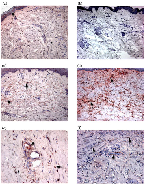
The mechanisms by which microvascular damage leads to dermal fibrosis in diffuse cutaneous systemic sclerosis (dcSSc) are unclear. We hypothesized that microvascular pericytes constitute a cellular link between microvascular damage and fibrosis by transdifferentiating into myofibroblasts. We used a combination of immunohistochemistry and double immunofluorescence labelling of frozen skin biopsies taken from normal and dcSSc patients to determine whether a phenotypic link between pericytes and myofibroblasts exists in dcSSc. Using α-smooth muscle actin, the ED-A splice variant of fibronectin (ED-A FN) and Thy-1 to identify myofibroblasts, we demonstrated the presence of myofibroblasts in fibrotic dcSSc skin. Myofibroblasts were totally absent from control skin, atrophic stage dcSSc skin and non-lesional skin. Using double immunofluorescence labelling, both myofibroblasts and pericytes were shown to express ED-A FN and Thy-1 in dcSSc skin but not in control skin. Proliferating cell nuclear antigen was also expressed by myofibroblasts and pericytes in dcSSc skin while being absent in control skin. These observations suggest that the presence of myofibroblasts may represent a transitional phase during the fibrotic stages of dcSSc and that Thy-1 +ve pericytes participate in the fibrogenic development of dcSSc by synthesizing ED-A FN, which may be associated with a proliferation and transition of pericytes and fibroblasts to myofibroblasts, thus linking microvascular damage and fibrosis.
Overview
The claims and statistics of this paper haven’t been checked yet.
Free: it reads the open-access text and checks the first 5 claims that cite a source we can check. Create a free account to check every claim.
Integrity signals
Registry
Cites
Statistics
Figures



Verification
Claims the paper makes
The claims and statistics of this paper haven’t been checked yet.
Free: it reads the open-access text and checks the first 5 claims that cite a source we can check. Create a free account to check every claim.
Claims other papers make about this one
Each is checked against this paper’s own text: does it say what the citing paper says it does?
The source backs part of the claim, not all of it — or backs it for a different population, condition or comparison.
In the citing paper
“Pericytes already express αSMA, and can become myofibroblasts if they leave their cellular niche and start to express proteins such as collagen type I and FN1-EDA.”
From Unraveling SSc Pathophysiology; The Myofibroblast (2018) · cites this paper as [27]
Experimental — accuracy not yet measuredIn the citing paper
“However, of note, in late stage fibrotic atrophic SSc skin these cells can no longer be detected (27).”
From Unraveling SSc Pathophysiology; The Myofibroblast (2018) · cites this paper as [27]
In the citing paper
“That this process occurs in SSc is suggested by a study that shows that pericytes in SSc skin, but not in healthy skin, express FN1-EDA and other myofibroblast markers (27).”
From Unraveling SSc Pathophysiology; The Myofibroblast (2018) · cites this paper as [27]
Self-check
Percentages, sums and reported differences re-computed from the paper’s own numbers.
The self-check needs the paper’s full text.
Citations
Cites
Myofibroblasts and mechano-regulation of connective tissue remodelling
Tomasek, J. J., Gabbiani, G., Hinz, B., Chaponnier, C., & Brown, R. A. (2002) · Nature Reviews Molecular Cell Biology · doi:10.1038/nrm809 · Cited by 4,290 · cited 1×
“Originally described in wound tissue, the primary role of myofibroblasts is contraction of early granulation tissue [6].”
BackgroundScleroderma (systemic sclerosis): classification, subsets and pathogenesis
(2001) · Classic Papers in Rheumatology · doi:10.3109/9780203214237-95 · Cited by 3,950
Self-citationThe myofibroblast in wound healing and fibrocontractive diseases
Gabbiani, G. (2003) · The Journal of Pathology · doi:10.1002/path.1427 · Cited by 1,630 · cited 1×
“Like pericytes, myofibroblasts express alpha smooth muscle actin (αSMA) and are strongly associated with fibrotic tissue [5].”
BackgroundDesmoulière, A., Redard, M., Darby, I. A., & Gabbiani, G. (1995) · Europe PMC (PubMed Central) · Cited by 1,448
PCNA: structure, functions and interactions
Kelman, Z. (1997) · Oncogene · doi:10.1038/sj.onc.1200886 · Cited by 929 · cited 1×
“Proliferating cells were labelled with a rabbit polyclonal antibody against proliferating cell nuclear antigen (PCNA) (Abcam, UK) [34].”
MethodSerini, G., Bochaton-Piallat, M.-L., Ropraz, P., Geinoz, A., Borsi, L., Zardi, L., & Gabbiani, G. (1998) · The Journal of Cell Biology · doi:10.1083/jcb.142.3.873 · Cited by 847 · cited 1×
“Thus, the de novo synthesis of ED-A FN appears to be a pre-requisite of α-SMA expression and myofibroblast differentiation [ 10 ].”
BackgroundBone marrow–derived progenitor cells in pulmonary fibrosis
Hashimoto, N., Jin, H., Liu, T., Chensue, S. W., & Phan, S. H. (2004) · Journal of Clinical Investigation · doi:10.1172/jci18847 · Cited by 720 · cited 1×
“This corroborates two recent studies of murine lung fibrosis in which collagen-synthesizing cells were found to be distinct from α-SMA+ve myofibroblasts [ 42 ,43] and a previous analysis of dcSSc skin, in which the presence of myofibroblasts did not correlate with α1(I) procollagen mRNA [14].”
ResultHinz, B., Mastrangelo, D., Iselin, C. E., Chaponnier, C., & Gabbiani, G. (2001) · The American Journal of Pathology · doi:10.1016/s0002-9440(10)61776-2 · Cited by 640 · cited 1×
“As ED-A FN is a pre-requisite for myofibroblast formation [9], the expression of ED-A FN by pericytes is likely to be of significance in the differentiation of perivascular fibroblasts and pericytes into myofibroblasts, and may represent a key step in linking microvascular damage and fibrosis.”
BackgroundBone marrow–derived progenitor cells in pulmonary fibrosis
Hashimoto, N., Jin, H., Liu, T., et al. (2004) · Journal of Clinical Investigation · doi:10.1172/jci200418847 · Cited by 621
Nailfold videocapillaroscopy assessment of microvascular damage in systemic sclerosis.
Houtman, N. M. (2000) · PubMed · Cited by 601
Research works printed but not linked: 1
- Jelaska A, Korn JH: Role of apoptosis and transforming growth factor beta1 in fibroblast selection and activation in systemic sclerosis. Arthritis Rheum. 2000, 43: 2230-2239. 10.1002/1529-0131(200010)43:10<2230::AID-ANR10>3.0.CO;2-8. · doi:10.1002/1529-0131(200010)43:10<2230::aid-anr10>3.0.co;2-8
Cited by
Hinz, B., Phan, S. H., Thannickal, V. J., Galli, A., Bochaton-Piallat, M.-L., & Gabbiani, G. (2007) · The American Journal of Pathology · doi:10.2353/ajpath.2007.070112 · Cited by 2,068
Formation and function of the myofibroblast during tissue repair
Hinz, B. (2007) · Journal of Investigative Dermatology · doi:10.1038/sj.jid.5700613 · Cited by 1,658
“Pericytes and SMC from the vasculature have been proposed as another local myofibroblast source in scleroderma (Rajkumar et al., 2005) as well as liver and glomerular fibrosis (Desmouliere et al., 2003).”
BackgroundFate Tracing Reveals the Pericyte and Not Epithelial Origin of Myofibroblasts in Kidney Fibrosis
Humphreys, B. D., Lin, S.-L., Kobayashi, A., Hudson, T. E., Nowlin, B. T., Bonventre, J. V., Valerius, M. T., McMahon, A. P., & Duffield, J. S. (2010) · The American Journal of Pathology · doi:10.2353/ajpath.2010.090517 · Cited by 1,442
Gabrielli, A., Avvedimento, E. V., & Krieg, T. (2009) · New England Journal of Medicine · doi:10.1056/nejmra0806188 · Cited by 1,384
Recent Developments in Myofibroblast Biology
Hinz, B., Phan, S. H., Thannickal, V. J., Prunotto, M., Desmoulière, A., Varga, J., De Wever, O., Mareel, M., & Gabbiani, G. (2012) · The American Journal of Pathology · doi:10.1016/j.ajpath.2012.02.004 · Cited by 1,193
“The extradomain A fibronectin splice variant, which is critical for myofibroblast differentiation, shows increased deposition.(93) Other structural ECM molecules that are aberrantly expressed in scleroderma include fibronectin, proteoglycans, fibrillin, and elastin fibrils.”
BackgroundSystemic sclerosis: a prototypic multisystem fibrotic disorder
Varga, J., & Abraham, D. (2007) · Journal of Clinical Investigation · doi:10.1172/jci31139 · Cited by 1,163
Self-citationLin, S.-L., Kisseleva, T., Brenner, D. A., & Duffield, J. S. (2008) · The American Journal of Pathology · doi:10.2353/ajpath.2008.080433 · Cited by 845
Evasion of apoptosis by myofibroblasts: a hallmark of fibrotic diseases
Hinz, B., & Lagares, D. (2020) · Nature Reviews Rheumatology · doi:10.1038/s41584-019-0324-5 · Cited by 614
Kisseleva, T., & Brenner, D. A. (2008) · Experimental Biology and Medicine · doi:10.3181/0707-mr-190 · Cited by 503
“Several other tissue resident cell types also contribute to fibrosis (73) Pericytes and vascular smooth muscle cells (SMC) are an additional source of local myofibroblasts in scleroderma, liver, and glomerular fibrosis (84, 85).”
BackgroundStromal vascular progenitors in adult human adipose tissue
Zimmerlin, L., Donnenberg, V. S., Pfeifer, M. E., Meyer, E. M., Péault, B., Rubin, J. P., & Donnenberg, A. D. (2010) · Cytometry Part A · doi:10.1002/cyto.a.20813 · Cited by 496
Pathogenesis of Systemic Sclerosis
Pattanaik, D., Brown, M., Postlethwaite, B. C., & Postlethwaite, A. E. (2015) · Frontiers in Immunology · doi:10.3389/fimmu.2015.00272 · Cited by 405
“The myofibroblasts in SSc lesional skin contain αSMA and fibronectin ED-A splice variant, the latter being a requirement for TGF-β1 to induce myofibroblast formation (275, 351).”
BackgroundRegulation of angiogenesis: Wound healing as a model
EMING, S., BRACHVOGEL, B., ODORISIO, T., & KOCH, M. (2007) · Progress in Histochemistry and Cytochemistry · doi:10.1016/j.proghi.2007.06.001 · Cited by 392
Molecular Subsets in the Gene Expression Signatures of Scleroderma Skin
Milano, A., Pendergrass, S. A., Sargent, J. L., George, L. K., McCalmont, T. H., Connolly, M. K., & Whitfield, M. L. (2008) · PLoS ONE · doi:10.1371/journal.pone.0002696 · Cited by 386
“The existence of a proliferation signature is consistent with reports demonstrating that a subset of cells in dSSc skin biopsies show high levels of tritiated thymidine uptake indicative of cells undergoing DNA replication [50,51]; a more recent study has shown increased expression of the cell cycle-regulated gene PCN…”
ResultUnderstanding fibrosis in systemic sclerosis: shifting paradigms, emerging opportunities
Bhattacharyya, S., Wei, J., & Varga, J. (2012) · Nature Reviews Rheumatology · doi:10.1038/nrrheum.2011.149 · Cited by 381
“In SSc, pericytes are increased in number in the dermis and show features of myofibroblast differentiation, including expression of fibronectin extra domain A (Fn EDA )”
Wick, G., Grundtman, C., Mayerl, C., Wimpissinger, T.-F., Feichtinger, J., Zelger, B., Sgonc, R., & Wolfram, D. (2013) · Annual Review of Immunology · doi:10.1146/annurev-immunol-032712-095937 · Cited by 327
“This hypothesis is supported by a microarray study on gene expression patterns in SSc skin that showed reduced TGF-β2 expression in diseased skin biopsies compared to healthy controls (81).”
InfluentialThe reactive stroma microenvironment and prostate cancer progression
Barron, D. A., & Rowley, D. R. (2012) · Endocrine-Related Cancer · doi:10.1530/erc-12-0085 · Cited by 329
“These seem to play an important role in blood vessel repair and in the pathogenesis of diseases such as scleroderma (Rajkumar et al. 2005, Hao et al. 2006).”
The Pathogenesis of Systemic Sclerosis
Katsumoto, T. R., Whitfield, M. L., & Connolly, M. K. (2011) · Annual Review of Pathology: Mechanisms of Disease · doi:10.1146/annurev-pathol-011110-130312 · Cited by 313
Fibrosis—a lethal component of systemic sclerosis
Ho, Y. Y., Lagares, D., Tager, A. M., & Kapoor, M. (2014) · Nature Reviews Rheumatology · doi:10.1038/nrrheum.2014.53 · Cited by 306
The pathogenesis, diagnosis and treatment of Raynaud phenomenon
Herrick, A. L. (2012) · Nature Reviews Rheumatology · doi:10.1038/nrrheum.2012.96 · Cited by 302
Mechanisms of pathological scarring: Role of myofibroblasts and current developments
Sarrazy, V., Billet, F., Micallef, L., Coulomb, B., & Desmoulière, A. (2011) · Wound Repair and Regeneration · doi:10.1111/j.1524-475x.2011.00708.x · Cited by 288
Pericytes: Pluripotent Cells of the Blood Brain Barrier
Dore-Duffy, P. (2008) · Current Pharmaceutical Design · doi:10.2174/138161208784705469 · Cited by 285
“[154] Rajkumar VS, Howell K, Csiszar K, Denton CP, Black CM, Abraham DJ.”
BackgroundScleroderma: from cell and molecular mechanisms to disease models
Abraham, D. J., & Varga, J. (2005) · Trends in Immunology · doi:10.1016/j.it.2005.09.004 · Cited by 283
Self-citationOverview of pathogenesis of systemic sclerosis
Abraham, D. J., Krieg, T., Distler, J., & Distler, O. (2006) · Rheumatology · doi:10.1093/rheumatology/ken481 · Cited by 242
“They may also constitute a cellular link between microvascular damage and fibrosis by transdifferentiating into myofibroblasts [40].”
BackgroundSelf-citationFibronectin EDA Promotes Chronic Cutaneous Fibrosis Through Toll-Like Receptor Signaling
Bhattacharyya, S., Tamaki, Z., Wang, W., Hinchcliff, M., Hoover, P., Getsios, S., White, E. S., & Varga, J. (2014) · Science Translational Medicine · doi:10.1126/scitranslmed.3008264 · Cited by 234
“Previous studies demonstrated aberrant Fn deposition in scleroderma lesional skin (51), and in lungs from patients with idiopathic pulmonary fibrosis (24), suggesting persistent tissue accumulation of Fn as a hallmark of fibrosis.”
BackgroundPang, C., Gao, Z., Yin, J., Zhang, J., Jia, W., & Ye, J. (2008) · American Journal of Physiology-Endocrinology and Metabolism · doi:10.1152/ajpendo.90296.2008 · Cited by 229
Morphology and Properties of Pericytes
Dore-Duffy, P., & Cleary, K. (2011) · Methods in Molecular Biology · doi:10.1007/978-1-60761-938-3_2 · Cited by 212
Cellular and molecular aspects of vascular dysfunction in systemic sclerosis
Trojanowska, M. (2010) · Nature Reviews Rheumatology · doi:10.1038/nrrheum.2010.102 · Cited by 217
“Previous studies have identified proteins that are expressed at abnormal levels in endothelial cells and pericytes from SSc patients”
Milam, J. E., Keshamouni, V. G., Phan, S. H., Hu, B., Gangireddy, S. R., Hogaboam, C. M., Standiford, T. J., Thannickal, V. J., & Reddy, R. C. (2008) · American Journal of Physiology-Lung Cellular and Molecular Physiology · doi:10.1152/ajplung.00333.2007 · Cited by 211
“It is the myofibroblast that secretes collagen and extracellular matrix components characteristic of clinical fibrosis (13, 14, 48, 51, 71).”
Kramann, R., DiRocco, D. P., & Humphreys, B. D. (2013) · The Journal of Pathology · doi:10.1002/path.4253 · Cited by 211
“During the last decade, indirect evidence has implicated another resident mesenchymal cell as a myofibroblast progenitor—the pericyte, which is abundant throughout the body [11,53,54].”
BackgroundScleroderma pathogenesis: a pivotal role for fibroblasts as effector cells
Gilbane, A. J., Denton, C. P., & Holmes, A. M. (2013) · Arthritis Research & Therapy · doi:10.1186/ar4230 · Cited by 196
“Studies from our own group have further highlighted a cellular link between microvascular damage and fi brosis via pericyte transdiff erentiating into myofi broblasts (pericyte to mesenchymal transition (PeMT)) [27,28].”
BackgroundSelf-citationEndothelial Fli1 Deficiency Impairs Vascular Homeostasis
Asano, Y., Stawski, L., Hant, F., Highland, K., Silver, R., Szalai, G., Watson, D. K., & Trojanowska, M. (2010) · The American Journal of Pathology · doi:10.2353/ajpath.2010.090593 · Cited by 194
“Activation of microvascular PCs has also been reported in early SSc and autoimmune Raynaud’s phenomenon.(13,14) In SSc lesions, PCs were characterized by the expression of platelet-derived growth factor (PDGF) receptors and high molecular weight-melanoma-associated antigen, a marker for activated PCs.”
BackgroundPericytes, mesenchymal stem cells and their contributions to tissue repair
Wong, S.-P., Rowley, J. E., Redpath, A. N., Tilman, J. D., Fellous, T. G., & Johnson, J. R. (2015) · Pharmacology & Therapeutics · doi:10.1016/j.pharmthera.2015.03.006 · Cited by 183
“In disease states, pericyte differentiation into osteoblasts has been linked to ectopic calcification (Collett and Canfield, 2005), and in fibrotic skin conditions, pericytes have been identified as the primary source of scar-forming fibroblasts (Rajkumar et al., 2005).”
Mechanisms and consequences of fibrosis in systemic sclerosis
Denton, C. P., Black, C. M., & Abraham, D. J. (2006) · Nature Clinical Practice Rheumatology · doi:10.1038/ncprheum0115 · Cited by 181
Self-citationThe immunology of fibrosis: innate and adaptive responses
Wick, G., Backovic, A., Rabensteiner, E., Plank, N., Schwentner, C., & Sgonc, R. (2010) · Trends in Immunology · doi:10.1016/j.it.2009.12.001 · Cited by 179
“• A cellular link between the microvascular damage and fibrogenesis could be the pericytes, α-SMA expressing mural cells of capillaries and venules that can synthesize ECM proteins and fibroblast activating cytokines and have been suggested as precursor cells for myofibroblasts in SSc [95].”
BackgroundCombined Effects of Pericytes in the Tumor Microenvironment
Ribeiro, A. L., & Okamoto, O. K. (2015) · Stem Cells International · doi:10.1155/2015/868475 · Cited by 168
“In fact, pericytes have been shown to differentiate into collagen-producing fibroblasts [75] and myofibroblasts [76], twomajor components of the heterogeneous population of cancer-associated fibroblasts.”
BackgroundNew developments in fibroblast and myofibroblast biology: Implications for fibrosis and scleroderma
Abraham, D. J., Eckes, B., Rajkumar, V., & Krieg, T. (2007) · Current Rheumatology Reports · doi:10.1007/s11926-007-0008-z · Cited by 166
“Interestingly, other groups have also reported increased expression of ED-A FN such as in SSc [ 13 ], liver fibrosis, and tumor stroma.”
BackgroundSelf-citationUnraveling SSc Pathophysiology; The Myofibroblast
van Caam, A., Vonk, M., van den Hoogen, F., van Lent, P., & van der Kraan, P. (2018) · Frontiers in Immunology · doi:10.3389/fimmu.2018.02452 · Cited by 153
“However, of note, in late stage fibrotic atrophic SSc skin these cells can no longer be detected (27).”
Multipotent stromal cells: One name, multiple identities
Soliman, H., Theret, M., Scott, W., Hill, L., Underhill, T. M., Hinz, B., & Rossi, F. M. V. (2021) · Cell Stem Cell · doi:10.1016/j.stem.2021.09.001 · Cited by 147
Epithelial–mesenchymal transition in the skin
Nakamura, M., & TOKURA, Y. (2010) · Journal of Dermatological Science · doi:10.1016/j.jdermsci.2010.11.015 · Cited by 156
Tokumura, A., Carbone, L. D., Yoshioka, Y., Morishige, J., Kikuchi, M., Postlethwaite, A., & Watsky, M. A. (2009) · International Journal of Medical Sciences · doi:10.7150/ijms.6.168 · Cited by 129
“A similar differentiation step is critical to the pathogenesis of SSc, in that myofibroblasts are one of the primary sources of SSc fibrotic tissue (10, 11).”
BackgroundMatrix‐dependent perturbation of TGFβ signaling and disease
Doyle, J. J., Gerber, E. E., & Dietz, H. C. (2012) · FEBS Letters · doi:10.1016/j.febslet.2012.05.027 · Cited by 127
Popova, A. P., Bozyk, P. D., Goldsmith, A. M., Linn, M. J., Lei, J., Bentley, J. K., & Hershenson, M. B. (2010) · American Journal of Physiology-Lung Cellular and Molecular Physiology · doi:10.1152/ajplung.00347.2009 · Cited by 126
“Other sources include pericytes and smooth muscle cells of the vascular intima (12, 31) and bone marrow-derived CD34-, CD45-positive circulating cells known as fibrocytes (1, 25, 27, 38).”
BackgroundClinical Relevance of Fascial Tissue and Dysfunctions
Klingler, W., Velders, M., Hoppe, K., Pedro, M., & Schleip, R. (2014) · Current Pain and Headache Reports · doi:10.1007/s11916-014-0439-y · Cited by 146
Vascular Disease in Scleroderma
Wigley, F. M. (2008) · Clinical Reviews in Allergy & Immunology · doi:10.1007/s12016-008-8106-x · Cited by 138
“Microvascular pericytes may transform into fibroblast or myofibroblast, thus linking cellular reactions to vascular injury and tissue fibrosis [242, 243].”
BackgroundMyofibroblasts and hyalinized collagen as markers of skin disease in systemic sclerosis
Kissin, E. Y., Merkel, P. A., & Lafyatis, R. (2006) · Arthritis & Rheumatism · doi:10.1002/art.22186 · Cited by 123
Dermal White Adipose Tissue: A Newly Recognized Layer of Skin Innate Defense
Chen, S. X., Zhang, L.-J., & Gallo, R. L. (2019) · Journal of Investigative Dermatology · doi:10.1016/j.jid.2018.12.031 · Cited by 120
“However, myofibroblasts in wounds have also been traced to pericytes, peripheral blood fibrocytes, and smooth muscle cells, forming by way of epithelial-to-myofibroblast and endothelial-to-myofibroblast transitions (Abe et al., 2001; Rajkumar et al., 2005).”
BackgroundVascular involvement in systemic sclerosis (scleroderma)
Postlethwaite, Pattanaik, D., & Brown, M. (2011) · Journal of Inflammation Research · doi:10.2147/jir.s18145 · Cited by 119
“The predominant fibroblast type in lesional fibrotic SSc skin and tissue is the myofibroblast which expresses α smooth muscle actin, contracts matrix, and expresses the ED-A splice variant of fibronectin.(71) ED-A-containing polymerized fibronectin is necessary for the induction of the myofibroblastic phenotype by TGF…”
Svegliati‐Baroni, G., De Minicis, S., & Marzioni, M. (2008) · Liver International · doi:10.1111/j.1478-3231.2008.01825.x · Cited by 119
Targeting ADAM‐17/notch signaling abrogates the development of systemic sclerosis in a murine model
Kavian, N., Servettaz, A., Mongaret, C., Wang, A. A., Nicco, C., Chéreau, C., Grange, P., Vuiblet, V., Birembaut, P. L., Diébold, M.-D., et al. (2010) · Arthritis & Rheumatism · doi:10.1002/art.27626 · Cited by 113
Causes and consequences of endoplasmic reticulum stress in rheumatic disease
Navid, F., & Colbert, R. A. (2016) · Nature Reviews Rheumatology · doi:10.1038/nrrheum.2016.192 · Cited by 121
Manzo, A., Bugatti, S., Caporali, R. F., Prevo, R., Jackson, D. G., Uguccioni, M., Buckley, C. D., Montecucco, C., & Pitzalis, C. (2007) · American Journal Of Pathology · doi:10.2353/ajpath.2007.061275 · Cited by 116
Fibrosis in Systemic Sclerosis
Varga, J., & Trojanowska, M. (2008) · Rheumatic Disease Clinics of North America · doi:10.1016/j.rdc.2007.11.002 · Cited by 119
“The presence of alpha smoothmuscle actin-positive myofibroblasts expressing Thy-1 is associated strongly with fibrotic disorders and SSc, but is absent from normal skin [18,19].”
BackgroundThe Role of Pericyte Detachment in Vascular Rarefaction
Schrimpf, C., Teebken, O. E., Wilhelmi, M., & Duffield, J. S. (2014) · Journal of Vascular Research · doi:10.1159/000365149 · Cited by 101
Isolation and characterization of porcine adult muscle‐derived progenitor cells
Wilschut, K. J., Jaksani, S., van den Dolder, J., Haagsman, H. P., & Roelen, B. A. J. (2008) · Journal of Cellular Biochemistry · doi:10.1002/jcb.21921 · Cited by 103
“THY-1 or CD90, which is expressed in mesenchymal stem cells and on myo(fibro)blasts [Fiegel et al., 2004; Rajkumar et al., 2005; Bosch et al., 2006], was additionally detected by RT-PCR in MDPCs-LP and -HP.”
BackgroundPericytes: A newly recognized player in wound healing
Bodnar, R. J., Satish, L., Yates, C. C., & Wells, A. (2016) · Wound Repair and Regeneration · doi:10.1111/wrr.12415 · Cited by 97
“Strong evidence shows convergence of microvascular pericytes and resident fibroblasts to a myofibroblast lineage and thereby contributing to SSc by synthesizing excessive ECM components.(101) Bodnar et al.”
BackgroundPathogenesis of systemic sclerosis associated interstitial lung disease
Nihtyanova, S. I., & Denton, C. P. (2020) · Journal of Scleroderma and Related Disorders · doi:10.1177/2397198320903867 · Cited by 90
Self-citationCXCL4 drives fibrosis by promoting several key cellular and molecular processes
Affandi, A. J., Carvalheiro, T., Ottria, A., de Haan, J. J., Brans, M. A. D., Brandt, M. M., Tieland, R. G., Lopes, A. P., Malvar‐Fernández, B., Bekker, C. P. J., et al. (2022) · Cell Reports · doi:10.1016/j.celrep.2021.110189 · Cited by 93
Ko, J., Noviani, M., Chellamuthu, V. R., Albani, S., & Low, A. H. L. (2023) · International Journal of Molecular Sciences · doi:10.3390/ijms241814287 · Cited by 85
“This hypothesis is further supported by the overlapping cellular markers of pericytes and my-ofibroblasts in SSc, such as alpha-smooth muscle cell actin ( α SMA), fibronectin (FN), and Thy-1 [65].”
ResultMyofibroblasts work best under stress
Wipff, P., & Hinz, B. (2008) · Journal of Bodywork and Movement Therapies · doi:10.1016/j.jbmt.2008.04.031 · Cited by 86
“and lung (Phan, 2002) and fibrosis reducing life quality in scleroderma (Rajkumar et al., 2005), hypertrophic scars (Desmouliere et al.”
BackgroundThe Pathogenesis of Systemic Sclerosis Revisited
Geyer, M., & Müller‐Ladner, U. (2010) · Clinical Reviews in Allergy & Immunology · doi:10.1007/s12016-009-8193-3 · Cited by 79
“Of utmost additional interest, pericytes can take a smooth muscle-like phenotype by transdifferentiation into myofibroblasts, facilitating to modify features of blood flow and to synthesize extracellular matrix constituents which are then deposited perivascularly [44].”
BackgroundInfluentialFarina, G. A., Lemaire, R., Pancari, P. A., Bayle, J., Widom, R. L., & Lafyatis, R. (2008) · Annals of the Rheumatic Diseases · doi:10.1136/ard.2007.086850 · Cited by 76
Higashiyama, R., Nakao, S., Shibusawa, Y., Ishikawa, O., Moro, T., Mikami, K., Fukumitsu, H., Ueda, Y., Minakawa, K., Tabata, Y., et al. (2010) · Journal of Investigative Dermatology · doi:10.1038/jid.2010.314 · Cited by 79
Endothelin in Pulmonary Fibrosis
Fonseca, C., Abraham, D., & Renzoni, E. A. (2011) · American Journal of Respiratory Cell and Molecular Biology · doi:10.1165/rcmb.2009-0388tr · Cited by 70
Self-citationTransforming growth factor-β signaling in systemic sclerosis
Nolan, B., Chenming, S., & Chen, S. (2018) · Journal of Biomedical Research · doi:10.7555/jbr.31.20170034 · Cited by 71
Oxidative stress and the pathogenesis of scleroderma: the Murrell’s hypothesis revisited
Gabrielli, A., Svegliati, S., Moroncini, G., Pomponio, G., Santillo, M., & Avvedimento, E. V. (2008) · Seminars in Immunopathology · doi:10.1007/s00281-008-0125-4 · Cited by 71
“The link between PDGF signaling and systemic sclerosis is also reinforced by: (1) the identification of PDGF-dependent pericyte transition to fibroblasts as an important process contributing to dermal fibrosis [106] and (2) the use of selective small molecule pharmacological inhibitors of PDGFR signaling for successfu…”
BackgroundIntegrin β 1: A Mechanosignaling Sensor Essential for Connective Tissue Deposition by Fibroblasts
Leask, A. (2013) · Advances in Wound Care · doi:10.1089/wound.2012.0365 · Cited by 67
“The presence of the myofibroblast is what distinguished clinically defined scar tissue from neighboring healthy tissue.(5,6) The myofibroblasts themselves inherently exhibit enhanced mechanotransductive abilities as evidenced by their elevated abilities to adhere to and contract the extracellular matrix (ECM), and hen…”
Brief Report: Lysyl Oxidase Is a Potential Biomarker of Fibrosis in Systemic Sclerosis
Rimar, D., Rosner, I., Nov, Y., Slobodin, G., Rozenbaum, M., Halasz, K., Haj, T., Jiries, N., Kaly, L., Boulman, N., et al. (2013) · Arthritis & Rheumatology · doi:10.1002/art.38277 · Cited by 70
“LOX has been evaluated in patients with diffuse cutaneous SSc (dcSSc) by immune staining of the skin and was found to be increased in interstitial fibroblastic cells as compared with normal skin, but was not increased in SSc patients with skin atrophy (6,7).”
BackgroundWatsky, M. A., Weber, K. T., Sun, Y., & Postlethwaite, A. E. (2010) · International review of cytology · doi:10.1016/s1937-6448(10)82004-0 · Cited by 71
Shi-wen, X., Thompson, K., Khan, K., Liu, S., Murphy-Marshman, H., Baron, M., Denton, C. P., Leask, A., & Abraham, D. J. (2012) · Rheumatology · doi:10.1093/rheumatology/kes234 · Cited by 65
Self-citationThe role of mechanobiology in progression of rotator cuff muscle atrophy and degeneration
Gibbons, M. C., Singh, A., Engler, A. J., & Ward, S. R. (2017) · Journal of Orthopaedic Research® · doi:10.1002/jor.23662 · Cited by 64
Exosomes in Systemic Sclerosis: Messengers Between Immune, Vascular and Fibrotic Components?
Colletti, M., Galardi, A., Santis, M. D., Guidelli, G. M., Giannatale, A. D., Luigi, L. D., & Antinozzi, C. (2019) · International Journal of Molecular Sciences · doi:10.3390/ijms20184337 · Cited by 64
“In these cells, there is a high expression of alpha-smooth muscle actin (SMA) fibers, an extra-domain variant form of fibronectin (FnEDA), which elicits potent toll-like receptor (TLR)-dependent fibrotic responses and maintains fibroblast activation [60], a lower CD-34 antigen expression, and a high expression of the…”
BackgroundAltered Dermal Fibroblasts in Systemic Sclerosis Display Podoplanin and CD90
Nazari, B., Rice, L. M., Stifano, G., Barron, A., Wang, Y. M., Korndorf, T., Lee, J. E., Bhawan, J., Lafyatis, R., & Browning, J. L. (2016) · American Journal Of Pathology · doi:10.1016/j.ajpath.2016.06.020 · Cited by 66
Relevance of the stroma and epithelial-mesenchymal transition (EMT) for the rheumatic diseases.
Zvaifler, N. J. (2006) · Arthritis Research & Therapy · doi:10.1186/ar1963 · Cited by 75
“British workers favor a link between vascular damage (an essential requirement in any scheme of SSc pathogenesis) and the formation of myofibroblasts from pericytes [ 53 ].”
BackgroundHayden, M. R., Karuparthi, P. R., Habibi, J., Lastra, G., Patel, K., Wasekar, C., Manrique, C. M., Özerdem, U., Stas, S. N., & Sowers, J. R. (2008) · Experimental Biology and Medicine · doi:10.3181/0709-rm-251 · Cited by 61
“a-SMA antibody staining was utilized to specifically identify islet pericytes–myofibroblast-like precursor cells in tissue as described (19, 20).”
MethodIshiguro, S., Akasaka, Y., Kiguchi, H., Suzuki, T., Imaizumi, R., Ishikawa, Y., Ito, K., & Ishii, T. (2009) · Wound Repair and Regeneration · doi:10.1111/j.1524-475x.2009.00511.x · Cited by 59
“Previous studies have postulated that myofibroblasts display a pericyte origin or macrophage derivation.(30) However, recent studies have provided increased evidence that myofibroblasts are derived from resting fibroblasts in the wound margin, which is consistent with the present findings, where a-SMA-positive fibrobl…”
ResultArif, S., Attiogbe, E., & Moulin, V. J. (2021) · Wound Repair and Regeneration · doi:10.1111/wrr.12919 · Cited by 62
“Pericytes and smooth muscle cells from the surrounding vessels are reported to contribute to the myofibroblast population.(5) Bone”
BackgroundTargeting Pericyte Differentiation as a Strategy to Modulate Kidney Fibrosis in Diabetic Nephropathy
Humphreys, B. D. (2012) · Seminars in Nephrology · doi:10.1016/j.semnephrol.2012.07.009 · Cited by 59
Martins, V., Gonzalez De Los Santos, F., Wu, Z., Capelozzi, V., Phan, S. H., & Liu, T. (2015) · The American Journal of Pathology · doi:10.1016/j.ajpath.2015.06.005 · Cited by 52
The role of adipokines in systemic sclerosis: a missing link?
Żółkiewicz, J., Stochmal, A., & Rudnicka, L. (2019) · Archives of Dermatological Research · doi:10.1007/s00403-019-01893-1 · Cited by 53
Back to the future: targeting the extracellular matrix to treat systemic sclerosis
Leask, A., Naik, A., & Stratton, R. J. (2023) · Nature Reviews Rheumatology · doi:10.1038/s41584-023-01032-1 · Cited by 53
“Thus, SSc fibroblasts are constitutively activated, resemble α-SMA-expressing (micro)vascular pericytes 45,46 , and inherently show elevated adhesive signalling, which consequently exerts increased contractile pressure on the surrounding ECM (in a process known as mechanotransduction).”
Origin of fibrosing cells in systemic sclerosis
Ebmeier, S., & Horsley, V. (2015) · Current Opinion in Rheumatology · doi:10.1097/bor.0000000000000217 · Cited by 48
Castelino, F. V., & Varga, J. (2014) · Current Opinion in Rheumatology · doi:10.1097/bor.0000000000000110 · Cited by 45
“Pericytes are normally found within the walls of small blood vessels, but in SSc, they accumulate in the lesional dermis and show features of myofibroblast differentiation [9].”
BackgroundHayden, M. R., Karuparthi, P. R., Habibi, J., Wasekar, C., Lastra, G., Manrique, C., Stas, S. N., & Sowers, J. R. (2007) · PubMed · Cited by 52
Pathophysiology of systemic sclerosis
Thoreau, B., Chaigne, B., Renaud, A., & Mouthon, L. (2021) · La Presse Médicale · doi:10.1016/j.lpm.2021.104087 · Cited by 62
Magalon, J., Velier, M., Simoncini, S., François, P., Bertrand, B., Daumas, A., Benyamine, A., Boissier, R., Arnaud, L., Lyonnet, L., et al. (2019) · Annals of the Rheumatic Diseases · doi:10.1136/annrheumdis-2018-214218 · Cited by 46
“Of note, pericytes involved in fibrosis 31 were also not altered in SSc-SVF.”
BackgroundEndothelial cells and endothelial progenitor cells in the pathogenesis of systemic sclerosis
Ota, Y., & Kuwana, M. (2020) · European Journal of Rheumatology · doi:10.5152/eurjrheum.2019.19158 · Cited by 48
“The altered function of microvascular pericytes has also been reported in patients with early SSc, which resulted from interactions with dysregulated ECs (34, 35).”
BackgroundThe contribution of mesenchymal transitions to the pathogenesis of systemic sclerosis
Rosa, I., Romano, E., Fioretto, B. S., & Manetti, M. (2020) · European Journal of Rheumatology · doi:10.5152/eurjrheum.2019.19081 · Cited by 44
“Indeed, they already express α-SMA and can differentiate into myofibroblasts when starting to express proteins, such as type I collagen and ED-A fibronectin splice variant, as demonstrated in the fibrotic skin of SSc patients with the diffuse cutaneous subset (21).”
BackgroundGoss, G., Rognoni, E., Salameti, V., & Watt, F. M. (2021) · Frontiers in Cell and Developmental Biology · doi:10.3389/fcell.2021.675080 · Cited by 46
Rossi, F. W., Napolitano, F., Pesapane, A., Mascolo, M., Staibano, S., Matucci‐Cerinic, M., Guiducci, S., Ragno, P., Spigna, G. D., Postiglione, L., et al. (2015) · The Journal of Immunology · doi:10.4049/jimmunol.1402819 · Cited by 44
Huang, M., Cai, G., Baugh, L. M., Liu, Z., Smith, A., Watson, M. C., Popovich, D., Zhang, T., Stawski, L., Trojanowska, M., et al. (2019) · Arthritis & Rheumatology · doi:10.1002/art.41163 · Cited by 41
“LOXs have been evaluated in the skin of SSc patients using immunohistochemical staining and, while LOXs were increased in interstitial fibroblasts compared to normal skin, LOX expression was not augmented in SSc patients with skin atrophy (17,18).”
BackgroundCardiac fibroblast diversity in health and disease
Soliman, H., & Rossi, F. M. V. (2020) · Matrix Biology · doi:10.1016/j.matbio.2020.05.003 · Cited by 39
Siegert, E., Uruha, A., Goebel, H., Preuße, C., Casteleyn, V., Kleefeld, F., Alten, R., Burmester, G. R., Schneider, U., Höppner, J., et al. (2021) · Acta Neuropathologica · doi:10.1007/s00401-021-02305-3 · Cited by 41
“Pericytes are known to guide angiogenesis of vessels, and abnormal features of these cells are known to occur in patients with early SSc [31] and have been suggested as a link between vasculopathy and fibrosis [30].”
BackgroundHuman Microvascular Pericyte Basement Membrane Remodeling Regulates Neutrophil Recruitment
Sava, P., Cook, I. O., Mahal, R. S., & Gonzalez, A. L. (2014) · Microcirculation · doi:10.1111/micc.12173 · Cited by 45
“Fibronectin Increases EC ICAM-1 Expression and Localization to EC–EC Borders To determine the relative effects of individual proteins that comprise the PC-deposited matrix, we cultured EC on gelatin, collagen I and fibronectin, matrix proteins demonstrated to be alternatively expressed in our PC-activated ECM and comm…”
BackgroundMethodResultInfluentialLkb1 is required for TGFβ-mediated myofibroblast differentiation
Vaahtomeri, K., Ventelä, E., Laajanen, K., Katajisto, P., Wipff, P., Hinz, B., Vallenius, T., Tiainen, M., & Mäkelä, T. P. (2008) · Journal of Cell Science · doi:10.1242/jcs.032706 · Cited by 49
“following vascular injury (Rajkumar et al., 2005) and in severe”
BackgroundInfluentialPatel, J. R., Joel, M. Z., Lee, K. K., Kambala, A., Cornman, H. L., Oladipo, O. O., Taylor, M. T., Imo, B. U., Ma, E. Z., Manjunath, J., et al. (2024) · Journal of Investigative Dermatology · doi:10.1016/j.jid.2023.12.021 · Cited by 45
Hegner, B., Schaub, T., Catar, R. A., Kusch, A., Wagner, P., Essin, K., Lange, C., Riemekasten, G., & Dragun, D. (2016) · PLoS ONE · doi:10.1371/journal.pone.0153101 · Cited by 40
“In SSc, pericytes/MSCs have been linked to the development of fibrosis [28] and they may also be involved in tissue calcification [29].”
BackgroundTargeting Systemic Sclerosis from Pathogenic Mechanisms to Clinical Manifestations: Why IL-6?
Cardoneanu, A., Burlui, A., Macovei, L. A., Bratoiu, I., Richter, P., & Rezuş, E. (2022) · Biomedicines · doi:10.3390/biomedicines10020318 · Cited by 42
“Myofibroblasts were found in the dermis in the early and progressive SS forms, with histological analysis indicating their disappearance in late (atrophic) forms of the disease [65].”
BackgroundIntracellular Signaling of Cardiac Fibroblasts
Roche, P. L., Filomeno, K. L., Bagchi, R. A., & Czubryt, M. P. (2015) · Comprehensive physiology · doi:10.1002/cphy.c140044 · Cited by 44
Niclosamide Prevents Systemic Sclerosis in a Reactive Oxygen Species–Induced Mouse Model
Morin, F., Kavian, N., Nicco, C., Cerles, O., Chéreau, C., & Batteux, F. (2016) · The Journal of Immunology · doi:10.4049/jimmunol.1502482 · Cited by 39
Thy-1 plays a pathogenic role and is a potential biomarker for skin fibrosis in scleroderma
Marangoni, R. G., Datta, P., Paine, A., Duemmel, S., Nuzzo, M., Sherwood, L., Varga, J., Ritchlin, C. T., & Korman, B. D. (2022) · JCI Insight · doi:10.1172/jci.insight.149426 · Cited by 36
“Our finding that Thy-1 is upregulated in SSc skin and dermal fibroblasts validates previous reports (21, 36), and we identified an important correlation between Thy-1 expression and the MRSS.”
Result
Metadata
- Authors
- Rajkumar, V. S., Howell, K., Csiszar, K., Denton, C. P., Black, C. M., & Abraham, D. J.
- Affiliations
- The Royal Free Hospital (GB)
- Corresponding author
- Abraham, David J
- Venue
- Arthritis Research & Therapy
- Published
- 2005-07-21
- Volume
- 7
- Issue
- 5
- Pages
- R1113-23
- ISSN
- 1478-6354 (ISSN-L)
- PMC
- PMC1257439
- OpenAlex
- W2138211373
- Cited by
- 242
- References
- 48
- Citations by year
- 2026: 6 · 2025: 4 · 2024: 8 · 2023: 9 · 2022: 14 · 2021: 16 · 2020: 9 · 2019: 13 · 2018: 9 · 2017: 6 · 2016: 14 · 2015: 18 · 2014: 14 · 2013: 13 · 2012: 13
- FWCI
- 7.04
- Type
- journal article
- Language
- en
- Open access
- gold
- doi.org
- Licence
- cc-by
- creativecommons.org/licenses/by/2.0/ (tdm)
- creativecommons.org/licenses/by/2.0/ (vor)
- Locations
- Arthritis Research & Therapy (publishedVersion, cc-by)
- PubMed Central (publishedVersion)
- PubMed (publishedVersion)
- UCL Discovery (University College London) (submittedVersion)
- Funders
- Rosetrees Trust
- Jean Shanks Foundation
- National Institutes of Health
- National Institute of Arthritis and Musculoskeletal and Skin Diseases (S11AR047713, AR47713)
- Topics
- Health Sciences › Medicine › Rheumatology › Systemic Sclerosis and Related Diseases
- Health Sciences › Medicine › Rheumatology › Dupuytren's Contracture and Treatments
- Life Sciences › Biochemistry, Genetics and Molecular Biology › Genetics › Connective tissue disorders research
- Keywords
- diffuse cutaneous systemic sclerosis, myofibroblasts, pericytes, fibroblasts, skin fibrosis, microvascular damage, ED-A fibronectin, CD90, alpha-smooth muscle actin, proliferating cell nuclear antigen
- MeSH terms
- Actins, Adult, Aged, Collagen, Female, Fibroblasts, Fibronectins, Fibrosis, Humans, Male, Middle Aged, Phenotype, Scleroderma, Systemic, Skin, Biomarkers, Thy-1 Antigens, Proliferating Cell Nuclear Antigen, Cell Lineage, Pericytes, Myoblasts
About these checks
Registry, retraction, Cites and statistics results are computed, not judged, with one exception: a printed reference that no index lists is read once by a model before it counts as a citation error. Claims are extracted by a model and kept only when their quote appears verbatim in the text; each cited source is then checked, with a model’s help, for whether it says what the claim relies on. Those verdicts can be wrong — read the claim’s quote and the cited source before relying on one.
Statistics and arithmetic are recomputed, not judged.
Confidence bands are shown only for fields calibrated on our annotated test set. None is calibrated yet, so no band is shown.
What counts as a claim ›How we measure confidence ›How we check this ›
Are you an author, or do you see a problem with this page? Request a review
Data sources
Bibliographic data: Semantic Scholar, OpenAlex, Crossref.
Contains information from the Semantic Scholar Academic Graph Datasets, made available under the ODC Attribution License (ODC-BY 1.0).
Semantic Scholar’s data is described in: Kinney, R., et al. (2023). The Semantic Scholar Open Data Platform. arXiv:2301.10140.
