

The DOI matches its Crossref record; 11 of the 11 references it cites were found in a registry or an index.
Associations between sensory processing and electrophysiological and neurochemical measures in children with ASD: an EEG-MRS study
Open accessAbstract
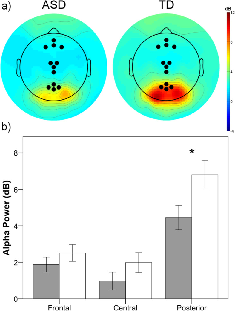
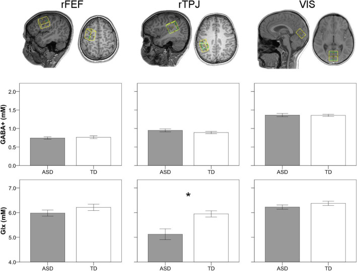
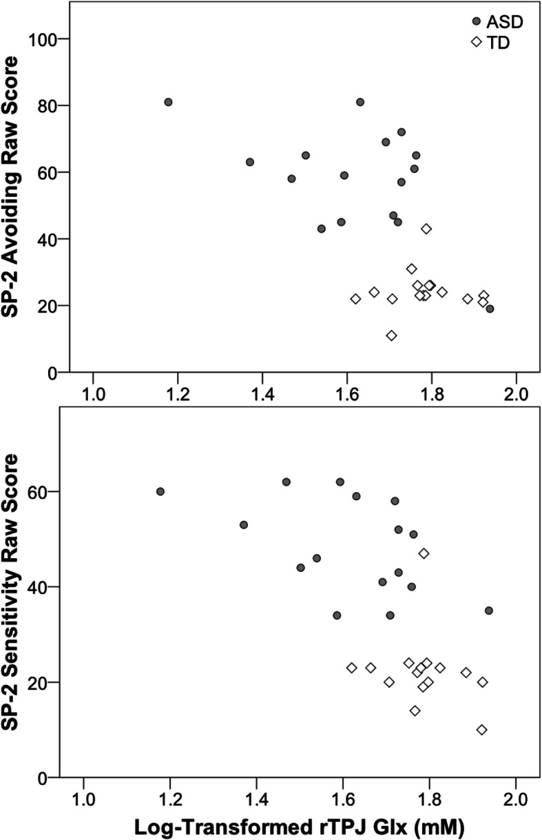
Background: Autism spectrum disorder (ASD) is associated with hyper- and/or hypo-sensitivity to sensory input. Spontaneous alpha power, which plays an important role in shaping responsivity to sensory information, is reduced across the lifespan in individuals with ASD. Furthermore, an excitatory/inhibitory imbalance has also been linked to sensory dysfunction in ASD and has been hypothesized to underlie atypical patterns of spontaneous brain activity. The present study examined whether resting-state alpha power differed in children with ASD as compared to TD children, and investigated the relationships between alpha levels, concentrations of excitatory and inhibitory neurotransmitters, and atypical sensory processing in ASD. Methods: Participants included thirty-one children and adolescents with ASD and thirty-one age- and IQ-matched typically developing (TD) participants. Resting-state electroencephalography (EEG) was used to obtain measures of alpha power. A subset of participants (ASD = 16; TD = 16) also completed a magnetic resonance spectroscopy (MRS) protocol in order to measure concentrations of excitatory (glutamate + glutamine; Glx) and inhibitory (GABA) neurotransmitters. Results: Children with ASD evidenced significantly decreased resting alpha power compared to their TD peers. MRS estimates of GABA and Glx did not differ between groups with the exception of Glx in the temporal-parietal junction. Inter-individual differences in alpha power within the ASD group were not associated with region-specific concentrations of GABA or Glx, nor were they associated with sensory processing differences. However, atypically decreased Glx was associated with increased sensory impairment in children with ASD. Conclusions: Although we replicated prior reports of decreased alpha power in ASD, atypically reduced alpha was not related to neurochemical differences or sensory symptoms in ASD. Instead, reduced Glx in the temporal-parietal cortex was associated with greater hyper-sensitivity in ASD. Together, these findings may provide insight into the neural underpinnings of sensory processing differences present in ASD.
Overview
The claims and statistics of this paper haven’t been checked yet.
Run the check — free, no account needed. It reads the open-access text and checks the sources it cites.
Integrity signals
Registry
Cites
Statistics
Figures
Verification
Claims the paper makes
The claims and statistics of this paper haven’t been checked yet.
Run the check — free, no account needed. It reads the open-access text and checks the sources it cites.
Claims other papers make about this one
Each is checked against this paper’s own text: does it say what the citing paper says it does?
The source backs part of the claim, not all of it — or backs it for a different population, condition or comparison.
In the citing paper
“A different study combining EEG and MRS showed reduced glutamine in the temporal–parietal cortex associated with greater hypersensibility to sensory input detection (Pierce et al. 2021).”
From Autism Spectrum Disorder and auditory sensory alterations: a systematic review on the integrity of cognitive and neuronal functions related to auditory processing (2023) · cites this paper as Pierce et al. 2021
Experimental — accuracy not yet measuredThe source backs part of the claim, not all of it — or backs it for a different population, condition or comparison.
In the citing paper
“Given that auditory-related gamma and alpha powers also seem to be altered in ASD subjects along development (Port et al. 2016; Pierce et al. 2021; Stefano et al. 2019), it would be interesting to assess these measures together with social, cognitive, and language abilities, as potential predictors of later outcomes in ASD.”
From Autism Spectrum Disorder and auditory sensory alterations: a systematic review on the integrity of cognitive and neuronal functions related to auditory processing (2023) · cites this paper as Pierce et al. 2021
Experimental — accuracy not yet measured
Self-check
Percentages, sums and reported differences re-computed from the paper’s own numbers.
The self-check needs the paper’s full text.
Citations
Cites
Maenner, M. J., Shaw, K. A., Baio, J., EdS1, Washington, A., Patrick, M., DiRienzo, M., Christensen, D. L., Wiggins, L. D., Pettygrove, S., et al. (2020) · MMWR. Surveillance Summaries · doi:10.15585/mmwr.ss6904a1 · Cited by 3,780
Wechsler Abbreviated Scale of Intelligence--Second Edition
Wechsler, D. (2018) · PsycTESTS Dataset · doi:10.1037/t15171-000 · Cited by 1,153
Laufs, H., Krakow, K., Sterzer, P., Eger, E., Beyerle, A., Salek-Haddadi, A., & Kleinschmidt, A. (2003) · Proceedings of the National Academy of Sciences · doi:10.1073/pnas.1831638100 · Cited by 1,028
Baranek, G. T., David, F. J., Poe, M. D., Stone, W. L., & Watson, L. R. (2006) · Journal of Child Psychology and Psychiatry · doi:10.1111/j.1469-7610.2005.01546.x · Cited by 975 · cited 2×
“a mixed pattern of hypo- and hyper-sensitivity to sensory input [3].”
BackgroundA multimodal cortical network for the detection of changes in the sensory environment
Downar, J., Crawley, A. P., Mikulis, D. J., & Davis, K. D. (2000) · Nature Neuroscience · doi:10.1038/72991 · Cited by 947
Precise minds in uncertain worlds: Predictive coding in autism.
Van de Cruys, S., Evers, K., Van der Hallen, R., Van Eylen, L., Boets, B., de-Wit, L., & Wagemans, J. (2014) · Psychological Review · doi:10.1037/a0037665 · Cited by 885
An aberrant precision account of autism
Lawson, R. P., Rees, G., & Friston, K. J. (2014) · Frontiers in Human Neuroscience · doi:10.3389/fnhum.2014.00302 · Cited by 641
Autism as a disorder of prediction
Sinha, P., Kjelgaard, M. M., Gandhi, T. K., Tsourides, K., Cardinaux, A. L., Pantazis, D., Diamond, S. P., & Held, R. M. (2014) · Proceedings of the National Academy of Sciences · doi:10.1073/pnas.1416797111 · Cited by 563
Baum, S. H., Stevenson, R. A., & Wallace, M. T. (2015) · Progress in Neurobiology · doi:10.1016/j.pneurobio.2015.09.007 · Cited by 370
Baranek, G. T., Watson, L. R., Boyd, B. A., Poe, M. D., David, F. J., & McGuire, L. (2013) · Development and Psychopathology · doi:10.1017/s0954579412001071 · Cited by 307
Research works printed but not linked: 72
- Wolff JJ, Dimian AF, Botteron KN, Dager SR, Elison JT, Estes AM, et al. A longitudinal study of parent-reported sensory responsiveness in toddlers at-risk for autism. J Child Psychol Psychiatry. 2019;60(3):314–24. · DOI
- Harris KD, Thiele A. Cortical state and attention. Nat Rev Neurosci. 2011;12(9):509–23. · DOI
- Herrmann CS, Struber D, Helfrich RF, Engel AK. EEG oscillations: from correlation to causality. Int J Psychophysiol. 2016;103:12–21. · DOI · Paper page
- Foxe JJ, Snyder AC. The role of alpha-band brain oscillations as a sensory suppression mechanism during selective attention. Front Psychol. 2011;2:154. · DOI · Paper page
- Klimesch W. Alpha-band oscillations, attention, and controlled access to stored information. Trends Cogn Sci. 2012;16(12):606–17. · DOI · Paper page
- Hanslmayr S, Aslan A, Staudigl T, Klimesch W, Herrmann CS, Bauml KH. Prestimulus oscillations predict visual perception performance between and within subjects. Neuroimage. 2007;37(4):1465–73. · DOI
- Zhang Y, Ding M. Detection of a weak somatosensory stimulus: role of the prestimulus mu rhythm and its top-down modulation. J Cogn Neurosci. 2010;22(2):307–22. · DOI
- Mathewson KE, Gratton G, Fabiani M, Beck DM, Ro T. To see or not to see: prestimulus alpha phase predicts visual awareness. J Neurosci. 2009;29(9):2725–32. · DOI
- Jensen O, Mazaheri A. Shaping functional architecture by oscillatory alpha activity: gating by inhibition. Front Hum Neurosci. 2010;4:186. · DOI · Paper page
- Mathewson KE, Lleras A, Beck DM, Fabiani M, Ro T, Gratton G. Pulsed out of awareness: EEG alpha oscillations represent a pulsed-inhibition of ongoing cortical processing. Front Psychol. 2011;2:99. · DOI
- Wang J, Barstein J, Ethridge LE, Mosconi MW, Takarae Y, Sweeney JA. Resting state EEG abnormalities in autism spectrum disorders. J Neurodev Disord. 2013;5(1):24. · DOI
- Levin AR, Varcin KJ, O'Leary HM, Tager-Flusberg H, Nelson CA. EEG power at 3 months in infants at high familial risk for autism. J Neurodev Disord. 2017;9(1):34. · DOI · Paper page
- Tierney AL, Gabard-Durnam L, Vogel-Farley V, Tager-Flusberg H, Nelson CA. Developmental trajectories of resting EEG power: an endophenotype of autism spectrum disorder. PLoS One. 2012;7(6):e39127. · DOI · Paper page
- Keehn B, Westerfield M, Müller R-A, Townsend J. Autism, attention, and alpha oscillations: an electrophysiological study of attentional capture. Biol Psychiatry Cogn Neurosci Neuroimaging. 2017. · DOI
- DiStefano C, Dickinson A, Baker E, Jeste SS. EEG data collection in children with ASD: the role of state in data quality and spectral power. Res Autism Spectr Disord. 2019;57:132–44. · DOI
- Shephard E, Tye C, Ashwood KL, Azadi B, Asherson P, Bolton PF, et al. Resting-state neurophysiological activity patterns in young people with ASD, ADHD, and ASD + ADHD. J Autism Dev Disord. 2018;48(1):110–22. · DOI
- Dawson G, Klinger LG, Panagiotides H, Lewy A, Castelloe P. Subgroups of autistic children based on social behavior display distinct patterns of brain activity. J Abnorm Child Psychol. 1995;23(5):569–83. · DOI
- Murias M, Webb SJ, Greenson J, Dawson G. Resting state cortical connectivity reflected in EEG coherence in individuals with autism. Biol Psychiatry. 2007;62(3):270–3. · DOI
- Cornew L, Roberts TP, Blaskey L, Edgar JC. Resting-state oscillatory activity in autism spectrum disorders. J Autism Dev Disord. 2012;42(9):1884–94. · DOI
- Mathewson KJ, Jetha MK, Drmic IE, Bryson SE, Goldberg JO, Schmidt LA. Regional EEG alpha power, coherence, and behavioral symptomatology in autism spectrum disorder. Clin Neurophysiol. 2012;123(9):1798–809. · DOI
- Sutton SK, Burnette CP, Mundy PC, Meyer J, Vaughan A, Sanders C, et al. Resting cortical brain activity and social behavior in higher functioning children with autism. J Child Psychol Psychiatry. 2005;46(2):211–22. · DOI · Paper page
- Coben R, Clarke AR, Hudspeth W, Barry RJ. EEG power and coherence in autistic spectrum disorder. Clin Neurophysiol. 2008;119(5):1002–9. · DOI
- Simon DM, Damiano CR, Woynaroski TG, Ibanez LV, Murias M, Stone WL, et al. Neural correlates of sensory hyporesponsiveness in toddlers at high risk for autism spectrum disorder. J Autism Dev Disord. 2017;47(9):2710–22. · DOI
- Damiano-Goodwin CR, Woynaroski TG, Simon DM, Ibanez LV, Murias M, Kirby A, et al. Developmental sequelae and neurophysiologic substrates of sensory seeking in infant siblings of children with autism spectrum disorder. Dev Cogn Neurosci. 2018;29:41–53. · DOI
- Simon DM, Wallace MT. Dysfunction of sensory oscillations in autism spectrum disorder. Neurosci Biobehav Rev. 2016;68:848–61. · DOI
- Hussman JP. Suppressed GABAergic inhibition as a common factor in suspected etiologies of autism. Journal of Autism and Developmental Disorders. 2001;31(2):247–8. · DOI · Paper page
- Rubenstein JL, Merzenich MM. Model of autism: increased ratio of excitation/inhibition in key neural systems. Genes, Brain and Behavior. 2003;2(5):255–67. · DOI · Paper page
- Ajram LA, Pereira AC, Durieux AMS, Velthius HE, Petrinovic MM, McAlonan GM. The contribution of [1H] magnetic resonance spectroscopy to the study of excitation-inhibition in autism. Prog Neuro-Psychopharmacol Biol Psychiatry. 2019;89:236–44. · DOI
- Puts NAJ, Wodka EL, Harris AD, Crocetti D, Tommerdahl M, Mostofsky SH, et al. Reduced GABA and altered somatosensory function in children with autism spectrum disorder. Autism Res. 2017;10(4):608–19. · DOI
- Sapey-Triomphe LA, Lamberton F, Sonie S, Mattout J, Schmitz C. Tactile hypersensitivity and GABA concentration in the sensorimotor cortex of adults with autism. Autism Res. 2019;12(4):562–75. · DOI
- Umesawa Y, Atsumi T, Chakrabarty M, Fukatsu R, Ide M. GABA concentration in the left ventral premotor cortex associates with sensory hyper-responsiveness in autism spectrum disorders without intellectual disability. Front Neurosci. 2020;14:482. · DOI
- Elliott C. Differential abilities scales - section edition. Assessment H, editor. San Antonio, TX2007.
- Lord C, Rutter M, DiLavore PC, Risi S, Gotham K, Bishop S. Autism diagnostic Obervation schedule. Second ed. Western Psychological Services: Torrance, CA; 2012.
- Rutter M, Bailey A, Lord C. Social communication questionnaire (SCQ). Western Psychological Services: Los Angeles, CA; 2003.
- Delorme A, Mullen T, Kothe C, Akalin Acar Z, Bigdely-Shamlo N, Vankov A, et al. EEGLAB, SIFT, NFT, BCILAB, and ERICA: new tools for advanced EEG processing. Comput Intell Neurosci. 2011;2011:130714. · DOI
- Chaumon M, Bishop DV, Busch NA. A practical guide to the selection of independent components of the electroencephalogram for artifact correction. J Neurosci Methods. 2015;250:47–63. · DOI
- Edmondson DA, Xia P, McNally Keehn R, Dydak U, Keehn B. A magnetic resonance spectroscopy study of superior visual search abilities in children with autism spectrum disorder. Autism Res. 2020;13(4):550–62. · DOI
- Andreychenko A, Boer VO. Arteaga de Castro CS, Luijten PR, Klomp DW. Efficient spectral editing at 7 T: GABA detection with MEGA-sLASER. Magn Reson Med. 2012;68(4):1018–25. · DOI
- Marjanska M, Lehericy S, Valabregue R, Popa T, Worbe Y, Russo M, et al. Brain dynamic neurochemical changes in dystonic patients: a magnetic resonance spectroscopy study. Mov Disord. 2013;28(2):201–9. · DOI
- Provencher SW. Estimation of metabolite concentrations from localized in vivo proton NMR spectra. Magn Reson Med. 1993;30(6):672–9. · DOI
- Kaiser LG, Young K, Matson GB. Elimination of spatial interference in PRESS-localized editing spectroscopy. Magn Reson Med. 2007;58(4):813–8. · DOI
- Harris AD, Puts NA, Edden RA. Tissue correction for GABA-edited MRS: considerations of voxel composition, tissue segmentation, and tissue relaxations. J Magn Reson Imaging. 2015;42(5):1431–40. · DOI
- Dunn W. Sensory profile - 2. San Antonio, TX: Pearson Publishing; 2014.
- Corcoran AW, Alday PM, Schlesewsky M, Bornkessel-Schlesewsky I. Toward a reliable, automated method of individual alpha frequency (IAF) quantification. Psychophysiology. 2018;55(7):e13064. · DOI
- Sadaghiani S, Scheeringa R, Lehongre K, Morillon B, Giraud AL, Kleinschmidt A. Intrinsic connectivity networks, alpha oscillations, and tonic alertness: a simultaneous electroencephalography/functional magnetic resonance imaging study. J Neurosci. 2010;30(30):10243–50. · DOI · Paper page
- Mathewson KE, Beck DM, Ro T, Maclin EL, Low KA, Fabiani M, et al. Dynamics of alpha control: preparatory suppression of posterior alpha oscillations by frontal modulators revealed with combined EEG and event-related optical signal. J Cogn Neurosci. 2014;26(10):2400–15. · DOI
- Sumner P, Edden RA, Bompas A, Evans CJ, Singh KD. More GABA, less distraction: a neurochemical predictor of motor decision speed. Nat Neurosci. 2010;13(7):825–7. · DOI
- Schreckenberger M, Lange-Asschenfeldt C, Lochmann M, Mann K, Siessmeier T, Buchholz HG, et al. The thalamus as the generator and modulator of EEG alpha rhythm: a combined PET/EEG study with lorazepam challenge in humans. Neuroimage. 2004;22(2):637–44. · DOI
- Lorincz ML, Kekesi KA, Juhasz G, Crunelli V, Hughes SW. Temporal framing of thalamic relay-mode firing by phasic inhibition during the alpha rhythm. Neuron. 2009;63(5):683–96. · DOI
- Edgar JC, Heiken K, Chen YH, Herrington JD, Chow V, Liu S, et al. Resting-state alpha in autism spectrum disorder and alpha associations with thalamic volume. J Autism Dev Disord. 2015;45(3):795–804. · DOI
- Cerliani L, Mennes M, Thomas RM, Di Martino A, Thioux M, Keysers C. Increased functional connectivity between subcortical and cortical resting-state networks in autism spectrum disorder. JAMA Psychiatry. 2015;72(8):767–77. · DOI · Paper page
- Woodward ND, Giraldo-Chica M, Rogers B, Cascio CJ. Thalamocortical dysconnectivity in autism spectrum disorder: an analysis of the autism brain imaging data exchange. Biol Psychiatry Cogn Neurosci Neuroimaging. 2017;2(1):76–84.
- Mash LE, Keehn B, Linke AC, Liu TT, Helm JL, Haist F, et al. Atypical relationships between spontaneous EEG and fMRI activity in autism. Brain Connect. 2020;10(1):18–28. · DOI
- Fung LK, Flores RE, Gu M, Sun KL, James D, Schuck RK, et al. Thalamic and prefrontal GABA concentrations but not GABAA receptor densities are altered in high-functioning adults with autism spectrum disorder. Mol Psychiatry. 2020. · DOI
- Hardan AY, Minshew NJ, Melhem NM, Srihari S, Jo B, Bansal R, et al. An MRI and proton spectroscopy study of the thalamus in children with autism. Psychiatry Res. 2008;163(2):97–105. · DOI
- Bernardi S, Anagnostou E, Shen J, Kolevzon A, Buxbaum JD, Hollander E, et al. In vivo 1H-magnetic resonance spectroscopy study of the attentional networks in autism. Brain Res. 2011;1380:198–205. · DOI
- Tebartz van Elst L, Maier S, Fangmeier T, Endres D, Mueller GT, Nickel K, et al. Disturbed cingulate glutamate metabolism in adults with high-functioning autism spectrum disorder: evidence in support of the excitatory/inhibitory imbalance hypothesis. Mol Psychiatry. 2014;19(12):1314–25. · DOI
- DeVito TJ, Drost DJ, Neufeld RW, Rajakumar N, Pavlosky W, Williamson P, et al. Evidence for cortical dysfunction in autism: a proton magnetic resonance spectroscopic imaging study. Biol Psychiatry. 2007;61(4):465–73. · DOI
- Horder J, Lavender T, Mendez MA, O'Gorman R, Daly E, Craig MC, et al. Reduced subcortical glutamate/glutamine in adults with autism spectrum disorders: a [(1)H]MRS study. Transl Psychiatry. 2014;4:e364. · DOI
- Horder J, Petrinovic MM, Mendez MA, Bruns A, Takumi T, Spooren W, et al. Glutamate and GABA in autism spectrum disorder-a translational magnetic resonance spectroscopy study in man and rodent models. Transl Psychiatry. 2018;8(1):106. · DOI
- Schallmo MP, Kolodny T, Kale AM, Millin R, Flevaris AV, Edden RAE, et al. Weaker neural suppression in autism. Nat Commun. 2020;11(1):2675. · DOI
- Patel GH, Sestieri C, Corbetta M. The evolution of the temporoparietal junction and posterior superior temporal sulcus. Cortex. 2019;118:38–50. · DOI
- Matsuhashi M, Ikeda A, Ohara S, Matsumoto R, Yamamoto J, Takayama M, et al. Multisensory convergence at human temporo-parietal junction - epicortical recording of evoked responses. Clin Neurophysiol. 2004;115(5):1145–60. · DOI
- Downar J, Crawley AP, Mikulis DJ, Davis KD. A cortical network sensitive to stimulus salience in a neutral behavioral context across multiple sensory modalities. J Neurophysiol. 2002;87(1):615–20. · DOI · Paper page
- Brandwein AB, Foxe JJ, Butler JS, Frey HP, Bates JC, Shulman LH, et al. Neurophysiological indices of atypical auditory processing and multisensory integration are associated with symptom severity in autism. J Autism Dev Disord. 2015;45(1):230–44. · DOI · Paper page
- Courchesne E, Allen G. Prediction and preparation, fundamental functions of the cerebellum. Learn Mem. 1997;4(1):1–35. · DOI
- Friston K. A theory of cortical responses. Philos Trans R Soc Lond Ser B Biol Sci. 2005;360(1456):815–36. · DOI · Paper page
- Kompus K, Westerhausen R, Craven AR, Kreegipuu K, Poldver N, Passow S, et al. Resting-state glutamatergic neurotransmission is related to the peak latency of the auditory mismatch negativity (MMN) for duration deviants: an (1)H-MRS-EEG study. Psychophysiology. 2015;52(9):1131–9. · DOI
- Kompus K, Volehaugen V, Craven A, Specht K. Glutamatergic modulation of auditory cortex connectivity with attentional brain networks in unpredictable perceptual environment. bioRxiv. 2019:2019.12.20.884049. · DOI
- Schwartz S, Shinn-Cunningham B, Tager-Flusberg H. Meta-analysis and systematic review of the literature characterizing auditory mismatch negativity in individuals with autism. Neurosci Biobehav Rev. 2018;87:106–17. · DOI · Paper page
- Matsuzaki J, Kagitani-Shimono K, Sugata H, Hanaie R, Nagatani F, Yamamoto T, et al. Delayed mismatch field latencies in autism spectrum disorder with abnormal auditory sensitivity: a magnetoencephalographic study. Front Hum Neurosci. 2017;11:446. · DOI
- Ludlow A, Mohr B, Whitmore A, Garagnani M, Pulvermuller F, Gutierrez R. Auditory processing and sensory behaviours in children with autism spectrum disorders as revealed by mismatch negativity. Brain Cogn. 2014;86:55–63. · DOI
Cited by
Sorted by citations
Gonçalves, A. M., & Monteiro, P. (2023) · Journal of Neural Transmission · doi:10.1007/s00702-023-02595-9 · Cited by 88
Halliday, A. R., Vucic, S. N., Georges, B., LaRoche, M., Mendoza Pardo, M. A., Swiggard, L. O., McDonald, K., Olofsson, M., Menon, S. N., Francis, S. M., et al. (2024) · Frontiers in Psychiatry · doi:10.3389/fpsyt.2024.1474003 · Cited by 20
Metadata
- Authors
- Pierce, S., Kadlaskar, G., Edmondson, D. A., McNally Keehn, R., Dydak, U., & Keehn, B.
- Affiliations
- Purdue University West Lafayette (US)
- Cincinnati Children's Hospital Medical Center (US)
- Corresponding author
- Keehn, Brandon
- Venue
- Journal of Neurodevelopmental Disorders
- Published
- 2021-01-06
- Volume
- 13
- Issue
- 1
- Pages
- 5-5
- ISSN
- 1866-1947 (ISSN-L)
- 1866-1955
- PMC
- PMC7788714
- OpenAlex
- W3119381096
- Cited by
- 42
- References
- 11
- Citations by year
- 2026: 4 · 2025: 9 · 2024: 12 · 2023: 5 · 2022: 8 · 2021: 4
- FWCI
- 3.81
- Type
- journal article
- Language
- en
- Open access
- gold
- doi.org
- Locations
- Journal of Neurodevelopmental Disorders (publishedVersion, cc-by)
- Europe PMC (PubMed Central) (publishedVersion, other-oa)
- PubMed Central (publishedVersion, cc-by)
- PubMed (publishedVersion)
- DOAJ (DOAJ: Directory of Open Access Journals) (submittedVersion, cc-by-sa)
- IUScholarWorks (Indiana University) (submittedVersion, cc-by)
- Funders
- National Institute of Mental Health (R21 MH114095)
- NIH Office of the Director (S10 OD012336)
- Purdue Institute for Integrative Neuroscience (#209257)
- Topics
- Life Sciences › Neuroscience › Cognitive Neuroscience › Autism Spectrum Disorder Research
- Life Sciences › Biochemistry, Genetics and Molecular Biology › Genetics › Genetics and Neurodevelopmental Disorders
- Health Sciences › Medicine › Psychiatry and Mental health › Child Nutrition and Feeding Issues
- Keywords
- autism spectrum disorder, sensory processing, glutamate, glutamine, GABA, excitatory-inhibitory imbalance, sensory hypersensitivity, temporal-parietal cortex, children
- MeSH terms
- Autism Spectrum Disorder, Adolescent, Child, Electroencephalography, Female, Humans, Magnetic Resonance Imaging, Male, Magnetic Resonance Spectroscopy, Perception
About these checks
Registry, retraction, Cites and statistics results are computed, not judged, with one exception: a printed reference that no index lists is read once by a model before it counts as a citation error. Claims are extracted by a model and kept only when their quote appears verbatim in the text; each cited source is then checked, with a model’s help, for whether it says what the claim relies on. Those verdicts can be wrong — read the claim’s quote and the cited source before relying on one.
Statistics and arithmetic are recomputed, not judged.
Confidence bands are shown only for fields calibrated on our annotated test set. None is calibrated yet, so no band is shown.
What counts as a claim ›How we measure confidence ›How we check this ›
Are you an author, or do you see a problem with this page? Request a review
