

The DOI matches its Crossref record; 52 of the 52 references it cites were found in a registry or an index.
Exosomes derived from gemcitabine-resistant cells transfer malignant phenotypic traits via delivery of miRNA-222-3p
Open accessAbstract
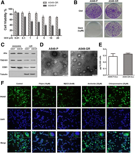
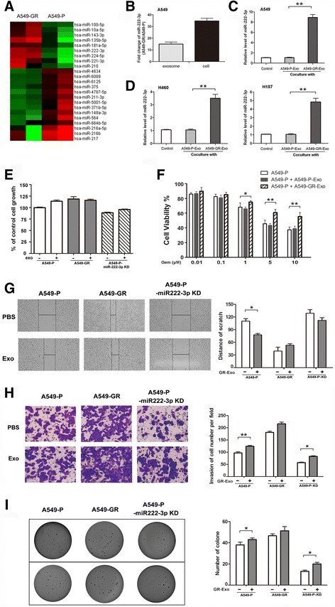
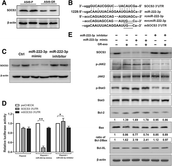
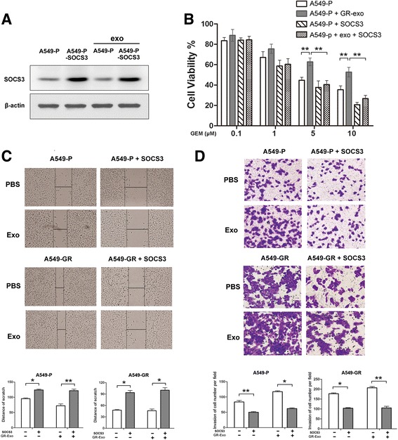
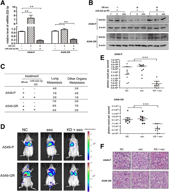
BACKGROUND: Although gemcitabine-based chemotherapy has been established as a core multimodal therapy for non-small cell lung cancer (NSCLC) treatment, its clinical efficacy remains limited by the development of acquired resistance following tumor metastasis and relapse. In this study, we investigated how gemcitabine-resistant (GR) cells contribute to the development of NSCLC tumor malignancy via exosome-mediated transfer of microRNAs. METHODS: We first studied the mechanism of exosome internalization via PKH-67 staining and an immunofluorescence assay, then confirmed our finding by transmission electron microscopy and western blot analysis. Candidate miRNAs were identified through microarray analysis. Thereafter, RT-PCR, MTS, Transwell and soft agar assays were performed to assess the role of exosomic miR-222-3p in vitro. A 3' untranslated region reporter assay was applied to identify the target of miR-222-3p. A lung metastasis mouse model was constructed to evaluate tumor growth and metastasis in vivo. Finally, clinical samples were used for correlation analysis between exosomic miR-222-3p levels and patients' response to gemcitabine. RESULTS: A549-GR-derived exosomes were internalized by receipt cells via caveolin- and lipid raft-dependent endocytosis, which allowed the transfer of miR-222-3p. Exosomic miR-222-3p enhanced the proliferation, gemcitabine resistance, migration, invasion, and anti-anoikis of parental sensitive cells by directly targeting the promoter of SOCS3. In addition, a higher level of exosomic miR-222-3p in sera usually predicted worse prognosis in NSCLC patients. CONCLUSION: Our data demonstrate that exosomic-miR-222-3p functions as a principal regulator of gemcitabine resistance and malignant characteristics by targeting SOCS3. The exosomic miR-222-3p level in sera may be a potential prognostic biomarker for predicting gemcitabine sensitivity in NSCLC patients.
Overview
The claims and statistics of this paper haven’t been checked yet.
Run the check — free, no account needed. It reads the open-access text and checks the sources it cites.
Integrity signals
Registry
Cites
Statistics
Figures
Verification
Claims the paper makes
The claims and statistics of this paper haven’t been checked yet.
Run the check — free, no account needed. It reads the open-access text and checks the sources it cites.
Claims other papers make about this one
Each is checked against this paper’s own text: does it say what the citing paper says it does?
In the citing paper
“However, another study conducted in A549-P cells showed that endocytosis of exosomal miRNAs is mediated by caveolae- and lipid raft-dependent pathways (143).”
From Overview of MicroRNA Biogenesis, Mechanisms of Actions, and Circulation (2018) · cites this paper as [143]
Self-check
Percentages, sums and reported differences re-computed from the paper’s own numbers.
The self-check needs the paper’s full text.
Citations
Cites
6 retracted
RETRACTED: MicroRNA signatures of tumor-derived exosomes as diagnostic biomarkers of ovarian cancer
Taylor, D. D., & Gerçel-Taylor, Ç. (2008) · Gynecologic Oncology · doi:10.1016/j.ygyno.2008.04.033 · Cited by 2,333
RetractedGarofalo, M., Leva, G. D., Romano, G., Nuovo, G. J., Suh, S., Ngankeu, A., Taccioli, C., Pichiorri, F., Alder, H., Secchiero, P., et al. (2009) · Cancer Cell · doi:10.1016/j.ccr.2009.10.014 · Cited by 743
RetractedZhao, J.-J., Lin, J., Yang, H., Kong, W. W., He, L., Ma, X., Coppola, D., & Cheng, J. Q. (2008) · Journal of Biological Chemistry · doi:10.1074/jbc.m806041200 · Cited by 524
RetractedHuang, L., Hu, B., Ni, J., Wu, J., Jiang, W., Chen, C., Yang, L., Zeng, Y., Wan, R., Hu, G., et al. (2016) · Journal of Experimental & Clinical Cancer Research · doi:10.1186/s13046-016-0301-7 · Cited by 86
RetractedLi, J., Yu, J. Q., Zhang, H., Wang, B., Guo, H., Bai, J., Wang, J., Dong, Y., Zhao, Y., & Wang, Y. (2016) · Cellular Physiology and Biochemistry · doi:10.1159/000443111 · Cited by 44
RetractedZhu, G., Huang, Q., Zheng, W., Huang, Y., Hua, J., Yang, S., Zhuang, J., Wang, J., Chang, J., Xu, J., et al. (2016) · Cellular Physiology and Biochemistry · doi:10.1159/000447893 · Cited by 10
RetractedValadi, H., Ekström, K., Bossios, A., Sjöstrand, M., Lee, J. J., & Lötvall, J. O. (2007) · Nature Cell Biology · doi:10.1038/ncb1596 · Cited by 12,834
Exosomes: composition, biogenesis and function
Théry, C., Zitvogel, L., & Amigorena, S. (2002) · Nature reviews. Immunology · doi:10.1038/nri855 · Cited by 5,478
Skog, J., Würdinger, T., van Rijn, S., Meijer, D. H., Gainche, L., Curry, W. T., Carter, B. S., Krichevsky, A. M., & Breakefield, X. O. (2008) · Nature Cell Biology · doi:10.1038/ncb1800 · Cited by 5,001
Exosomes: Extracellular organelles important in intercellular communication
Mathivanan, S., Ji, H. L., & Simpson, R. J. (2010) · Journal of Proteomics · doi:10.1016/j.jprot.2010.06.006 · Cited by 2,581
Cited by
Sorted by citations
Overview of MicroRNA Biogenesis, Mechanisms of Actions, and Circulation
O'Brien, J., Hayder, H., Zayed, Y., & Peng, C. (2018) · Frontiers in Endocrinology · doi:10.3389/fendo.2018.00402 · Cited by 5,287
Exosomes: composition, biogenesis, and mechanisms in cancer metastasis and drug resistance
Mashouri, L., Yousefi, H., Aref, A. R., Ahadi, A. M., Molaei, F., & Alahari, S. K. (2019) · Molecular Cancer · doi:10.1186/s12943-019-0991-5 · Cited by 1,363
Exosomes: key players in cancer and potential therapeutic strategy
Dai, J., Su, Y., Zhong, S., Cong, L., Liu, B., Yang, J., Tao, Y., He, Z., Chen, C., & Jiang, Y. (2020) · Signal Transduction and Targeted Therapy · doi:10.1038/s41392-020-00261-0 · Cited by 1,186
Exosomes and Ectosomes in Intercellular Communication
Meldolesi, J. (2018) · Current Biology · doi:10.1016/j.cub.2018.01.059 · Cited by 944
Brennan, K., Martin, K., Fitzgerald, S. P., O’Sullivan, J., Wu, Y., Blanco, A., Richardson, C., & Gee, M. M. M. (2020) · Scientific Reports · doi:10.1038/s41598-020-57497-7 · Cited by 943
Application of exosomes as liquid biopsy in clinical diagnosis
Zhou, B., Xu, K., Zheng, X., Chen, T., Wang, J., Song, Y., Shao, Y., & Zheng, S. (2020) · Signal Transduction and Targeted Therapy · doi:10.1038/s41392-020-00258-9 · Cited by 685
Maacha, S., Bhat, A. A., Jimenez, L., Raza, A., Haris, M., Shahab Uddin, M. S., & Grivel, J. (2019) · Molecular Cancer · doi:10.1186/s12943-019-0965-7 · Cited by 583
Emerging role of exosomes in cancer progression and tumor microenvironment remodeling
Paskeh, M. D. A., Entezari, M., Mirzaei, S., Zabolian, A., Saleki, H., Naghdi, M. J., Sabet, S., Khoshbakht, M. A., Hashemi, M., Hushmandi, K., et al. (2022) · Journal of Hematology & Oncology · doi:10.1186/s13045-022-01305-4 · Cited by 591
Circulating microRNAs as potential cancer biomarkers: the advantage and disadvantage
Wang, H., Peng, R., Wang, J., Qin, Z., & Xue, L. (2018) · Clinical Epigenetics · doi:10.1186/s13148-018-0492-1 · Cited by 540
Extracellular vesicles as biomarkers and therapeutic targets for cancer
Urabe, F., Kosaka, N., Ito, K., Kimura, T., Egawa, S., & Ochiya, T. (2019) · American Journal of Physiology-Cell Physiology · doi:10.1152/ajpcell.00280.2019 · Cited by 413
Exosomal miRNAs and miRNA dysregulation in cancer-associated fibroblasts
Yang, F., Ning, Z., Ma, L., Liu, W., Shao, C., Shu, Y., & Shen, H. (2017) · Molecular Cancer · doi:10.1186/s12943-017-0718-4 · Cited by 333
Extracellular vesicles in the tumor microenvironment: old stories, but new tales
Han, L., Lam, E. W. ‐., & Sun, Y. (2019) · Molecular Cancer · doi:10.1186/s12943-019-0980-8 · Cited by 301
Extracellular vesicles and anti-cancer drug resistance
McNamee, N., & O’Driscoll, L. (2018) · Biochimica et Biophysica Acta (BBA) - Reviews on Cancer · doi:10.1016/j.bbcan.2018.07.003 · Cited by 280
Song, Z., Jia, G., Ma, P., & Cang, S. (2021) · Life Sciences · doi:10.1016/j.lfs.2021.119399 · Cited by 261
Liquid biopsy and tumor heterogeneity in metastatic solid tumors: the potentiality of blood samples
Russano, M., Napolitano, A., Ribelli, G., Iuliani, M., Simonetti, S., Citarella, F., Pantano, F., Dell’Aquila, E., Anesi, C., Silvestris, N., et al. (2020) · Journal of Experimental & Clinical Cancer Research · doi:10.1186/s13046-020-01601-2 · Cited by 267
Chen, S., Zhu, S., Pei, X., Qiu, B., Xiong, D., Long, X., Lin, K., Lü, F., Xu, J., & Wu, Y. (2021) · Molecular Cancer · doi:10.1186/s12943-021-01448-x · Cited by 263
Li, Q., Li, B., Li, Q., Wei, S., He, Z., Huang, X., Wang, L., Xia, Y., Xu, Z., Li, Z., et al. (2018) · Cell Death & Disease · doi:10.1038/s41419-018-0928-8 · Cited by 219
Self-citationFu, X., Liu, M., Qu, S., Ma, J., Zhang, Y., Shi, T., Wen, H., Yang, Y., Wang, S., Wang, J., et al. (2018) · Journal of Experimental & Clinical Cancer Research · doi:10.1186/s13046-018-0677-7 · Cited by 217
RetractedHu, W., Liu, C., Bi, Z., Zhou, Q. Y., Zhang, H., Li, L., Zhang, J., Zhu, W., Song, Y., Zhang, F., et al. (2020) · Molecular Cancer · doi:10.1186/s12943-020-01199-1 · Cited by 215
Tumor-associated exosomes promote lung cancer metastasis through multiple mechanisms
Jiang, C., Zhang, N., Hu, X., & Wang, H. (2021) · Molecular Cancer · doi:10.1186/s12943-021-01411-w · Cited by 210
The emerging role of exosome-derived non-coding RNAs in cancer biology
Fan, Q., Yang, L., Zhang, X., Peng, X., Wei, S., Su, D., Zhai, Z., Hua, X., & Li, H. (2017) · Cancer Letters · doi:10.1016/j.canlet.2017.10.040 · Cited by 209
The roles of exosomal miRNAs and lncRNAs in lung diseases
Yang, L., Yin, Z., Fan, J., Zhang, S., & Yang, W.-B. (2019) · Signal Transduction and Targeted Therapy · doi:10.1038/s41392-019-0080-7 · Cited by 190
The role of exosomes in liquid biopsy for cancer diagnosis and prognosis prediction
Li, S., Yi, M., Dong, B., Tan, X., Luo, S., & Wu, K. (2020) · International Journal of Cancer · doi:10.1002/ijc.33386 · Cited by 181
He, Y., Rodrigues, R. M., Wang, X., Seo, W., Ma, J., Hwang, S., Fu, Y., Trojnár, E., Mátyás, C., Zhao, S., et al. (2021) · Journal of Clinical Investigation · doi:10.1172/jci141513 · Cited by 165
Insight into Extracellular Vesicle-Cell Communication: From Cell Recognition to Intracellular Fate
Ginini, L., Billan, S., Fridman, E., & Gil, Z. (2022) · Cells · doi:10.3390/cells11091375 · Cited by 188
Role of exosomal microRNAs in lung cancer biology and clinical applications
Hu, C., Meiners, S., Lukas, C., Stathopoulos, G. T., & Chen, J. (2020) · Cell Proliferation · doi:10.1111/cpr.12828 · Cited by 153
Mechanisms of resistance to chemotherapy in non-small cell lung cancer
Min, H., & Lee, H. (2021) · Archives of Pharmacal Research · doi:10.1007/s12272-021-01312-y · Cited by 163
Vasconcelos, M. H., Caires, H. R., Ābols, A., Xavier, C. P. R., & Linē, A. (2019) · Drug Resistance Updates · doi:10.1016/j.drup.2019.100647 · Cited by 150
Extracellular Vesicles in Cancer Drug Resistance: Roles, Mechanisms, and Implications
Yang, Q., Xu, J. B., Gu, J., Shi, H., Zhang, J., Zhang, J., Chen, Z., Fang, X., Zhu, T., & Zhang, X. (2022) · Advanced Science · doi:10.1002/advs.202201609 · Cited by 148
Extracellular vesicles and particles impact the systemic landscape of cancer
Lucotti, S., Kenific, C. M., Zhang, H., & Lyden, D. C. (2022) · The EMBO Journal · doi:10.15252/embj.2021109288 · Cited by 145
Functional miRNAs in breast cancer drug resistance
Hu, W., Tan, C., He, Y., Zhang, G., Xu, Y., & Tang, J. (2018) · OncoTargets and Therapy · doi:10.2147/ott.s152462 · Cited by 126
Wang, M., Qiu, R., Yu, S., Xu, X., Li, G., Gu, R., Tan, C., Zhu, W., & Shen, B. (2018) · International Journal of Oncology · doi:10.3892/ijo.2018.4601 · Cited by 124
Anoikis-Associated Lung Cancer Metastasis: Mechanisms and Therapies
Wang, J., Luo, Z., Lin, L., Sui, X., Yu, L., Xu, C., Zhang, R., Zhao, Z., Zhu, Q., An, B., et al. (2022) · Cancers · doi:10.3390/cancers14194791 · Cited by 115
Liu, X., Lü, Y., Xu, Y., Hou, S., Huang, J., Wang, B., Zhao, J., Xia, S., Fan, S., Yu, X., et al. (2019) · Cancer Letters · doi:10.1016/j.canlet.2019.05.035 · Cited by 112
Intratumoral heterogeneity and drug resistance in cancer
Fu, Y.-C., Liang, S.-B., Luo, M., & Wang, X. (2025) · Cancer Cell International · doi:10.1186/s12935-025-03734-w · Cited by 129
The biological functions and clinical applications of exosomes in lung cancer
Chen, R., Xu, X., Qian, Z., Zhang, C., Niu, Y., Wang, Z., Sun, J., Zhang, X., & Yu, Y. (2019) · Cellular and Molecular Life Sciences · doi:10.1007/s00018-019-03233-y · Cited by 105
The roles of exosomes in cancer drug resistance and its therapeutic application
Li, S., Yi, M., Dong, B., Jiao, Y., Luo, S., & Wu, K. (2020) · Clinical and Translational Medicine · doi:10.1002/ctm2.257 · Cited by 99
Role of miR-221/222 in Tumor Development and the Underlying Mechanism
Song, Q., An, Q., Niu, B., Lu, X., Zhang, N., & Cao, X. (2019) · Journal of Oncology · doi:10.1155/2019/7252013 · Cited by 96
Potential roles and prognostic significance of exosomes in cancer drug resistance
Mostafazadeh, M., Samadi, N., Kahroba, H., Baradaran, B., Haiaty, S., & Nouri, M. (2021) · Cell & Bioscience · doi:10.1186/s13578-020-00515-y · Cited by 95
Seo, H. A., Moeng, S., Sim, S., Kuh, H. J., Choi, S. Y., & Park, J. K. (2019) · Cells · doi:10.3390/cells9010029 · Cited by 93
The role of extracellular vesicles in the transfer of drug resistance competences to cancer cells
Xavier, C. P. R., Belisario, D. C., Rebelo, R., Assaraf, Y. G., Giovannetti, E., Kopecka, J., & Vasconcelos, M. H. (2022) · Drug Resistance Updates · doi:10.1016/j.drup.2022.100833 · Cited by 98
Fan, L., Lei, H., Zhang, S., Peng, Y., Fu, C., Shu, G., & Yin, G. (2020) · Theranostics · doi:10.7150/thno.43198 · Cited by 86
Yuan, F., Peng, W., Yang, Y., Xu, J., Liu, Y., Xie, Y., Huang, T., Shi, C., Ding, Y., Li, C., et al. (2023) · Journal of Neuroinflammation · doi:10.1186/s12974-023-02833-7 · Cited by 86
Role of T cell-derived exosomes in immunoregulation
Lu, J., Wu, J., Tian, J., & Wang, S. (2018) · Immunologic Research · doi:10.1007/s12026-018-9000-0 · Cited by 86
The Role of Exosomal microRNA in Cancer Drug Resistance
Guo, Q.-R., Wang, H., Yan, Y.-D., Liu, Y., Su, C., Chen, H., Yan, Y., Adhikari, R., Wu, Q., & Zhang, J. (2020) · Frontiers in Oncology · doi:10.3389/fonc.2020.00472 · Cited by 85
Exosomes: A New Pathway for Cancer Drug Resistance
Zhong, Y., Li, H., Li, P., Chen, Y., Zhang, M., Yuan, Z., Zhang, Y., Xu, Z., Luo, G. Q., Fang, Y., et al. (2021) · Frontiers in Oncology · doi:10.3389/fonc.2021.743556 · Cited by 77
Huang, Y., Kanada, M., Ye, J., Deng, Y., He, Q., Lei, Z., Chen, Y., Li, Y., Qin, P., Zhang, J., et al. (2022) · Cancer Letters · doi:10.1016/j.canlet.2022.215796 · Cited by 76
Doghish, A. S., Ismail, A., Elrebehy, M. A., Elbadry, A. M. M., Mahmoud, H. H., Farouk, S. M., Serea, G. A. A., Elghany, R. A. A., El-Halwany, K. K., Alsawah, A. O., et al. (2022) · Pathology - Research and Practice · doi:10.1016/j.prp.2022.154053 · Cited by 77
Exosomal transfer of miR-214 mediates gefitinib resistance in non-small cell lung cancer
Zhang, Y., Li, M., & Hu, C. (2018) · Biochemical and Biophysical Research Communications · doi:10.1016/j.bbrc.2018.11.061 · Cited by 76
Extracellular Vesicles: Emerging Modulators of Cancer Drug Resistance
Fontana, F., Carollo, E., Melling, G. E., & Carter, D. R. F. (2021) · Cancers · doi:10.3390/cancers13040749 · Cited by 75
Liu, X., Jiang, T., Li, X., Zhao, C., Li, J., Zhou, F., Zhang, L., Zhao, S., Jia, Y., Shi, J., et al. (2020) · Journal of Cellular and Molecular Medicine · doi:10.1111/jcmm.14838 · Cited by 74
Exosome-mediated miRNA delivery promotes liver cancer EMT and metastasis.
Lin, Q., Zhou, C., Bai, M., Zhu, D., Chen, J., Wang, H., Li, M., Wu, C., Li, Z., & Huang, M. (2020) · PubMed Central · Cited by 72
Ferroptosis: At the Crossroad of Gemcitabine Resistance and Tumorigenesis in Pancreatic Cancer
Yang, J., Xu, J., Zhang, B., Tan, Z., Meng, Q., Hua, J., Liu, J., Wang, W., Shi, S., Yu, X., et al. (2021) · International Journal of Molecular Sciences · doi:10.3390/ijms222010944 · Cited by 69
Perez‐Añorve, I. X., la Rosa, C. H. G.-D., Soto‐Reyes, E., Beltrán‐Anaya, F. O., Moral-Hernández, Ó. D., Salgado-Albarrán, M., Angeles‐Zaragoza, O., González-Barrios, J. A., Landero‐Huerta, D. A., Chávez‐Saldaña, M., et al. (2019) · Molecular Oncology · doi:10.1002/1878-0261.12483 · Cited by 72
Exosomal Transfer of Macrophage-Derived miR-223 Confers Doxorubicin Resistance in Gastric Cancer
Gao, H., Ma, J., Cheng, Y., & Zheng, P. (2020) · OncoTargets and Therapy · doi:10.2147/ott.s283542 · Cited by 69
Host Immune Responses to Salivary Components - A Critical Facet of Tick-Host Interactions
Ali, A., Zeb, I., Alouffi, A., Zahid, H., Almutairi, M. M., Alshammari, F. A., Alrouji, M., Termignoni, C., Vaz, I. D. S., & Tanaka, T. (2022) · Frontiers in Cellular and Infection Microbiology · doi:10.3389/fcimb.2022.809052 · Cited by 68
Exosomes: natural nanoparticles as bio shuttles for RNAi delivery
Darband, S. G., Mirza‐Aghazadeh‐Attari, M., Kaviani, M., Mihanfar, A., Sadighparvar, S., Yousefi, B., & Majidinia, M. (2018) · Journal of Controlled Release · doi:10.1016/j.jconrel.2018.10.001 · Cited by 69
Lin, H., Zhang, L., Zhang, C., & Liu, P. (2020) · Journal of Cellular and Molecular Medicine · doi:10.1111/jcmm.15524 · Cited by 56
Frontiers of MicroRNA Signature in Non-small Cell Lung Cancer
Zhu, X., Kudo, M., Huang, X., Sui, H., Tian, H., Croce, C. M., & Cui, R. (2021) · Frontiers in Cell and Developmental Biology · doi:10.3389/fcell.2021.643942 · Cited by 62
He, H., Huang, B., Fu, G., Xiong, D., Xu, Z., Wu, X., & Pan, X. (2018) · Water Research · doi:10.1016/j.watres.2018.03.019 · Cited by 60
Novel exosomal miR-46146 transfer oxaliplatin chemoresistance in colorectal cancer
Xu, Y., & Zhu, M. (2019) · Clinical & Translational Oncology · doi:10.1007/s12094-019-02237-1 · Cited by 61
Xiao, P., Liu, Y.-K., Han, W., Hu, Y. W., Zhang, B., & Liu, W. (2021) · Molecular Cancer Research · doi:10.1158/1541-7786.mcr-20-0541 · Cited by 59
Smart exosomes enhance PDAC targeted therapy
Creeden, J. F., Sevier, J., Zhang, J., Lapitsky, Y., Brunicardi, F. C., Jin, G., Nemunaitis, J. J., Liu, J., Nestor‐Kalinoski, A. L., Rao, D., et al. (2024) · Journal of Controlled Release · doi:10.1016/j.jconrel.2024.02.037 · Cited by 62
Gayosso-Gómez, L. V., & Ortíz-Quintero, B. (2021) · Diagnostics · doi:10.3390/diagnostics11030421 · Cited by 61
Zhu, Y., Tao, Z., Chen, Y., Lin, S., Zhu, M., Ji, W., Liu, X., Li, T., & Hu, X. (2022) · Breast Cancer Research and Treatment · doi:10.1007/s10549-022-06514-6 · Cited by 59
Zhang, P., Wu, P., Khan, U. Z., Zhou, Z., Sui, X., Li, C., Dong, K., Liu, Y.-J., Qing, L., & Tang, J. (2023) · Journal of Nanobiotechnology · doi:10.1186/s12951-023-02087-8 · Cited by 62
Cai, L., Gong, C., Li, W., Zhu, J., Li, F., Qi, B., Wei, Y. X., Chen, S., Zhou, G., Lu, X., et al. (2020) · Aging · doi:10.18632/aging.103084 · Cited by 57
Exosomal circRNAs in cancer: Implications for therapy resistance and biomarkers
Lin, Z., Ji, Y., Zhou, J., Li, G., Wu, Y., Liu, W., Li, Z., & Liu, T. (2023) · Cancer Letters · doi:10.1016/j.canlet.2023.216245 · Cited by 57
Serum Exosomal miR-1290 is a Potential Biomarker for Lung Adenocarcinoma
Wu, Y., Wei, J., Zhang, W., Xie, M., Wang, X., & Xu, J. (2020) · OncoTargets and Therapy · doi:10.2147/ott.s263934 · Cited by 56
Extracellular Vesicle-Associated miRNAs and Chemoresistance: A Systematic Review
Campos, A. V., Sharma, S., Obermair, A., & Salomón, C. (2021) · Cancers · doi:10.3390/cancers13184608 · Cited by 55
Roles of exosomes in the carcinogenesis and clinical therapy of non-small cell lung cancer
Liu, S., Zhan, Y., Luo, J., Feng, J., Lu, J., Zheng, H., Wen, Q., & Fan, S. (2018) · Biomedicine & Pharmacotherapy · doi:10.1016/j.biopha.2018.12.088 · Cited by 53
Emerging roles of suppressor of cytokine signaling 3 in human cancers
Dai, L., Li, Z., Tao, Y., Liang, W., Hu, W., Zhou, S., Fu, X., & Wang, X. (2021) · Biomedicine & Pharmacotherapy · doi:10.1016/j.biopha.2021.112262 · Cited by 53
Hisakane, K., Seike, M., Sugano, T., Yoshikawa, A., Matsuda, K., Takano, N., Takahashi, S., Noro, R., & Gemma, A. (2021) · Thoracic Cancer · doi:10.1111/1759-7714.13943 · Cited by 50
A new approach for COVID-19 treatment by micro-RNA
El‐Nabi, S. H., Elhiti, M., & El-Sheekh, M. M. (2020) · Medical Hypotheses · doi:10.1016/j.mehy.2020.110203 · Cited by 51
The Emerging Role of Exosomes in Cancer Chemoresistance
Li, J., Gao, N., Gao, Z., Liu, W., Pang, B., Dong, X., Li, Y., & Fan, T. (2021) · Frontiers in Cell and Developmental Biology · doi:10.3389/fcell.2021.737962 · Cited by 52
The Evolving Role of Caveolin-1: A Critical Regulator of Extracellular Vesicles
Ni, K., Wang, C., Carnino, J. M., & Jin, Y. (2020) · Medical Sciences · doi:10.3390/medsci8040046 · Cited by 52
Gao, L., Shao, T., Zheng, W., & Ding, J. (2021) · Clinical & Translational Oncology · doi:10.1007/s12094-020-02531-3 · Cited by 47
Emerging roles and mechanisms of microRNA‑222‑3p in human cancer (Review)
Wang, D., Sang, Y., Sun, T., Kong, P., Zhang, L., Dai, Y., Cao, Y., Tao, Z., & Liu, W. (2021) · International Journal of Oncology · doi:10.3892/ijo.2021.5200 · Cited by 47
Zhao, S., Pan, T., Deng, J., Cao, L., Vicencio, J. M., Liu, J., Zhou, G., Ng, T., & Zhang, J. (2022) · British Journal of Cancer · doi:10.1038/s41416-022-02077-x · Cited by 47
Exosomal miR-92b-3p Promotes Chemoresistance of Small Cell Lung Cancer Through the PTEN/AKT Pathway
Li, M., Shan, W., Hua, Y., Chao, F., Cui, Y., Lv, L., Dou, X., Bian, X., Zou, J., Li, H., et al. (2021) · Frontiers in Cell and Developmental Biology · doi:10.3389/fcell.2021.661602 · Cited by 46
Extracellular vesicles: Toward a clinical application in urological cancer treatment
Urabe, F., Kosaka, N., Kimura, T., Egawa, S., & Ochiya, T. (2018) · International Journal of Urology · doi:10.1111/iju.13594 · Cited by 45
Advances in exosomes utilization for clinical applications in cancer
Batista, I. A., Machado, J. C., & Melo, S. A. (2024) · Trends in cancer · doi:10.1016/j.trecan.2024.07.010 · Cited by 49
Predictive and Prognostic Biomarkers for Lung Cancer Bone Metastasis and Their Therapeutic Value
Chai, X., Yinwang, E., Wang, Z., Wang, Z., Xue, Y., Li, B., Zhou, H., Zhang, W., Wang, S., Zhang, Y., et al. (2021) · Frontiers in Oncology · doi:10.3389/fonc.2021.692788 · Cited by 43
Sun, M., Xu, B., Wu, Q., Cai, S., Chen, W., Li, D., Zhang, H., & Tang, Q. (2021) · Frontiers in Cell and Developmental Biology · doi:10.3389/fcell.2021.618899 · Cited by 43
Ahmed, S. H., Espinoza‐Sánchez, N. A., El‐Damen, A., Fahim, S. A., Badawy, M. A., Greve, B., El‐Shinawi, M., Götte, M., & Ibrahim, S. (2021) · PLoS ONE · doi:10.1371/journal.pone.0250642 · Cited by 42
Kashyap, D., Tuli, H. S., Garg, V. K., Goel, N., & Bishayee, A. (2018) · Molecular Diagnosis & Therapy · doi:10.1007/s40291-018-0316-1 · Cited by 41
Exosomal microRNAs and long noncoding RNAs: Novel mediators of drug resistance in lung cancer
Taghvimi, S., Vakili, O., Fard, E. S., Khatami, S. H., Karami, N., Taheri‐Anganeh, M., Salehi, M., Negahdari, B., Ghasemi, H., & Movahedpour, A. (2022) · Journal of Cellular Physiology · doi:10.1002/jcp.30697 · Cited by 43
Diagnostic and Therapeutic Implications of microRNAs in Non-Small Cell Lung Cancer
Ahn, Y., & Ko, Y. H. (2020) · International Journal of Molecular Sciences · doi:10.3390/ijms21228782 · Cited by 40
Guyon, N., Garnier, D., Briand, J., Nadaradjane, A., Bougras-Cartron, G., Raimbourg, J., Campone, M., Heymann, D., Vallette, F. M., Frenel, J., et al. (2020) · Cell Death and Disease · doi:10.1038/s41419-020-03224-z · Cited by 40
Exosomal microRNAs in non-small cell lung cancer
Duréndez-Saéz, E., Torres‐Martínez, S., Fariñas, S. C., Meri-Abad, M., Ferrero, M., & Camps, C. (2021) · Translational Cancer Research · doi:10.21037/tcr-20-2815 · Cited by 41
Hao, Y., Zhu, G., Li, Y., Ren, Z., Zhang, P., Zhu, J. H., & Cao, S. (2022) · Osteoarthritis and Cartilage · doi:10.1016/j.joca.2022.08.009 · Cited by 40
Guo, Y., Wu, H., Xiong, J., Gou, S., Cui, J., & Peng, T. (2022) · Cell Biology and Toxicology · doi:10.1007/s10565-022-09736-y · Cited by 39
Exosomal microRNAs in regulation of tumor cells resistance to apoptosis
Salehi, M., Kamali, M. J., Arab, D., Safaeian, N., Ashuori, Z., Maddahi, M., Latifi, N., & Jahromi, A. M. (2024) · Biochemistry and Biophysics Reports · doi:10.1016/j.bbrep.2024.101644 · Cited by 38
Extracellular vesicles as a promising biomarker resource in liquid biopsy for cancer
Tamura, T., Yoshioka, Y., Sakamoto, S., Ichikawa, T., & Ochiya, T. (2021) · Extracellular Vesicles and Circulating Nucleic Acids · doi:10.20517/evcna.2021.06 · Cited by 39
Mir, R., Baba, S. K., Elfaki, I., Algehainy, N. A., Alanazi, M. A., Altemani, F. H., Tayeb, F. J., Barnawi, J., Husain, E., Bedaiwi, R. I., et al. (2024) · Journal of Cancer · doi:10.7150/jca.98426 · Cited by 39
Urabe, F., Kosaka, N., Yoshioka, Y., Egawa, S., & Ochiya, T. (2017) · Clinical and Translational Medicine · doi:10.1186/s40169-017-0176-z · Cited by 40
Ritter, A., Hirschfeld, M., Berner, K., Rücker, G., J�ger, M., Weiß, D., Medl, M., N�thling, C., Gassner, S., Asberger, J., et al. (2019) · International Journal of Oncology · doi:10.3892/ijo.2019.4920 · Cited by 39
Exosomal transfer of miR-429 confers chemoresistance in epithelial ovarian cancer.
Li, T., Li, L., Liu, Q., Gao, W., Chen, L., Sha, C., Chen, Q., Xu, W., Li, Y., & Zhu, X. (2021) · PubMed Central · Cited by 36
CCL18 promotes breast cancer progression by exosomal miR-760 activation of ARF6/Src/PI3K/Akt pathway
Huang, X., Lai, S., Qu, F., Li, Z., Fu, X., Li, Q., Zhong, X., Wang, C., & Li, H. (2022) · Molecular Therapy — Oncolytics · doi:10.1016/j.omto.2022.03.004 · Cited by 36
Mechanisms of extracellular vesicle uptake and implications for the design of cancer therapeutics
Jackson, S. R., Flemming, J. P., Karagoz, K., Wermuth, P. J., & Mahoney, M. G. (2024) · Journal of Extracellular Biology · doi:10.1002/jex2.70017 · Cited by 42
Metadata
- Authors
- Wei, F., Ma, C., Zhou, T., Dong, X., Luo, Q., Geng, L., Ding, L., Zhang, Y., Zhang, L., Li, N., Li, Y., & Liu, Y.
- Affiliations
- First Hospital of Jilin University (CN)
- Academy of Military Medical Sciences (CN)
- Jilin University (CN)
- Corresponding author
- Liu, Yan
- Venue
- Molecular Cancer
- Published
- 2017-07-25
- Volume
- 16
- Issue
- 1
- Pages
- 132-132
- ISSN
- 1476-4598 (ISSN-L)
- PMC
- PMC5526308
- OpenAlex
- W2736949652
- Cited by
- 245
- References
- 52
- Citations by year
- 2026: 11 · 2025: 19 · 2024: 26 · 2023: 25 · 2022: 32 · 2021: 45 · 2020: 43 · 2019: 23 · 2018: 17 · 2017: 4
- FWCI
- 9.72
- Type
- journal article
- Language
- en
- Update notices
- correction (2021-02-17)
- Open access
- gold
- doi.org
- Locations
- Molecular Cancer (publishedVersion, cc-by)
- PubMed Central (publishedVersion, cc-by)
- PubMed (publishedVersion)
- DOAJ (DOAJ: Directory of Open Access Journals) (submittedVersion, cc-by-sa)
- Funders
- National Natural Science Foundation of China (81301884, 81401883, 81302173)
- Natural Science Foundation of Jilin Province (20140520036JH, 2016041052GH, 20140414027GH, 20160101053JC)
- Jilin University (450060521279)
- China Postdoctoral Science Foundation (2016M590264)
- Topics
- Life Sciences › Biochemistry, Genetics and Molecular Biology › Molecular Biology › Extracellular vesicles in disease
- Life Sciences › Biochemistry, Genetics and Molecular Biology › Cancer Research › MicroRNA in disease regulation
- Physical Sciences › Materials Science › Biomaterials › Nanoparticle-Based Drug Delivery
- Keywords
- exosomes, gemcitabine resistance, non-small cell lung cancer, miR-222-3p, SOCS3, caveolin-dependent endocytosis, lipid rafts, metastasis, anoikis resistance, cell proliferation and invasion, serum biomarkers, prognostic factors, chemotherapy response
- MeSH terms
- Gemcitabine, Animals, Carcinoma, Non-Small-Cell Lung, Cell Movement, Deoxycytidine, Female, Humans, Lung Neoplasms, Male, Middle Aged, Neoplasm Recurrence, Local, Gene Expression Regulation, Neoplastic, Mice, SCID, 3' Untranslated Regions, MicroRNAs, Cell Line, Tumor, Cell Proliferation, Mice, Exosomes
About these checks
Registry, retraction, Cites and statistics results are computed, not judged, with one exception: a printed reference that no index lists is read once by a model before it counts as a citation error. Claims are extracted by a model and kept only when their quote appears verbatim in the text; each cited source is then checked, with a model’s help, for whether it says what the claim relies on. Those verdicts can be wrong — read the claim’s quote and the cited source before relying on one.
Statistics and arithmetic are recomputed, not judged.
Confidence bands are shown only for fields calibrated on our annotated test set. None is calibrated yet, so no band is shown.
What counts as a claim ›How we measure confidence ›How we check this ›
Are you an author, or do you see a problem with this page? Request a review
