

The DOI matches its Crossref record; 4 of the 4 references it cites were found in a registry or an index.
Precision nanomedicine: navigating the tumor microenvironment for enhanced cancer immunotherapy and targeted drug delivery
Overview
The claims and statistics of this paper haven’t been checked yet.
Run the check — free, no account needed. It reads the open-access text and checks the sources it cites.
Integrity signals
Registry
Cites
Statistics
Figures
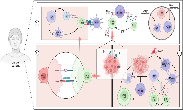
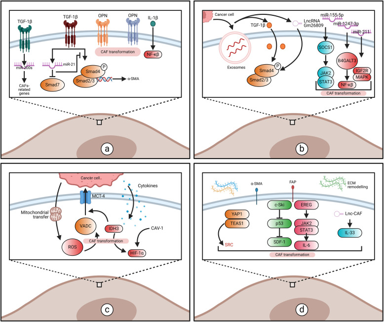
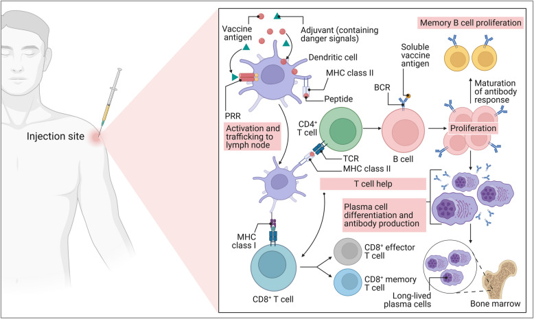
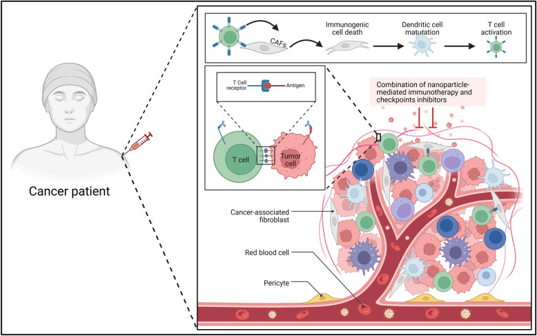
Verification
Claims the paper makes
The claims and statistics of this paper haven’t been checked yet.
Run the check — free, no account needed. It reads the open-access text and checks the sources it cites.
Claims other papers make about this one
Each is checked against this paper’s own text: does it say what the citing paper says it does?
In the citing paper
“Many elegant platforms still stall at the same three failure points: perivascular sequestration, stromal drag and incomplete or misplaced activation [10].”
From Overcoming tumor microenvironment barriers: transformable and bioinspired nanomedicine strategies for deep tumor penetration (2026) · cites this paper as [10]
Self-check
Percentages, sums and reported differences re-computed from the paper’s own numbers.
The self-check needs the paper’s full text.
Citations
Cites
Principles of nanoparticle design for overcoming biological barriers to drug delivery
Blanco, E., Shen, H., & Ferrari, M. (2015) · Nature Biotechnology · doi:10.1038/nbt.3330 · Cited by 7,002
mRNA-lipid nanoparticle COVID-19 vaccines: structure and stability
Schoenmaker, L., Witzigmann, D., Kulkarni, J. A., Verbeke, R., Kersten, G., Jiskoot, W., & Crommelin, D. J. A. (2021) · International Journal of Pharmaceutics · doi:10.1016/j.ijpharm.2021.120586 · Cited by 1,566
Ning, S., Lyu, M., Zhu, D., Lam, J. W. Y., Huang, Q., Zhang, T., & Tang, B. Z. (2023) · ACS Nano · doi:10.1021/acsnano.3c00326 · Cited by 293
Role of the tumor microenvironment in cancer hallmarks and targeted therapy (Review)
Naser, R., Fakhoury, I., El‑Fouani, A., Abi‑Habib, R., & El‑Sibai, M. (2022) · International Journal of Oncology · doi:10.3892/ijo.2022.5471 · Cited by 61
Research works printed but not linked: 350
- Smith J, et al. Collagen deposition and hypoxia in NSCLC resistance mechanisms. Journal of Cancer Research. 2023;45(3):123–34.
- Zhang Y, et al. pH-responsive nanoparticles for targeted drug delivery in cancer therapy. Nanomedicine. 2022;17(8):567–79.
- Wang L, et al. Hypoxia-responsive nanocarriers for enhanced chemotherapy and radiotherapy. Adv Drug Deliv Rev. 2021;170:45–60.
- Li X, et al. Enzyme-responsive nanoparticles for cancer therapy. Biomaterials. 2020;250:120–35.
- Chen H, et al. Iron oxide nanoplatforms for combined photothermal and immunotherapy in pancreatic cancer. Nat Nanotechnol. 2023;18(4):456–67.
- Liu R, et al. Multifunctional nanocarriers for combination therapy in cancer treatment. ACS Nano. 2022;16(5):7890–905. · DOI
- Patel S, et al. Machine learning models for predicting immunotherapy response in cancer patients. Cancer Immunol Res. 2023;11(2):234–46.
- Kim T, et al. Mitophagy-related gene signatures as predictive biomarkers in rectal cancer. Clin Cancer Res. 2022;28(10):2015–27.
- Huang Y, et al. Overcoming biological barriers in the tumor microenvironment for effective drug delivery. J Control Release. 2021;330:714–28.
- Zhao W, et al. Iron oxide nanoparticles for repolarizing tumor-associated macrophages. Biomaterials Science. 2023;11(6):1234–45. · DOI
- Yan Y, et al. LBP-CD155 Liposome Nanovaccine Efficiently Resist Colorectal Cancer and Enhance ICB Therapy. Int J Nanomedicine. 2025;20:1047–63. · DOI
- Qiu X, Yang A, Poholek AC. Stayin’ Alive: Targeting Chromatin Regulators of Clonal Hematopoiesis Promotes CD8 T-cell Stemness. Cancer Res. 2025;85(2):200–2. · DOI
- Cai Y, et al. Comprehensive Analyses of PANoptosome with Potential Implications in Cancer Prognosis and Immunotherapy. Biochem Genet. 2025;63(1):331–53. · DOI
- Zhang S, et al. An ultra-long-acting L-asparaginase synergizes with an immune checkpoint inhibitor in starvation-immunotherapy of metastatic solid tumors. Biomaterials. 2025;312:122740. · DOI
- Wu Z, et al. IL-12 minicircle delivery via extracellular vesicles as immunotherapy for bladder cancer. Cell Prolif. 2025;58(1):e13739. · DOI
- Chen H, et al. Manganese-coordinated nanoparticle with high drug-loading capacity and synergistic photo-/immuno-therapy for cancer treatments. Biomaterials. 2025;312:122745. · DOI
- Gao S, et al. A precision intelligent nanomissile for inhibiting tumor metastasis, boosting energy deprivation and immunotherapy. Biomaterials. 2025;315:122953. · DOI
- Xia J, et al. Antigen self-presenting dendrosomes swallowing nanovaccines boost antigens and STING agonists codelivery for cancer immunotherapy. Biomaterials. 2025;316:122998. · DOI
- Guo C, et al. Precise HER2 Protein Degradation via Peptide-Conjugated Photodynamic Therapy for Enhanced Breast Cancer Immunotherapy. Adv Sci (Weinh). 2025;12(2):e2410778. · DOI
- Moon Y, et al. Self-Assembled Peptide-Derived Proteolysis-Targeting Chimera (PROTAC) Nanoparticles for Tumor-Targeted and Durable PD-L1 Degradation in Cancer Immunotherapy. Angew Chem Int Ed Engl. 2025;64(5):e202414146. · DOI
- Chu X, et al. Bioactive nanocomposite hydrogel enhances postoperative immunotherapy and bone reconstruction for osteosarcoma treatment. Biomaterials. 2025;312:122714. · DOI
- Yang W, et al. One-dimensional nanosonosensitizer boosted multiple branches of immune responses against MHC-deficient immune-evasive urologic tumor Sci Adv. 2025;11(5):eado7373.
- Yang Z, et al. Elucidating stearoyl metabolism and NCOA4-mediated ferroptosis in gastric cancer liver metastasis through multi-omics single-cell integrative mendelian analysis: advancing personalized immunotherapy strategies. Discov Oncol. 2025;16(1):46. · DOI
- Wang L, et al. Intestinal Subtype as a Biomarker of Response to Neoadjuvant Immunochemotherapy in Locally Advanced Gastric Adenocarcinoma: Insights from a Prospective Phase II Trial. Clin Cancer Res. 2025;31(1):74–86. · DOI
- Tan CL, et al. Prediction of tumor-reactive T cell receptors from scRNA-seq data for personalized T cell therapy. Nat Biotechnol. 2025;43(1):134–42. · DOI
- Liu B, et al. Impact of air pollution on the progress-free survival of non-small cell lung cancer patients with anti-PD-1/PD-L1 immunotherapy: A cohort study. Environ Pollut. 2025;368:125683. · DOI
- Valtis YK, et al. Cancer cachexia and weight loss before CAR T-cell therapy for lymphoma are independently associated with poor outcomes. Blood Adv. 2025;9(1):151–61. · DOI
- Lordick F, et al. Adjuvant immunotherapy in patients with resected gastric and oesophagogastric junction cancer following preoperative chemotherapy with high risk for recurrence (ypN+ and/or R1): European Organisation of Research and Treatment of Cancer (EORTC) 1707 VESTIGE study. Ann Oncol. 2025;36(2):197–207. · DOI
- Ren XH, et al. A Multiple Targeting Genome Editing System for Remodulation of Circulating Malignant Cells to Eliminate Cancer Immunosuppression and Restore Immune Responses. Adv Healthc Mater. 2025;14(1):e2401223. · DOI
- Logun M, et al. Patient-derived glioblastoma organoids as real-time avatars for assessing responses to clinical CAR-T cell therapy. Cell Stem Cell. 2025;32(2):181-190.e4. · DOI
- Cui JW, et al. Tumor immunotherapy resistance: Revealing the mechanism of PD-1 / PD-L1-mediated tumor immune escape. Biomed Pharmacother. 2024;171:116203. · DOI
- Lai DW, et al. The potential immunotherapy effect of Ginkgolide B thwarts oral squamous cell carcinoma progression by targeting the SREBP1/KLK8/CCL22 axis. Phytomedicine. 2025;136:156249. · DOI
- An Z, et al. The characterization of tumor immune microenvironment after neoadjuvant immunotherapy in head and neck squamous cell cancer using multiplex immunohistochemistry. Oral Oncol. 2025;161:107151. · DOI
- Feng Z, et al. Expression of extracellular HSP90α in pre- and post-treatment samples of driver-gene-negative non-small cell lung cancer. Int Immunopharmacol. 2025;146:113792. · DOI
- Liu Y, et al. Tumor-Selective Nano-Dispatcher Enforced Cancer Immunotherapeutic Effects via Regulating Lactate Metabolism and Activating Toll-Like Receptors. Small. 2025;21(1):e2406870. · DOI
- Vanacker H, et al. Brief Communication on MAGE-A4 and Coexpression of Cancer Testis Antigens in Metastatic Synovial Sarcomas: Considerations for Development of Immunotherapeutics. J Immunother. 2025;48(1):27–31. · DOI
- Wang Q, et al. HPK1 kinase inhibitor: a sufficient approach to target HPK1 to modulate T cell activation in cancer immunotherapy compared with degraders. Front Immunol. 2025;16:1449106. · DOI
- Huang D, et al. Pan-cancer and experimental analyses reveal the immunotherapeutic significance of CST2 and its association with stomach adenocarcinoma proliferation and metastasis. Front Immunol. 2024;15:1466806. · DOI
- Wang C, et al. Tumor-Associated Myeloid Cells Selective Delivery of a Therapeutic Tumor Nano-Vaccine for Overcoming Immune Barriers for Effective and Long-Term Cancer Immunotherapy. Adv Healthc Mater. 2024;13(26):e2401416.
- Li C, et al. Classical Monocytes Shuttling for Precise Delivery of Nanotherapeutics to Glioblastoma. Adv Healthc Mater. 2024;13(29):e2400925. · DOI
- Littrell CA, et al. Systemically targeting monocytic myeloid-derived suppressor cells using dendrimers and their cell-level biodistribution kinetics. J Control Release. 2024;374:181–93. · DOI
- Zhou Q, et al. Immunotherapy Strategy Targeting Programmed Cell Death Ligand 1 and CD73 with Macrophage-Derived Mimetic Nanovesicles to Treat Bladder Cancer. Mol Pharm. 2021;18(11):4015–28. · DOI
- Wang Y, et al. mRNA Delivery of a Bispecific Single-Domain Antibody to Polarize Tumor-Associated Macrophages and Synergize Immunotherapy against Liver Malignancies. Adv Mater. 2021;33(23):e2007603. · DOI
- Chen D, et al. MBene as a Theranostic Nanoplatform for Photocontrolled Intratumoral Retention and Drug Release. Adv Mater. 2021;33(16):e2008089. · DOI
- Liu X, et al. Pillar[6]arene-Based Supramolecular Nanocatalysts for Synergistically Enhanced Chemodynamic Therapy by the Intracellular Cascade Reaction. ACS Appl Mater Interfaces. 2021;13(45):53574–85. · DOI
- Zou J, et al. Glycoprotein Ib-regulated micro platelet ghost for biosafe distribution and photothermal oncotherapy. J Control Release. 2022;351:341–60. · DOI
- Meng Q, et al. 10-hydroxycamptothecin-loaded starch-based microcapsules with the stepwise responsive release strategy for targeted controlled release. Int J Biol Macromol. 2023;252:126424. · DOI
- Zhang BB, et al. Lipid/PAA-coated mesoporous silica nanoparticles for dual-pH-responsive codelivery of arsenic trioxide/paclitaxel against breast cancer cells. Acta Pharmacol Sin. 2021;42(5):832–42. · DOI
- Jiang X, et al. In situ tumor-triggered subcellular precise delivery of multi-drugs for enhanced chemo-photothermal-starvation combination antitumor therapy. Theranostics. 2020;10(26):12158–73. · DOI
- Xiong Z, et al. A Dual-Responsive Platform Based on Antifouling Dendrimer-CuS Nanohybrids for Enhanced Tumor Delivery and Combination Therapy. Small Methods. 2021;5(6):e2100204. · DOI
- Li J, et al. Rapid pH-responsive self-disintegrating nanoassemblies balance tumor accumulation and penetration for enhanced anti-breast cancer therapy. Acta Biomater. 2021;134:546–58. · DOI
- Zhang Y, et al. Cascade-Responsive 2-DG Nanocapsules Encapsulate aV-siCPT1C Conjugates to Inhibit Glioblastoma through Multiple Inhibition of Energy Metabolism. ACS Appl Mater Interfaces. 2023;15(8):10356–70. · DOI
- Chang R, et al. Controlled Dissociation of Polymeric Micelles in Response to Oxidative Stress. Biomacromol. 2024;25(2):1162–70. · DOI
- Chen X, et al. Construction of multi-program responsive vitamin E succinate-chitosan-histidine nanocarrier and its response strategy in tumor therapy. Int J Biol Macromol. 2023;246:125678. · DOI
- Priwitaningrum DL, et al. Apoptosis-inducing peptide loaded in PLGA nanoparticles induces anti-tumor effects in vivo. Int J Pharm. 2020;585:119535. · DOI
- Hu S, et al. Codelivery of CuS and DOX into Deep Tumors with Size and Charge-Switchable PAMAM Dendrimers for Chemo-photothermal Therapy. ACS Appl Mater Interfaces. 2023;15(46):53273–82. · DOI
- Hou L, et al. Transformable Honeycomb-Like Nanoassemblies of Carbon Dots for Regulated Multisite Delivery and Enhanced Antitumor Chemoimmunotherapy. Angew Chem Int Ed Engl. 2021;60(12):6581–92. · DOI
- Wang C, et al. A paclitaxel prodrug nanoparticles with glutathion/reactive oxygen species dual-responsive and CD206 targeting to improve the anti-tumour effect. IET Nanobiotechnol. 2023;17(5):406–19. · DOI
- Zhong S, et al. Herceptin-decorated paclitaxel-loaded poly(lactide-co-glycolide) nanobubbles: ultrasound-facilitated release and targeted accumulation in breast cancers. Pharm Dev Technol. 2020;25(4):454–63. · DOI
- Zhang Y, et al. Engineering a porphyrin COFs encapsulated by hyaluronic acid tumor-targeted nanoplatform for sequential chemo-photodynamic multimodal tumor therapy. Int J Biol Macromol. 2024;279(Pt 3):135328. · DOI
- Vaghasiya K, et al. Matrix Metalloproteinase-Responsive Mesoporous Silica Nanoparticles Cloaked with Cleavable Protein for “Self-Actuating” On-Demand Controlled Drug Delivery for Cancer Therapy. ACS Appl Bio Mater. 2020;3(8):4987–99. · DOI
- Gou S, et al. Glutathione-Responsive and Hydrogen Sulfide Self-Generating Nanocages Based on Self-Weaving Technology To Optimize Cancer Immunotherapy. ACS Nano. 2024;18(14):9871–85. · DOI
- Nourollahian T, et al. Targeted doxorubicin-loaded core-shell copper peroxide-mesoporous silica nanoparticles for combination of ferroptosis and chemotherapy of metastatic breast cancer. Int J Pharm. 2024;662:124487. · DOI
- Yuan P, et al. Modulating Elasticity of Liposome for Enhanced Cancer Immunotherapy. ACS Nano. 2024;18(34):23797–811. · DOI
- Huang Y, et al. Dynamic core crosslinked camptothecin prodrug micelles with reduction sensitivity and boronic acid-mediated enhanced endocytosis: An intelligent tumor-targeted delivery nanoplatform. Int J Pharm. 2020;580:119250. · DOI
- Singh S, Pal K. Folic-acid adorned alginate-polydopamine modified paclitaxel/Zn-CuO nanocomplex for pH triggered drug release and synergistic antitumor efficacy. Int J Biol Macromol. 2023;234:123602. · DOI
- Jalilian S, et al. A comparative study of sericin and gluten for magnetic nanoparticle-mediated drug delivery to breast cancer cell lines. Sci Rep. 2024;14(1):18150. · DOI
- Li Y, et al. ROS-Triggered Self-Assembled Nanoparticles Based on a Chemo-Sonodynamic Combinational Therapy Strategy for the Noninvasive Elimination of Hypoxic Tumors. ACS Appl Mater Interfaces. 2023;15(12):15893–906. · DOI
- Wu B, et al. Nanoparticle-Stabilized Oxygen Microcapsules Prepared by Interfacial Polymerization for Enhanced Oxygen Delivery. Angew Chem Int Ed Engl. 2021;60(17):9284–9. · DOI
- Mansur AAP, et al. Carboxymethylcellulose biofunctionalized ternary quantum dots for subcellular-targeted brain cancer nanotheranostics. Int J Biol Macromol. 2022;210:530–44. · DOI
- Chauhan M, et al. Polypropylene sulphide coating on magnetic nanoparticles as a novel platform for excellent biocompatible, stimuli-responsive smart magnetic nanocarriers for cancer therapeutics. Nanoscale. 2023;15(16):7384–402. · DOI
- Sun, Q., et al., pH-sensitive and redox-responsive poly(tetraethylene glycol) nanoparticle-based platform for cancer treatment. Nanotechnology. 2024;35(49). · DOI
- Zhang N, et al. Construction of hyperbranched and pH-responsive polymeric nanocarriers by yne-phenol click-reaction for tumor synergistic chemotherapy. Colloids Surf B Biointerfaces. 2021;204:111790. · DOI
- Joseph MM, et al. Targeted Theranostic Nano Vehicle Endorsed with Self-Destruction and Immunostimulatory Features to Circumvent Drug Resistance and Wipe-Out Tumor Reinitiating Cancer Stem Cells. Small. 2020;16(38):e2003309. · DOI
- Luo H, et al. Framework Nucleic Acid-Based Multifunctional Tumor Theranostic Nanosystem for miRNA Fluorescence Imaging and Chemo/Gene Therapy. ACS Appl Mater Interfaces. 2023;15(28):33223–38. · DOI
- Yang, Y., et al., Nanoparticles loaded withβ-Lapachone and Fe(3+)exhibit enhanced chemodynamic therapy by producing H(2)O(2)through cascaded amplification. Biomed Mater. 2024;19(2). · DOI
- Shi K, et al. Tumor microenvironment targeting with dual stimuli-responsive nanoparticles based on small heat shock proteins for antitumor drug delivery. Acta Biomater. 2020;114:369–83. · DOI
- Gong N, et al. Tumour-derived small extracellular vesicles act as a barrier to therapeutic nanoparticle delivery. Nat Mater. 2024;23(12):1736–47. · DOI
- Zhang W, et al. Tumor microenvironment-activated cancer cell membrane-liposome hybrid nanoparticle-mediated synergistic metabolic therapy and chemotherapy for non-small cell lung cancer. J Nanobiotechnology. 2021;19(1):339. · DOI
- Zhu Y, et al. Paclitaxel-loaded ginsenoside Rg3 liposomes for drug-resistant cancer therapy by dual targeting of the tumor microenvironment and cancer cells. J Adv Res. 2023;49:159–73. · DOI
- He L, et al. Tumor Microenvironment-Responsive Nanocapsule Delivery CRISPR/Cas9 to Reprogram the Immunosuppressive Microenvironment in Hepatoma Carcinoma. Adv Sci (Weinh). 2024;11(26):e2403858. · DOI
- Lin Y, et al. Immunostimulatory gene therapy combined with checkpoint blockade reshapes tumor microenvironment and enhances ovarian cancer immunotherapy. Acta Pharm Sin B. 2024;14(2):854–68. · DOI
- Su X, Wang Z, Duan S. Targeted drug delivery systems for pancreatic ductal adenocarcinoma: overcoming tumor microenvironment challenges with CAF-specific nanoparticles. J Natl Cancer Cent. 2023;3(4):306–9. · DOI
- Zhang Y, Han X, Nie G. Responsive and activable nanomedicines for remodeling the tumor microenvironment. Nat Protoc. 2021;16(1):405–30. · DOI
- Liu X, et al. Hypoxia responsive nano-drug delivery system based on angelica polysaccharide for liver cancer therapy. Drug Deliv. 2022;29(1):138–48. · DOI
- Zhang N, et al. Nano-drug delivery system with enhanced tumour penetration and layered anti-tumour efficacy. Nanomedicine. 2022;45:102592. · DOI
- Hu Z, et al. Construction of nano-drug delivery and antitumor system of stimuli-responsive polypeptides. Colloids Surf B Biointerfaces. 2023;226:113310. · DOI
- Badparvar F, et al. pH/redox responsive size-switchable intelligent nanovehicle for tumor microenvironment targeted DOX release. Sci Rep. 2023;13(1):22475. · DOI
- Ma T, et al. GSH/pH dual response drug delivery system for photothermal enhanced gene-immunotherapy. Nanoscale. 2023;15(42):16947–58. · DOI · Paper page
- Yang L, et al. Sequential responsive nano-PROTACs for precise intracellular delivery and enhanced degradation efficacy in colorectal cancer therapy. Signal Transduct Target Ther. 2024;9(1):275. · DOI
- Sun X, et al. A tumor microenvironment-activatable nanoplatform with phycocyanin-assisted in-situ nanoagent generation for synergistic treatment of colorectal cancer. Biomaterials. 2023;301:122263. · DOI
- Maeda H, Nakamura H, Fang J. Tumor-selective delivery of macromolecular drugs via the EPR effect: From the discovery to the novel concepts and applications. J Control Release. 2013;172(2):590–602.
- Danhier F. To exploit the tumor EPR effect: Fact or fiction. J Control Release. 2016;241:e138.
- Lammers T, et al. Tumour-targeted nanomedicines: Principles and recent progress. J Control Release. 2012;161(2):175–87. · DOI · Paper page
- Peer D, et al. Nanocarriers as an emerging platform for cancer therapy. Nat Nanotechnol. 2007;2(12):751–60. · DOI · Paper page
- Miele E, et al. Solid tumor targeting by nanoparticles: The EPR effect and beyond. Drug Discovery Today. 2017;22(6):687–97.
- Sun Q, et al. Stimuli-responsive nanoparticles for controlled drug release and cancer therapy. Biomaterials Science. 2019;7(1):10–25.
- Lee ES, et al. pH-sensitive polymeric micelles for delivery of doxorubicin. J Control Release. 2008;123(1):19–29. · DOI · Paper page
- Wang Y, et al. Stimuli-responsive drug delivery systems for cancer therapy. Asian J Pharm Sci. 2018;13(3):205–16.
- Shi J, et al. Inorganic nanocarriers for tumor-targeted drug delivery. Chem Rev. 2017;117(12):9373–465.
- Mahdavi Firouzabadi B, et al. Design of Nanoparticles in Cancer Therapy Based on Tumor Microenvironment Properties. Pharmaceutics. 2022;14(12):2708. · DOI
- Zein R, Sharrouf W, Selting KA. Physical Properties of Nanoparticles That Result in Improved Cancer Targeting. J Oncol. 2020;2020:5194780. · DOI
- Saboorian-Jooybari H, Chen Z. Surface charging parameters of charged particles in symmetrical electrolyte solutions. Phys Chem Chem Phys. 2020;22(35):20123–42. · DOI
- Ejigah V, et al. Approaches to Improve Macromolecule and Nanoparticle Accumulation in the Tumor Microenvironment by the Enhanced Permeability and Retention Effect. Polymers. 2022;14(13):2601. · DOI
- Duan S, et al. Detachable Dual-Targeting Nanoparticles for Improving the Antitumor Effect by Extracellular Matrix Depletion. ACS Biomater Sci Eng. 2023;9(3):1437–49. · DOI · Paper page
- He Y, et al. Enzyme-Triggered Size-Switchable Nanosystem for Deep Tumor Penetration and Hydrogen Therapy. ACS Appl Mater Interfaces. 2023;15(1):552–65. · DOI
- Souri M, et al. Stimuli-sensitive nano-drug delivery with programmable size changes to enhance accumulation of therapeutic agents in tumors. Drug Deliv. 2023;30(1):2186312. · DOI
- Qian X, et al. Strategies of engineering nanomedicines for tumor retention. J Control Release. 2022;346:193–211. · DOI
- Soprano E, et al. Biomimetic cell-derived nanocarriers in cancer research. J Nanobiotechnology. 2022;20(1):538. · DOI
- Hu T, et al. Biomimetic Cell-Derived Nanoparticles: Emerging Platforms for Cancer Immunotherapy. Pharmaceutics. 2023;15(7):1821. · DOI
- Chang, A., et al., Enrichment of surface charge contributes to the stability of surface nanobubbles. Particuology. 2023. · DOI
- Zhang P, et al. Charge reversal nano-systems for tumor therapy. J Nanobiotechnology. 2022;20(1):31s. · DOI
- Santra S, et al. Tumor acidity-induced surface charge modulation in covalent nanonetworks for activated cellular uptake: targeted delivery of anticancer drugs and selective cancer cell death. Biomater Sci. 2023;11(16):5549–59. · DOI
- Bilardo, R., et al., Influence of surface chemistry and morphology of nanoparticles on protein corona formation. Wiley Interdisciplinary Reviews-Nanomedicine and Nanobiotechnology. 2022. · DOI
- Yang W, et al. Nanoparticle Surface Engineering with Heparosan Polysaccharide Reduces Serum Protein Adsorption and Enhances Cellular Uptake. Nano Lett. 2022;22(5):2103–11. · DOI
- Fukuda S, Xu Y. A biomimetic anti-biofouling coating in nanofluidic channels. J Mater Chem B. 2022;10(14):2481–9. · DOI
- Liu Y, et al. Intrapleural nano-immunotherapy promotes innate and adaptive immune responses to enhance anti-PD-L1 therapy for malignant pleural effusion. Nat Nanotechnol. 2022;17(2):206–16. · DOI
- Baljon JJ, et al. A Cancer Nanovaccine for Co-Delivery of Peptide Neoantigens and Optimized Combinations of STING and TLR4 Agonists. ACS Nano. 2024;18(9):6845–62. · DOI
- Chen W, et al. Efficient pH-Responsive Nano-Drug Delivery System Based on Dynamic Boronic Acid/Ester Transformation. Molecules. 2023;28(11):4461. · DOI
- Li Y, et al. Ultrasound-Responsive Nanocarriers Delivering siRNA and Fe(3)O(4) Nanoparticles Reprogram Macrophages and Inhibit M2 Polarization for Enhanced NSCLC Immunotherapy. ACS Appl Mater Interfaces. 2024;16(42):56634–52. · DOI
- Chen X, et al. Dual-targeted delivery of temozolomide by multi-responsive nanoplatform via tumor microenvironment modulation for overcoming drug resistance to treat glioblastoma. J Nanobiotechnology. 2024;22(1):264. · DOI
- Sjoerdsma JN, et al. Combination non-targeted and sGRP78-targeted nanoparticle drug delivery outperforms either component to treat metastatic ovarian cancer. J Control Release. 2024;375:438–53. · DOI
- Zhang Q, et al. Comparative Efficacy of Tumor Microenvironment-responsive Nanotherapeutics Targeting PSD95/Discs-large/ZO-1 Binding Kinase in Different Histological Subgroups of Medulloblastoma. Int J Med Sci. 2024;21(15):3018–33. · DOI
- Guo F, et al. Enzyme-responsive nano-drug delivery system for combined antitumor therapy. Int J Biol Macromol. 2022;220:1133–45. · DOI
- Jiang X, et al. Tumor-Activatable Nanoparticles Target Low-Density Lipoprotein Receptor to Enhance Drug Delivery and Antitumor Efficacy. Adv Sci (Weinh). 2022;9(24):e2201614. · DOI
- Yang Y, et al. A multi-responsive targeting drug delivery system for combination photothermal/chemotherapy of tumor. J Biomater Sci Polym Ed. 2023;34(2):166–83. · DOI
- Zelepukin IV, et al. Flash drug release from nanoparticles accumulated in the targeted blood vessels facilitates the tumour treatment. Nat Commun. 2022;13(1):6910. · DOI
- Li L, et al. Quercetin-ferrum nanoparticles enhance photothermal therapy by modulating the tumor immunosuppressive microenvironment. Acta Biomater. 2022;154:454–66. · DOI
- An J, et al. ROS-augmented and tumor-microenvironment responsive biodegradable nanoplatform for enhancing chemo-sonodynamic therapy. Biomaterials. 2020;234:119761. · DOI
- Zhang Y, et al. Novel co-delivery nanomedicine for photodynamic enlarged immunotherapy by cascade immune activation and efficient Immunosuppression reversion. Bioorg Chem. 2024;153:107978. · DOI
- Yu Z, et al. Nano delivery of simvastatin targets liver sinusoidal endothelial cells to remodel tumor microenvironment for hepatocellular carcinoma. J Nanobiotechnology. 2022;20(1):9. · DOI · Paper page
- Koula G, et al. Integrin receptor-targeted, doxorubicin-loaded cerium oxide nanoparticles delivery to combat glioblastoma. Nanomedicine (Lond). 2024;19(15):1389–406. · DOI
- Burkert SC, et al. Nitrogen-Doped Carbon Nanotube Cups for Cancer Therapy. ACS Appl Nano Mater. 2022;5(10):13685–96. · DOI
- Singla P, et al. Double Imprinted Nanoparticles for Sequential Membrane-to-Nuclear Drug Delivery. Adv Sci (Weinh). 2024;11(36):e2309976. · DOI
- Jia Y, et al. Tumor Microenvironment-Responsive Nanoherb Delivery System for Synergistically Inhibition of Cancer Stem Cells. ACS Appl Mater Interfaces. 2023;15(13):16329–42. · DOI
- Wang HF, et al. Tumor-Microenvironment-on-a-Chip for Evaluating Nanoparticle-Loaded Macrophages for Drug Delivery. ACS Biomater Sci Eng. 2020;6(9):5040–50. · DOI
- Liu Y, et al. Hydrogel Microtumor Arrays to Evaluate Nanotherapeutics. Adv Healthc Mater. 2023;12(14):e2201696. · DOI
- Zhang K, et al. A Self-Disguised Nanospy for Improving Drug Delivery Efficiency via Decreasing Drug Protonation. Small. 2023;19(25):e2300060. · DOI
- Guo Y, et al. Polymeric nanocarrier via metabolism regulation mediates immunogenic cell death with spatiotemporal orchestration for cancer immunotherapy. Nat Commun. 2024;15(1):8586. · DOI
- Jiang G, et al. TMTP1-Modified, Tumor Microenvironment Responsive Nanoparticles Co-Deliver Cisplatin and Paclitaxel Prodrugs for Effective Cervical Cancer Therapy. Int J Nanomedicine. 2021;16:4087–104. · DOI
- Souri M, et al. Towards principled design of cancer nanomedicine to accelerate clinical translation. Materials Today Bio. 2022;13:100208. · DOI · Paper page
- Ahmed S, Rehman SU, Tabish M. Cancer nanomedicine: A step towards improving the drug delivery and enhanced efficacy of chemotherapeutic drugs. OpenNano. 2022;7:100051. · DOI
- Bhatia SN, et al. Cancer nanomedicine. Nat Rev Cancer. 2022;22(10):550–6. · DOI
- Hu R, et al. Liquid Nanoparticles for Nanocatalytic Cancer Therapy. Adv Mater. 2023;35(48):e2306469. · DOI
- Shi Y, et al. The EPR effect and beyond: Strategies to improve tumor targeting and cancer nanomedicine treatment efficacy. Theranostics. 2020;10(17):7921–4. · DOI · Paper page
- Fernandes C, et al. Scale-Up of Nanoparticle Manufacturing Process. In: Jindal AB, editor., et al., Pharmaceutical Process Engineering and Scale-up Principles. Cham: Springer Nature Switzerland; 2023. p. 173–203. · DOI
- Singh, S., et al., Chapter 20 - Regulatory pathway to introduce a nanomedicine product in the market at international level, in Theory and Applications of Nonparenteral Nanomedicines, P. Kesharwani, S. Taurin, and K. Greish, Editors. 2021, Academic Press. p. 489–499. · DOI
- Đorđević S, et al. Current hurdles to the translation of nanomedicines from bench to the clinic. Drug Deliv Transl Res. 2022;12(3):500–25. · DOI
- Tian X, Shi A, Wu J. Construction of Biomimetic-Responsive Nanocarriers and their Applications in Tumor Targeting. Anticancer Agents Med Chem. 2022;22(12):2255–73. · DOI
- Nasra S, Chaudhari R, Kumar A. Role of Nanomedicine for Cancer Immunotherapy. In: Malik A, Afaq S, Tarique M, editors. Nanomedicine for Cancer Diagnosis and Therapy. Singapore: Springer Singapore; 2021. p. 115–32. · DOI
- Martin JD, et al. Improving cancer immunotherapy using nanomedicines: progress, opportunities and challenges. Nat Rev Clin Oncol. 2020;17(4):251–66. · DOI
- Wang M, et al. Synergistic interventional photothermal therapy and immunotherapy using an iron oxide nanoplatform for the treatment of pancreatic cancer. Acta Biomater. 2022;138:453–62. · DOI
- Unnikrishnan BS, et al. Self-assembled drug loaded glycosyl-protein metal nanoconstruct: Detailed synthetic procedure and therapeutic effect in solid tumor treatment. Colloids Surf B Biointerfaces. 2020;193:111082. · DOI
- Zhang, H., et al., Artemisinin co-delivery system based on manganese oxide for precise diagnosis and treatment of breast cancer. Nanotechnology. 2021;32(32). · DOI
- Li J, et al. An oral bioactive chitosan-decorated doxorubicin nanoparticles/bacteria bioconjugates enhance chemotherapy efficacy in an in-situ breast cancer model. Int J Biol Macromol. 2024;267(Pt 1):131428. · DOI
- Snyder CS, et al. Co-Delivery Polymeric Poly(Lactic-Co-Glycolic Acid) (PLGA) Nanoparticles to Target Cancer Stem-Like Cells. Methods Mol Biol. 2024;2777:191–204. · DOI
- Iranpour S, et al. Improving anti-cancer drug delivery performance of magnetic mesoporous silica nanocarriers for more efficient colorectal cancer therapy. J Nanobiotechnology. 2021;19(1):314. · DOI
- Gao X, et al. A Virus-Mimicking Nucleic Acid Nanogel Reprograms Microglia and Macrophages for Glioblastoma Therapy. Adv Mater. 2021;33(9):e2006116. · DOI
- Kim HS, et al. A non-lipid nucleic acid delivery vector with dendritic cell tropism and stimulation. Theranostics. 2024;14(7):2934–45. · DOI
- Eldehna WM, et al. Development of potent nanosized carbonic anhydrase inhibitor for targeted therapy of hypoxic solid tumors. Int J Pharm. 2023;631:122537. · DOI
- Song N, et al. LyP-1-Modified Multifunctional Dendrimers for Targeted Antitumor and Antimetastasis Therapy. ACS Appl Mater Interfaces. 2020;12(11):12395–406. · DOI
- Su Y, et al. Amphiphilic Dendrimer Doping Enhanced pH-Sensitivity of Liposomal Vesicle for Effective Co-delivery toward Synergistic Ferroptosis-Apoptosis Therapy of Hepatocellular Carcinoma. Adv Healthc Mater. 2023;12(6):e2202663. · DOI
- Wang X, et al. Synergistic chemo-photothermal cancer therapy of pH-responsive polymeric nanoparticles loaded IR825 and DTX with charge-reversal property. Colloids Surf B Biointerfaces. 2022;209(Pt 2):112164. · DOI
- Badparvar F, et al. Dual pH/redox-responsive hyperbranched polymeric nanocarriers with TME-trigger size shrinkage and charge reversible ability for amplified chemotherapy of breast cancer. Sci Rep. 2024;14(1):8567. · DOI · Paper page
- Ding J, et al. Ultra pH-sensitive polymeric nanovesicles co-deliver doxorubicin and navitoclax for synergetic therapy of endometrial carcinoma. Biomater Sci. 2020;8(8):2264–73. · DOI
- Terracciano M, et al. Development of Surface Chemical Strategies for Synthesizing Redox-Responsive Diatomite Nanoparticles as a Green Platform for On-Demand Intracellular Release of an Antisense Peptide Nucleic Acid Anticancer Agent. Small. 2022;18(41):e2204732. · DOI
- Zhou Z, et al. ROS-responsive self-assembly nanoplatform overcomes hypoxia for enhanced photodynamic therapy. Biomater Sci. 2024;12(19):5105–14. · DOI
- Zhang J, et al. Dual-Prodrug-Based Hyaluronic Acid Nanoplatform Provides Cascade-Boosted Drug Delivery for Oxidative Stress-Enhanced Chemotherapy. ACS Appl Mater Interfaces. 2024;16(38):50459–73. · DOI
- Chen H, et al. Dual-targeting SERS-encoded graphene oxide nanocarrier for intracellular co-delivery of doxorubicin and 9-aminoacridine with enhanced combination therapy. Analyst. 2021;146(22):6893–901. · DOI
- Cheng Y, et al. Light-switchable diphtherin transgene system combined with losartan for triple negtative breast cancer therapy based on nano drug delivery system. Int J Pharm. 2022;618:121613. · DOI
- Peng N, et al. Sequential-targeting nanocarriers with pH-controlled charge reversal for enhanced mitochondria-located photodynamic-immunotherapy of cancer. Acta Biomater. 2020;105:223–38. · DOI
- Tsai MF, et al. Multi-Stimuli-Responsive DOX Released from Magnetosome for Tumor Synergistic Theranostics. Int J Nanomedicine. 2020;15:8623–39. · DOI
- Luo Y, et al. TME-activatable theranostic nanoplatform with ATP burning capability for tumor sensitization and synergistic therapy. Theranostics. 2020;10(15):6987–7001. · DOI
- Chen S, et al. Enhancing antitumor immunity with stimulus-responsive mesoporous silicon in combination with chemotherapy and photothermal therapy. Biomater Sci. 2024;12(15):3826–40. · DOI
- Yen YT, et al. Enzymatically responsive nanocarriers targeting PD-1 and TGF-β pathways reverse immunotherapeutic resistance and elicit robust therapeutic efficacy. J Nanobiotechnology. 2025;23(1):124. · DOI
- Song Y, et al. Biodegradable Imprinted Polymer Based on ZIF-8/DOX-HA for Synergistically Targeting Prostate Cancer Cells and Controlled Drug Release with Multiple Responses. ACS Appl Mater Interfaces. 2023;15(21):25339–53. · DOI
- Song X, et al. pH-Sensitive Biomimetic Nanosystem Based on Large-Pore Mesoporous Silica Nanoparticles with High Hyaluronidase Loading for Tumor Deep Penetration. ACS Appl Mater Interfaces. 2023;15(32):38294–308. · DOI
- Lai CM, et al. Three-pronged attacks by hybrid nanoassemblies involving a natural product, carbon dots, and Cu(2+) for synergistic HCC therapy. J Colloid Interface Sci. 2023;650(Pt A):526–40. · DOI
- Sui D, et al. Optimization design of sialic acid derivatives enhances the performance of liposomes for modulating immunosuppressive tumor microenvironments. Life Sci. 2022;310:121081. · DOI
- Das M, et al. Vitamin-H Channeled Self-Therapeutic P-gp Inhibitor Curcumin-Derived Nanomicelles for Targeting the Tumor Milieu by pH- and Enzyme-Triggered Hierarchical Disassembly. Bioconjug Chem. 2022;33(2):369–85. · DOI · Paper page
- Wang Y, et al. A Natural Membrane Vesicle Exosome-based Sinomenine Delivery Platform for Hepatic Carcinoma Therapy. Curr Top Med Chem. 2021;21(14):1224–34. · DOI
- Choi YS, et al. Tie2-mediated vascular remodeling by ferritin-based protein C nanoparticles confers antitumor and anti-metastatic activities. J Hematol Oncol. 2020;13(1):123. · DOI
- Da X, et al. Inhibition of growth of hepatocellular carcinoma by co-delivery of anti-PD-1 antibody and sorafenib using biomimetic nano-platelets. BMC Cancer. 2024;24(1):273. · DOI
- Liu X, et al. A Smart CA IX-Targeting and pH-Responsive Nano-Mixed Micelles for Delivery of FB15 with Superior Anti-Breast Cancer Efficacy. Int J Nanomedicine. 2024;19:10247–62. · DOI
- Wang, L., et al., Targeting Decidual CD16(+) Immune Cells with Exosome-Based Glucocorticoid Nanoparticles for Miscarriage. Adv Sci (Weinh). 2024:e2406370. · DOI
- Liu X, Chen L, Chen Z. Acid-triggered controlled release and fluorescence-switchable phthalocyanine nanoassemblies combined with O(2)-economizer for tumor imaging and collaborative photodynamic antitumor therapy. Bioorg Chem. 2024;143:106986. · DOI
- Chen M, et al. “Watson-Crick G[triple bond, length as m-dash]C”-inspired supramolecular nanodrug of methotrexate and 5-fluorouracil for tumor microenvironment-activatable self-recognizing synergistic chemotherapy. J Mater Chem B. 2020;8(17):3829–41. · DOI
- Zhang Q, et al. Biomineralized and metallized small extracellular vesicles encapsulated in hydrogels for mitochondrial-targeted synergistic tumor therapy. Acta Biomater. 2025;194:428–41. · DOI
- Xing C, et al. Two-Dimensional MXene (Ti(3)C(2))-Integrated Cellulose Hydrogels: Toward Smart Three-Dimensional Network Nanoplatforms Exhibiting Light-Induced Swelling and Bimodal Photothermal/Chemotherapy Anticancer Activity. ACS Appl Mater Interfaces. 2018;10(33):27631–43. · DOI
- D’Angelo E, et al. Tumor microenvironment-mimicking macrophage nanovesicles as a targeted therapy platform for colorectal cancer. Int J Pharm. 2025;670:125169. · DOI
- Huang W, et al. Immunometabolic reprogramming of macrophages with inhalable CRISPR/Cas9 nanotherapeutics for acute lung injury intervention. Acta Biomater. 2024;181:308–16. · DOI
- Li Z, Huang J, Wu J. pH-Sensitive nanogels for drug delivery in cancer therapy. Biomater Sci. 2021;9(3):574–89. · DOI
- Rayamajhi S, et al. Macrophage-derived exosome-mimetic hybrid vesicles for tumor targeted drug delivery. Acta Biomater. 2019;94:482–94. · DOI · Paper page
- Thambi T, et al. Hypoxia-responsive polymeric nanoparticles for tumor-targeted drug delivery. Biomaterials. 2014;35(5):1735–43. · DOI
- Fathi M, Safary A, Barar J. Therapeutic impacts of enzyme-responsive smart nanobiosystems. Bioimpacts. 2020;10(1):1–4. · DOI
- Song SJ, Choi JS. Enzyme-Responsive Amphiphilic Peptide Nanoparticles for Biocompatible and Efficient Drug Delivery. Pharmaceutics. 2022;14(1):143. · DOI
- Horvat NK, et al. Superparamagnetic Iron Oxide Nanoparticles Reprogram the Tumor Microenvironment and Reduce Lung Cancer Regrowth after Crizotinib Treatment. ACS Nano. 2024;18(17):11025–41. · DOI
- Liang C, et al. Tumor cell-targeted delivery of CRISPR/Cas9 by aptamer-functionalized lipopolymer for therapeutic genome editing of VEGFA in osteosarcoma. Biomaterials. 2017;147:68–85. · DOI
- Riley, R.S. and E.S. Day, Gold nanoparticle-mediated photothermal therapy: applications and opportunities for multimodal cancer treatment. Wiley Interdiscip Rev Nanomed Nanobiotechnol. 2017;9(4). · DOI
- Han H, et al. Biomimetic platelet membrane-coated nanoparticles for targeted therapy. Eur J Pharm Biopharm. 2022;172:1–15. · DOI
200 of 350 shown
Cited by
Sorted by citations
Sheng, J., Yuan, W., Zhang, M., Chen, Z., Qi, Y., Zhao, Y., & Zhang, S. (2026) · Journal of Nanobiotechnology · doi:10.1186/s12951-026-04028-7 · Cited by 35
He, J., Xue, W., & Li, Y. (2026) · International Journal of Nanomedicine · doi:10.2147/ijn.s560983 · Cited by 16
Metadata
- Authors
- Sabit, H., Pawlik, T. M., Radwan, F., Abdel-Hakeem, M., Abdel-Ghany, S., Wadan, A.-H. S., Elzawahri, M., El-Hashash, A., & Arneth, B.
- Venue
- Molecular Cancer
- Published
- 2025-06-03
- Volume
- 24
- Issue
- 1
- ISSN
- 1476-4598
- PMC
- PMC12131435
- Cited by
- 239
- References
- 4
- Language
- en
About these checks
Registry, retraction, Cites and statistics results are computed, not judged, with one exception: a printed reference that no index lists is read once by a model before it counts as a citation error. Claims are extracted by a model and kept only when their quote appears verbatim in the text; each cited source is then checked, with a model’s help, for whether it says what the claim relies on. Those verdicts can be wrong — read the claim’s quote and the cited source before relying on one.
Statistics and arithmetic are recomputed, not judged.
Confidence bands are shown only for fields calibrated on our annotated test set. None is calibrated yet, so no band is shown.
What counts as a claim ›How we measure confidence ›How we check this ›
Are you an author, or do you see a problem with this page? Request a review
