

The DOI matches its Crossref record; 3 of the 3 references it cites were found in a registry or an index.
Exosome-liposome hybrid nanoparticle codelivery of TP and miR497 conspicuously overcomes chemoresistant ovarian cancer
Abstract
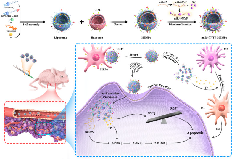
Background: Although cisplatin-based chemotherapy has been used as the first-line treatment for ovarian cancer (OC), tumor cells develop resistance to cisplatin during treatment, causing poor prognosis in OC patients. Studies have demonstrated that overactivation of the phosphatidylinositol 3-kinase/protein kinase B/mammalian target of rapamycin (PI3K/AKT/mTOR) pathway is involved in tumor chemoresistance and that overexpression of microRNA-497 (miR497) may overcome OC chemotherapy resistance by inhibiting the mTOR pathway. However, the low transcriptional efficiency and unstable chemical properties of miR497 limit its clinical application. Additionally, triptolide (TP) was confirmed to possess a superior killing effect on cisplatin-resistant cell lines, partially through inhibiting the mTOR pathway. Even so, the clinical applications of TP are restricted by serious systemic toxicity and weak water solubility. Results: Herein, whether the combined application of miR497 and TP could further overcome OC chemoresistance by synergically suppressing the mTOR signaling pathway was investigated. Bioinspired hybrid nanoparticles formed by the fusion of CD47-expressing tumor exosomes and cRGD-modified liposomes (miR497/TP-HENPs) were prepared to codeliver miR497 and TP. In vitro results indicated that the nanoparticles were efficiently taken up by tumor cells, thus significantly enhancing tumor cell apoptosis. Similarly, the hybrid nanoparticles were effectively enriched in the tumor areas and exerted significant anticancer activity without any negative effects in vivo. Mechanistically, they promoted dephosphorylation of the overactivated PI3K/AKT/mTOR signaling pathway, boosted reactive oxygen species (ROS) generation and upregulated the polarization of macrophages from M2 to M1 macrophages. Conclusion: Overall, our findings may provide a translational strategy to overcome cisplatin-resistant OC and offer a potential solution for the treatment of other cisplatin-resistant tumors. Graphical Abstract:
Overview
The claims and statistics of this paper haven’t been checked yet.
Run the check — free, no account needed. It reads the open-access text and checks the sources it cites.
Integrity signals
Registry
Cites
Statistics
Figures
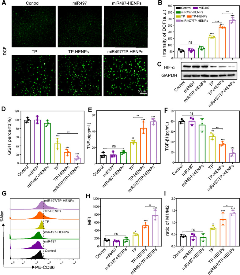
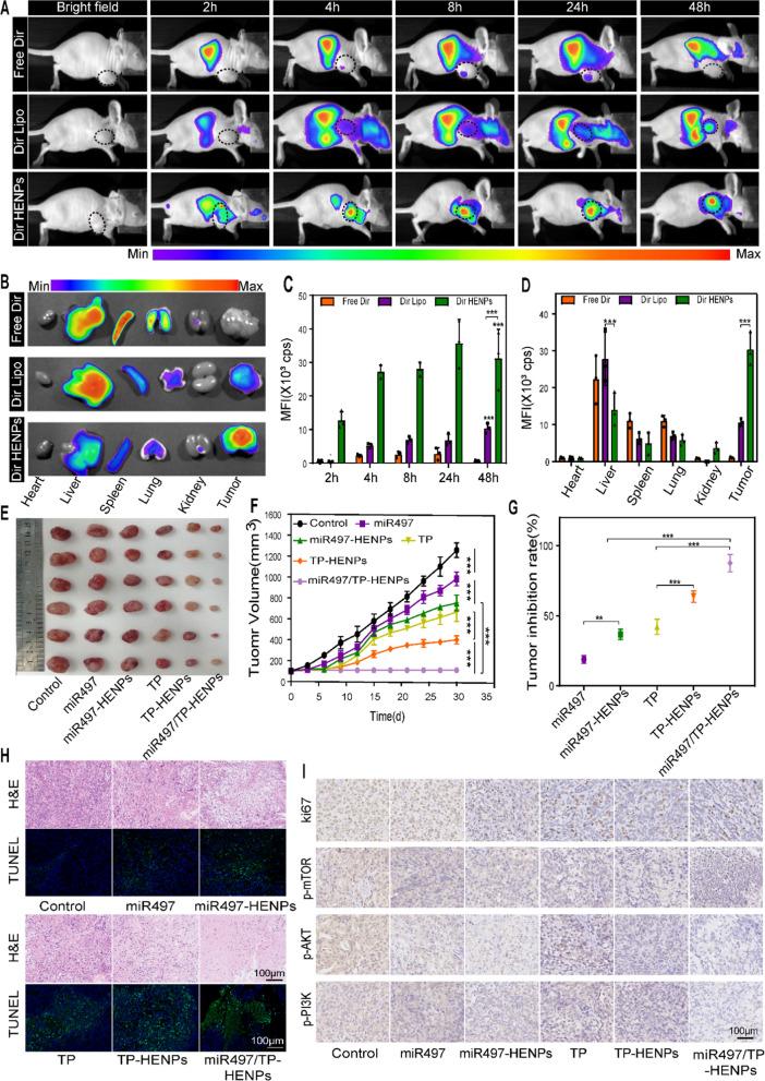
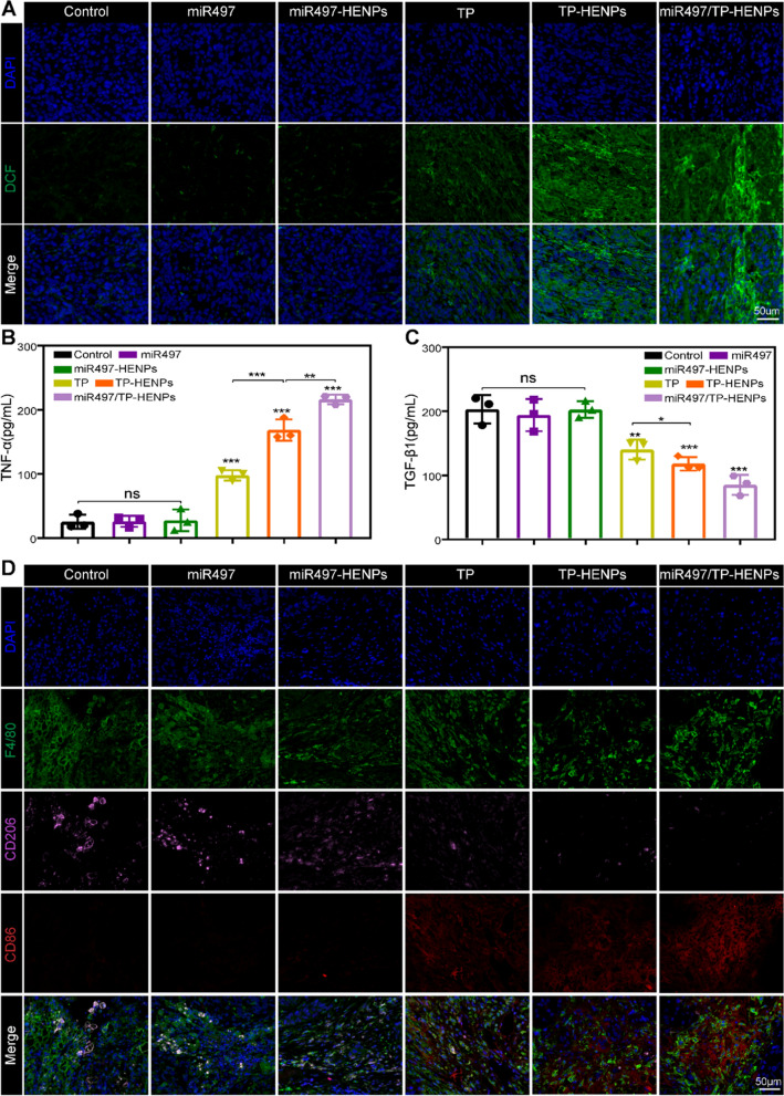
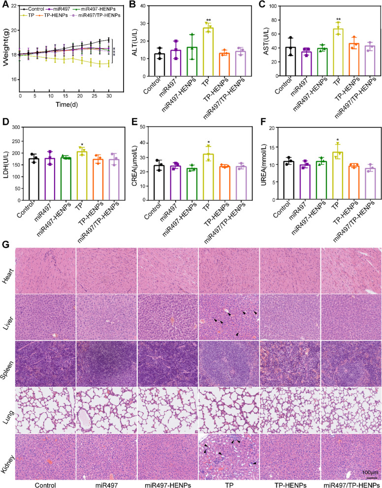
Verification
Claims the paper makes
The claims and statistics of this paper haven’t been checked yet.
Run the check — free, no account needed. It reads the open-access text and checks the sources it cites.
Claims other papers make about this one
Each is checked against this paper’s own text: does it say what the citing paper says it does?
In the citing paper
“Exosome-nanoparticle hybrid systems offer an innovative strategy by combining the natural benefits of exosomes with the versatility and enhanced features of synthetic nanoparticles (Table 2).”
From Emerging role of exosomes in cancer therapy: progress and challenges (2025) · cites this paper as [48]
In the citing paper
“By incorporating nanoparticles into or onto exosomes, researchers can create a delivery platform that offers improved stability, targeting specificity, and payload capacity [48].”
From Emerging role of exosomes in cancer therapy: progress and challenges (2025) · cites this paper as [48]
In the citing paper
“The use of these hybrids has shown significant potential in cancer therapy, where accuracy and effectiveness are crucial.”
From Emerging role of exosomes in cancer therapy: progress and challenges (2025) · cites this paper as [48]
Self-check
Percentages, sums and reported differences re-computed from the paper’s own numbers.
The self-check needs the paper’s full text.
Citations
Cites
Cisplatin: mode of cytotoxic action and molecular basis of resistance
Siddik, Z. H. (2003) · Oncogene · doi:10.1038/sj.onc.1206933 · Cited by 3,335
Molecular mechanisms of cisplatin resistance
Galluzzi, L., Senovilla, L., Vitale, I., Michels, J., Martins, I., Kepp, O., Castedo, M., & Kroemer, G. (2012) · Oncogene · doi:10.1038/onc.2011.384 · Cited by 2,760
MAST1 drives cisplatin resistance in human cancers by rewiring cRaf-independent MEK activation
Jin, L., Chun, J., Pan, C., Li, D., Lin, R., Alesi, G. N., Wang, X., Kang, H.-B., Song, L., Wang, D., et al. (2018) · Cancer Cell · doi:10.1016/j.ccell.2018.06.012 · Cited by 132
Research works printed but not linked: 57
- Siegel RL, Miller KD, Fuchs HE, Jemal A. Cancer Statistics, 2021. CA Cancer J Clin. 2021;71:7–33. · DOI · Paper page
- Yang J, Huang S, Cheng S, Jin Y, Zhang N, Wang Y. Application of ovarian cancer organoids in precision medicine: key challenges and current opportunities. Front Cell Dev Biol. 2021;9:701429.2.
- Lheureux S, Braunstein M, Oza AM. Epithelial ovarian cancer: evolution of management in the era of precision medicine. CA Cancer J Clin. 2019;69:280–304. · DOI
- Wang Y, Qiao X, Yang X, Yuan M, Xian S, Zhang L, Yang D, Liu S, Dai F, Tan Z, Cheng Y. The role of a drug-loaded poly (lactic co-glycolic acid) (PLGA) copolymer stent in the treatment of ovarian cancer. Cancer Biol Med. 2020;17:237–50. · DOI
- Spears N, Lopes F, Stefansdottir A, Rossi V, De Felici M, Anderson RA, Klinger FG. Ovarian damage from chemotherapy and current approaches to its protection. Hum Reprod Update. 2019;25:673–93. · DOI
- Mukherjee A, Huynh V, Gaines K, Reh WA, Vasquez KM. Targeting the high-mobility group box 3 protein sensitizes chemoresistant ovarian cancer cells to cisplatin. Cancer Res. 2019;79:3185–91. · DOI
- Tong L, Zhao Q, Datan E, Lin GQ, Minn I, Pomper MG, Yu B, Romo D, He QL, Liu JO. Triptolide: reflections on two decades of research and prospects for the future. Nat Prod Rep. 2021;38:843–60. · DOI
- Noel P, Von Hoff DD, Saluja AK, Velagapudi M, Borazanci E, Han H. Triptolide and its derivatives as cancer therapies. Trends Pharmacol Sci. 2019;40:327–41. · DOI · Paper page
- Hou ZY, Tong XP, Peng YB, Zhang BK, Yan M. Broad targeting of triptolide to resistance and sensitization for cancer therapy. Biomed Pharmacother. 2018;104:771–80. · DOI
- Gao J, Zhang Y, Liu X, Wu X, Huang L, Gao W. Triptolide: pharmacological spectrum, biosynthesis, chemical synthesis and derivatives. Theranostics. 2021;11:7199–221. · DOI
- Beglyarova N, Banina E, Zhou Y, Mukhamadeeva R, Andrianov G, Bobrov E, Lysenko E, Skobeleva N, Gabitova L, Restifo D, et al. Screening of conditionally reprogrammed patient-derived carcinoma cells identifies ERCC3-MYC interactions as a target in pancreatic cancer. Clin Cancer Res. 2016;22:6153–63. · DOI
- Wei J, Yan Y, Chen X, Qian L, Zeng S, Li Z, Dai S, Gong Z, Xu Z. The roles of plant-derived triptolide on non-small cell lung cancer. Oncol Res. 2019;27:849–58. · DOI
- Zhu J, Wang H, Chen F, Lv H, Xu Z, Fu J, Hou Y, Xu Y, Pi J. Triptolide enhances chemotherapeutic efficacy of antitumor drugs in non-small-cell lung cancer cells by inhibiting Nrf2-ARE activity. Toxicol Appl Pharmacol. 2018;358:1–9. · DOI
- Wang R, Ma X, Su S, Liu Y. Triptolide antagonized the cisplatin resistance in human ovarian cancer cell line A2780/CP70 via hsa-mir-6751. Future Med Chem. 2018;10:1947–55. · DOI
- Huang G, Hu H, Zhang Y, Zhu Y, Liu J, Tan B, Chen T. Triptolide sensitizes cisplatin-resistant human epithelial ovarian cancer by inhibiting the phosphorylation of AKT. J Cancer. 2019;10:3012–20. · DOI
- Ho JN, Byun SS, Lee S, Oh JJ, Hong SK, Lee SE, Yeon JS. Synergistic antitumor effect of triptolide and cisplatin in cisplatin resistant human bladder cancer cells. J Urol. 2015;193:1016–22. · DOI
- Chen YW, Lin GJ, Chuang YP, Chia WT, Hueng DY, Lin CK, Nieh S, Sytwu HK. Triptolide circumvents drug-resistant effect and enhances 5-fluorouracil antitumor effect on KB cells. Anticancer Drugs. 2010;21:502–13. · DOI
- Burris HA 3rd. Overcoming acquired resistance to anticancer therapy: focus on the PI3K/AKT/mTOR pathway. Cancer Chemother Pharmacol. 2013;71:829–42. · DOI
- Huang Y, Wu S, Zhang Y, Wang L, Guo Y. Antitumor effect of triptolide in T-cell lymphoblastic lymphoma by inhibiting cell viability, invasion, and epithelial-mesenchymal transition via regulating the PI3K/AKT/mTOR pathway. Onco Targets Ther. 2018;11:769–79. · DOI
- Kim SH, Kang JG, Kim CS, Ihm SH, Choi MG, Yoo HJ, Lee SJ. Synergistic cytotoxicity of BIIB021 with triptolide through suppression of PI3K/Akt/mTOR and NF-κB signal pathways in thyroid carcinoma cells. Biomed Pharmacother. 2016;83:22–32. · DOI
- Brozovic A, Majhen D, Roje V, Mikac N, Jakopec S, Fritz G, Osmak M, Ambriovic-Ristov A. alpha(v)beta(3) Integrin-mediated drug resistance in human laryngeal carcinoma cells is caused by glutathione-dependent elimination of drug-induced reactive oxidative species. Mol Pharmacol. 2008;74:298–306. · DOI
- Brozovic A, Ambriović-Ristov A, Osmak M. The relationship between cisplatin-induced reactive oxygen species, glutathione, and BCL-2 and resistance to cisplatin. Crit Rev Toxicol. 2010;40:347–59. · DOI
- Zhong YY, Chen HP, Tan BZ, Yu HH, Huang XS. Triptolide avoids cisplatin resistance and induces apoptosis via the reactive oxygen species/nuclear factor-κB pathway in SKOV3(PT) platinum-resistant human ovarian cancer cells. Oncol Lett. 2013;6:1084–92. · DOI
- Jiang X, Cao G, Gao G, Wang W, Zhao J, Gao C. Triptolide decreases tumor-associated macrophages infiltration and M2 polarization to remodel colon cancer immune microenvironment via inhibiting tumor-derived CXCL12. J Cell Physiol. 2021;236:193–204. · DOI
- Zheng Y, Zhang WJ, Wang XM. Triptolide with potential medicinal value for diseases of the central nervous system. CNS Neurosci Ther. 2013;19:76–82. · DOI
- Xi C, Peng S, Wu Z, Zhou Q, Zhou J. Toxicity of triptolide and the molecular mechanisms involved. Biomed Pharmacother. 2017;90:531–41. · DOI
- Wang S, Xu J, Guo Y, Cai Y, Ren X, Zhu W, Geng M, Meng L, Jiang C, Lu S. MicroRNA-497 reduction and increase of its family member microRNA-424 lead to dysregulation of multiple inflammation related genes in synovial fibroblasts with rheumatoid arthritis. Front Immunol. 2021;12: 619392.
- Nigita G, Distefano R, Veneziano D, Romano G, Rahman M, Wang K, Pass H, Croce CM, Acunzo M, Nana-Sinkam P. Tissue and exosomal miRNA editing in non-small cell lung cancer. Sci Rep. 2018;8:10222. · DOI
- Zhang T, Feng X, Zhou T, Zhou N, Shi X, Zhu X, Qiu J, Deng G, Qiu C. miR-497 induces apoptosis by the IRAK2/NF-κB axis in the canine mammary tumour. Vet Comp Oncol. 2021;19:69–78. · DOI
- Gui ZL, Wu TL, Zhao GC, Lin ZX, Xu HG. MicroRNA-497 suppress osteosarcoma by targeting MAPK/Erk pathway. Bratisl Lek Listy. 2017;118:449–52.
- Wang L, Ji XB, Wang LH, Qiu JG, Zhou FM, Liu WJ, Wan DD, Lin MC, Liu LZ, Zhang JY, Jiang BH. Regulation of microRNA-497-targeting AKT2 influences tumor growth and chemoresistance to cisplatin in lung cancer. Front Cell Dev Biol. 2020;8:840. · DOI
- Shao XJ, Miao MH, Xue J, Xue J, Ji XQ, Zhu H. The down-regulation of microRNA-497 contributes to cell growth and cisplatin resistance through PI3K/Akt pathway in osteosarcoma. Cell Physiol Biochem. 2015;36:2051–62. · DOI
- Xu S, Fu GB, Tao Z, OuYang J, Kong F, Jiang BH, Wan X, Chen K. MiR-497 decreases cisplatin resistance in ovarian cancer cells by targeting mTOR/P70S6K1. Oncotarget. 2015;6:26457–71. · DOI
- Chen X, Liang H, Zhang J, Zen K, Zhang CY. Secreted microRNAs: a new form of intercellular communication. Trends Cell Biol. 2012;22:125–32. · DOI · Paper page
- Guan W, Xu DW, Ji CH, Wang CN, Liu Y, Tang WQ, Gu JH, Chen YM, Huang J, Liu JF, Jiang B. Hippocampal miR-206–3p participates in the pathogenesis of depression via regulating the expression of BDNF. Pharmacol Res. 2021;174: 105932. · DOI
- Zhang L, Jing D, Wang L, Sun Y, Li JJ, Hill B, Yang F, Li Y, Lam KS. Unique photochemo-immuno-nanoplatform against orthotopic xenograft oral cancer and metastatic syngeneic breast cancer. Nano Lett. 2018;18:7092–103. · DOI
- Zhai Y, Ran W, Su J, Lang T, Meng J, Wang G, Zhang P, Li Y. Traceable bioinspired nanoparticle for the treatment of metastatic breast cancer via NIR-trigged intracellular delivery of methylene blue and cisplatin. Adv Mater. 2018. https://doi.org/10.1002/adma.201802378. · DOI
- Sailor MJ, Park JH. Hybrid nanoparticles for detection and treatment of cancer. Adv Mater. 2012;24:3779–802. · DOI
- Wang Y, Zhang Z, Zheng C, Zhao X, Zheng Y, Liu Q, Liu Y, Shi L. Multistage adaptive nanoparticle overcomes biological barriers for effective chemotherapy. Small. 2021;17: e2100578.
- Wang G, Zannikou M, Lofchy L, Li Y, Gaikwad H, Balyasnikova IV, Simberg D. Liposomal extravasation and accumulation in tumors as studied by fluorescence microscopy and imaging depend on the fluorescent label. ACS Nano. 2021. https://doi.org/10.1021/acsnano.1c02982. · DOI
- Song Y, Guo X, Fu J, He B, Wang X, Dai W, Zhang H, Zhang Q. Dual-targeting nanovesicles enhance specificity to dynamic tumor cells in vitro and in vivo via manipulation of αvβ3-ligand binding. Acta Pharm Sin B. 2020;10:2183–97. · DOI
- Gu Y, Lai S, Dong Y, Fu H, Song L, Chen T, Duan Y, Zhang Z. AZD9291 resistance reversal activity of a ph-sensitive nanocarrier dual-loaded with chloroquine and FGFR1 inhibitor in NSCLC. Adv Sci (Weinh). 2021;8:2002922. · DOI
- Cohen O, Betzer O, Elmaliach-Pnini N, Motiei M, Sadan T, Cohen-Berkman M, Dagan O, Popovtzer A, Yosepovich A, Barhom H, et al. “Golden” exosomes as delivery vehicles to target tumors and overcome intratumoral barriers: in vivo tracking in a model for head and neck cancer. Biomater Sci. 2021;9:2103–14. · DOI
- Pan S, Zhang Y, Huang M, Deng Z, Zhang A, Pei L, Wang L, Zhao W, Ma L, Zhang Q, Cui D. Urinary exosomes-based engineered nanovectors for homologously targeted chemo-chemodynamic prostate cancer therapy via abrogating EGFR/AKT/NF-kB/IkB signaling. Biomaterials. 2021;275: 120946.
- Wang C, Steinmetz NF. CD47 Blockade and Cowpea Mosaic Virus Nanoparticle In Situ Vaccination Triggers Phagocytosis and Tumor Killing. Adv Healthc Mater. 2019;8: e1801288.
- Lv Q, Cheng L, Lu Y, Zhang X, Wang Y, Deng J, Zhou J, Liu B, Liu J. Thermosensitive exosome-liposome hybrid nanoparticle-mediated chemoimmunotherapy for improved treatment of metastatic peritoneal cancer. Adv Sci (Weinh). 2020;7:2000515. · DOI
- Cheng L, Zhang X, Tang J, Lv Q, Liu J. Gene-engineered exosomes-thermosensitive liposomes hybrid nanovesicles by the blockade of CD47 signal for combined photothermal therapy and cancer immunotherapy. Biomaterials. 2021;275: 120964. · DOI
- Piffoux M, Silva AKA, Wilhelm C, Gazeau F, Tareste D. Modification of extracellular vesicles by fusion with liposomes for the design of personalized biogenic drug delivery systems. ACS Nano. 2018;12:6830–42. · DOI
- Yong T, Zhang X, Bie N, Zhang H, Zhang X, Li F, Hakeem A, Hu J, Gan L, Santos HA, Yang X. Tumor exosome-based nanoparticles are efficient drug carriers for chemotherapy. Nat Commun. 2019;10:3838. · DOI · Paper page
- He D, Xu X, Li L, Chen C, Gong K, Guo Q, Liu F, Wang Y, Duan Y, Li H. Functional exosome-mediated delivery of triptolide endowed with targeting properties as chemotherapy carriers for ovarian carcinoma. J Biomed Nanotechnol. 2021;17:426–38. · DOI
- Zhou BG, Wei CS, Zhang S, Zhang Z, Gao HM. Matrine reversed multidrug resistance of breast cancer MCF-7/ADR cells through PI3K/AKT signaling pathway. J Cell Biochem. 2018;119:3885–91. · DOI
- Musa F, Alard A, David-West G, Curtin JP, Blank SV, Schneider RJ. Dual mTORC1/2 inhibition as a novel strategy for the resensitization and treatment of platinum-resistant ovarian cancer. Mol Cancer Ther. 2016;15:1557–67. · DOI
- Zhang Y, Dong Y, Fu H, Huang H, Wu Z, Zhao M, Yang X, Guo Q, Duan Y, Sun Y. Multifunctional tumor-targeted PLGA nanoparticles delivering Pt(IV)/siBIRC5 for US/MRI imaging and overcoming ovarian cancer resistance. Biomaterials. 2021;269: 120478.
- Rudin CM, Yang Z, Schumaker LM, VanderWeele DJ, Newkirk K, Egorin MJ, Zuhowski EG, Cullen KJ. Inhibition of glutathione synthesis reverses Bcl-2-mediated cisplatin resistance. Cancer Res. 2003;63:312–8.
- Rodriguez-Garcia A, Lynn RC, Poussin M, Eiva MA, Shaw LC, O’Connor RS, Minutolo NG, Casado-Medrano V, Lopez G, Matsuyama T, Powell DJ Jr. CAR-T cell-mediated depletion of immunosuppressive tumor-associated macrophages promotes endogenous antitumor immunity and augments adoptive immunotherapy. Nat Commun. 2021;12:877. · DOI
- Wang H, Zheng Y, Sun Q, Zhang Z, Zhao M, Peng C, Shi S. Ginsenosides emerging as both bifunctional drugs and nanocarriers for enhanced antitumor therapies. J Nanobiotechnology. 2021;19:322. · DOI
- Ru D, Ding C, Liu L, Liu H, Shen M, Duan Y, Li H. pH sensitive triptolide-loaded liposome calcium phosphate nanoparticles exhibit enhanced anti-tumor activities against ovarian cancer without damaging the reproductive system. J Biomed Nanotechnol. 2017;13:1413–24. · DOI
Cited by
Sorted by citations
Recent advances in exosome-mediated nucleic acid delivery for cancer therapy
Zhang, Y., Liu, Q., Zhang, X., Huang, H., Tang, S., Chai, Y., Xu, Z., Li, M., Chen, X., Liu, J., et al. (2022) · Journal of Nanobiotechnology · doi:10.1186/s12951-022-01472-z · Cited by 306
Emerging role of exosomes in cancer therapy: progress and challenges
Li, J., Wang, J., & Chen, Z. (2025) · Molecular Cancer · doi:10.1186/s12943-024-02215-4 · Cited by 297
Liu, Q., Li, D., Pan, X., & Liang, Y. (2023) · Journal of Nanobiotechnology · doi:10.1186/s12951-023-02081-0 · Cited by 201
Liang, Y., Iqbal, Z., Lu, J., Wang, J., Zhang, H., Chen, X., Duan, L., & Xia, J. (2023) · Molecular Therapy · doi:10.1016/j.ymthe.2022.10.008 · Cited by 162
Programming assembly of biomimetic exosomes: An emerging theranostic nanomedicine platform
Xu, X., Xu, L., Wen, C., Xia, J., Zhang, Y., & Liang, Y. (2023) · Materials Today Bio · doi:10.1016/j.mtbio.2023.100760 · Cited by 101
Metadata
- Authors
- Li, L., He, D., Guo, Q., Zhang, Z., Ru, D., Wang, L., Gong, K., Liu, F., Duan, Y., & Li, H.
- Venue
- Journal of Nanobiotechnology
- Published
- 2022-01-25
- Volume
- 20
- Issue
- 1
- ISSN
- 1477-3155
- PMC
- PMC8787930
- Cited by
- 206
- References
- 3
- Language
- en
- Funders
- National Natural Science Foundation of China (81874479, 81773272, 81972886, 82174135)
About these checks
Registry, retraction, Cites and statistics results are computed, not judged, with one exception: a printed reference that no index lists is read once by a model before it counts as a citation error. Claims are extracted by a model and kept only when their quote appears verbatim in the text; each cited source is then checked, with a model’s help, for whether it says what the claim relies on. Those verdicts can be wrong — read the claim’s quote and the cited source before relying on one.
Statistics and arithmetic are recomputed, not judged.
Confidence bands are shown only for fields calibrated on our annotated test set. None is calibrated yet, so no band is shown.
What counts as a claim ›How we measure confidence ›How we check this ›
Are you an author, or do you see a problem with this page? Request a review
