

The NESH/Abi-3-based WAVE2 complex is functionally distinct from the Abi-1-based WAVE2 complex
Open accessThe DOI matches its Crossref record; 50 of the 50 references it cites were found in a registry or an index.
Abstract
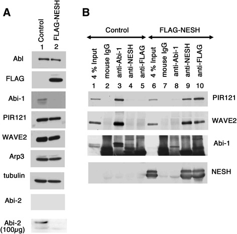
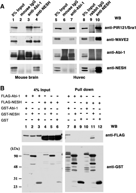
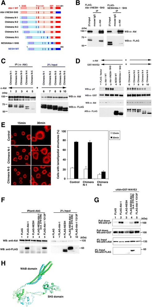
BACKGROUND: Abl interactor (Abi) family proteins play significant roles in actin cytoskeleton organization through participation in the WAVE complex. Mammals possess three Abi proteins: Abi-1, Abi-2, and NESH/Abi-3. Abi-1 and Abi-2 were originally identified as Abl tyrosine kinase-binding proteins. It has been disclosed that Abi-1 acts as a bridge between c-Abl and WAVE2, and c-Abl-mediated WAVE2 phosphorylation promotes actin remodeling. We showed previously that NESH/Abi-3 is present in the WAVE2 complex, but neither binds to c-Abl nor promotes c-Abl-mediated phosphorylation of WAVE2. RESULTS: In this study, we characterized NESH/Abi-3 in more detail, and compared its properties with those of Abi-1 and Abi-2. NESH/Abi-3 was ectopically expressed in NIH3T3 cells, in which Abi-1, but not NESH/Abi-3, is expressed. The expression of NESH/Abi-3 caused degradation of endogenous Abi-1, which led to the formation of a NESH/Abi-3-based WAVE2 complex. When these cells were plated on fibronectin-coated dishes, the translocation of WAVE2 to the plasma membrane was significantly reduced and the formation of peripheral lamellipodial structures was disturbed, suggesting that the NESH/Abi-3-based WAVE2 complex was unable to help produce lamellipodial protrusions. Next, Abi-1, Abi-2, or NESH/Abi-3 was expressed in v-src-transformed NIH3T3 cells. Only in NESH/Abi-3-expressed cells did treatment with an Abl kinase inhibitor, imatinib mesylate, or siRNA-mediated knockdown of c-Abl promote the formation of invadopodia, which are ventral membrane protrusions with extracellular matrix degradation activity. Structural studies showed that a linker region between the proline-rich regions and the Src homology 3 (SH3) domain of Abi-1 is crucial for its interaction with c-Abl and c-Abl-mediated phosphorylation of WAVE2. CONCLUSIONS: The NESH/Abi-3-based WAVE2 complex is functionally distinct from the Abi-1-based one, and NESH/Abi-3 may be involved in the formation of ventral protrusions under certain conditions.
Overview
The claims and statistics of this paper haven’t been checked yet.
Free: it reads the open-access text and checks the first 5 claims that cite a source we can check. Create a free account to check every claim.
Integrity signals
Registry
Cites
Statistics
Figures


Verification
Claims the paper makes
The claims and statistics of this paper haven’t been checked yet.
Free: it reads the open-access text and checks the first 5 claims that cite a source we can check. Create a free account to check every claim.
Claims other papers make about this one
Each is checked against this paper’s own text: does it say what the citing paper says it does?
The source backs part of the claim, not all of it — or backs it for a different population, condition or comparison.
In the citing paper
“ABI3 has been identified as a component of WASP-family verprolin homologous protein (WAVE) regulatory complex in several cancer cells (12, 13).”
From Deletion of Abi3 gene locus exacerbates neuropathological features of Alzheimer’s disease in a mouse model of Aβ amyloidosis (2021) · cites this paper as [12]
Experimental — accuracy not yet measuredThe source backs part of the claim, not all of it — or backs it for a different population, condition or comparison.
In the citing paper
“Both ABI1 and ABI2 but not ABI3 promote the c-ABL-mediated phosphorylation of WAVE2, since ABI3 does not directly interact with c-ABL (17,18).”
From Microglia express ABI3 in the brains of Alzheimer's disease and Nasu-Hakola disease (2017) · cites this paper as [18]
Experimental — accuracy not yet measuredThe source backs part of the claim, not all of it — or backs it for a different population, condition or comparison.
In the citing paper
“C terminally FLAG-tagged ABI3 has been previously shown to be functionally active [44].”
From Deletion of Abi3/Gngt2 influences age-progressive amyloid β and tau pathologies in distinctive ways (2022) · cites this paper as [44]
Experimental — accuracy not yet measuredThe cited source says what the claim attributes to it.
In the citing paper
“ABI3 plays a significant role in actin cytoskeleton organization through participation in the WAVE2 complex41, a complex that regulates multiple pathways leading to T-cell activation42.”
From Rare coding variants in PLCG2, ABI3, and TREM2 implicate microglial-mediated innate immunity in Alzheimer's disease (2017) · cites this paper as [41]
The cited source says what the claim attributes to it.
In the citing paper
“ABI3 has similarly been implicated in regulation of the immune response through interferon signaling [143] and also plays a role in actin cytoskeletal organization [144].”
From Genetic architecture of Alzheimer's disease (2020) · cites this paper as [144]
6 more claims about this paper
Create a free account to see every claim other papers make about this one.
Self-check
Percentages, sums and reported differences re-computed from the paper’s own numbers.
The self-check needs the paper’s full text.
Citations
Cites
Calcium's Role in Mechanotransduction during Muscle Development
Benavides Damm, T., & Egli, M. (2014) · Cellular Physiology and Biochemistry · doi:10.1159/000356667 · Cited by 14,502
Comparative Protein Modelling by Satisfaction of Spatial Restraints
Šali, A., & Blundell, T. L. (1993) · Journal of Molecular Biology · doi:10.1006/jmbi.1993.1626 · Cited by 12,977
Nobes, C. D., & Hall, A. (1995) · Cell · doi:10.1016/0092-8674(95)90370-4 · Cited by 4,412
Simons, K. T., Kooperberg, C., Huang, E., & Baker, D. (1997) · Journal of Molecular Biology · doi:10.1006/jmbi.1997.0959 · Cited by 1,396
Regulation of the actin cytoskeleton in cancer cell migration and invasion
Yamaguchi, H., & Condeelis, J. (2007) · Biochimica et Biophysica Acta (BBA) - Molecular Cell Research · doi:10.1016/j.bbamcr.2006.07.001 · Cited by 1,144
Structural Basis for the Autoinhibition of c-Abl Tyrosine Kinase
Nagar, B., Hantschel, O., Young, M. A., Scheffzek, K., Veach, D., Bornmann, W., Clarkson, B., Superti-Furga, G., & Kuriyan, J. (2003) · Cell · doi:10.1016/s0092-8674(03)00194-6 · Cited by 879
Toward High-Resolution de Novo Structure Prediction for Small Proteins
Bradley, P., Misura, K. M. S., & Baker, D. (2005) · Science · doi:10.1126/science.1113801 · Cited by 864
Mechanism of regulation of WAVE1-induced actin nucleation by Rac1 and Nck
Eden, S., Rohatgi, R., Podtelejnikov, A. V., Mann, M., & Kirschner, M. W. (2002) · Nature · doi:10.1038/nature00859 · Cited by 835 · cited 1×
“filament nucleation and actin network assembly in response to Rac GTPase [12–15].”
BackgroundStructure prediction for CASP8 with all‐atom refinement using Rosetta
Raman, S., Vernon, R., Thompson, J., Tyka, M., Sadreyev, R., Pei, J., Kim, D., Kellogg, E., DiMaio, F., Lange, O., et al. (2009) · Proteins: Structure, Function, and Bioinformatics · doi:10.1002/prot.22540 · Cited by 518
Structure and control of the actin regulatory WAVE complex
Chen, Z., Borek, D., Padrick, S. B., Gomez, T. S., Metlagel, Z., Ismail, A. M., Umetani, J., Billadeau, D. D., Otwinowski, Z., & Rosen, M. K. (2010) · Nature · doi:10.1038/nature09623 · Cited by 500 · cited 1×
“complex was reported [43].”
Cited by
Microglia in Alzheimer’s disease
Hansen, D. V., Hanson, J. E., & Sheng, M. (2018) · Journal of Cell Biology · doi:10.1083/jcb.201709069 · Cited by 1,935
ARUK Consortium, Sims, R., GERAD/PERADES, C., van der Lee, S. J., Naj, A. C., Bellenguez, C., Badarinarayan, N., Jakobsdottir, J., Kunkle, B. W., Boland, A., et al. (2017) · Nature Genetics · doi:10.1038/ng.3916 · Cited by 1,012
Microglia in Alzheimer’s disease: pathogenesis, mechanisms, and therapeutic potentials
Miao, J., Ma, H., Yang, Y., Liao, Y., Lin, C., Zheng, J., Yu, M., & Lan, J. (2023) · Frontiers in Aging Neuroscience · doi:10.3389/fnagi.2023.1201982 · Cited by 293
Genetic architecture of Alzheimer's disease
Neuner, S. M., TCW, J., & Goate, A. M. (2020) · Neurobiology of Disease · doi:10.1016/j.nbd.2020.104976 · Cited by 126
Conway, O. J., Carrasquillo, M. M., Wang, X., Bredenberg, J. M., Reddy, J. S., Strickland, S. L., Younkin, C. S., Burgess, J. D., Allen, M., Lincoln, S. J., et al. (2018) · Molecular Neurodegeneration · doi:10.1186/s13024-018-0289-x · Cited by 112
“ABI3 encodes the Abelson (Abl) interactor (Abi) family protein 3 (a.k.a. NESH), which is involved in actin cytoskeleton organization and functionally distinct from ABI1 or ABI2 [2].”
Background
Metadata
- Authors
- Sekino, S., Kashiwagi, Y., Kanazawa, H., Takada, K., Baba, T., Sato, S., Inoue, H., Kojima, M., & Tani, K.
- Affiliations
- Tokyo University of Pharmacy and Life Sciences (JP)
- Corresponding author
- Tani, Katsuko
- Venue
- Cell Communication and Signaling
- Published
- 2015-10-01
- Volume
- 13
- Issue
- 1
- Pages
- 41-41
- ISSN
- 1478-811X (ISSN-L)
- PMC
- PMC4589964
- OpenAlex
- W1905136210
- Cited by
- 43
- References
- 50
- Citations by year
- 2026: 2 · 2025: 2 · 2024: 1 · 2023: 6 · 2022: 16 · 2021: 4 · 2020: 1 · 2019: 2 · 2018: 3 · 2017: 6
- FWCI
- 1.60
- Type
- journal article
- Language
- en
- Open access
- gold
- doi.org
- Licence
- cc-by
- Locations
- Cell Communication and Signaling (publishedVersion, cc-by)
- Europe PMC (PubMed Central) (publishedVersion, cc-by)
- PubMed Central (publishedVersion, cc-by)
- PubMed (publishedVersion)
- Funders
- Ministry of Education, Culture, Sports, Science and Technology
- Japan Society for the Promotion of Science (26440105)
- Topics
- Life Sciences › Biochemistry, Genetics and Molecular Biology › Cell Biology › Cellular Mechanics and Interactions
- Life Sciences › Biochemistry, Genetics and Molecular Biology › Molecular Biology › Protein Kinase Regulation and GTPase Signaling
- Life Sciences › Biochemistry, Genetics and Molecular Biology › Cell Biology › Caveolin-1 and cellular processes
- Keywords
- Abi-1, c-Abl, actin cytoskeleton remodeling, lamellipodia, invadopodia, extracellular matrix degradation, imatinib mesylate, v-Src
- MeSH terms
- Animals, Cell Movement, Cytoskeletal Proteins, Humans, Pseudopodia, Protein Transport, NIH 3T3 Cells, Adaptor Proteins, Signal Transducing, Wiskott-Aldrich Syndrome Protein Family, Mice, HEK293 Cells, Human Umbilical Vein Endothelial Cells
About these checks
Registry, retraction, Cites and statistics results are computed, not judged, with one exception: a printed reference that no index lists is read once by a model before it counts as a citation error. Claims are extracted by a model and kept only when their quote appears verbatim in the text; each cited source is then checked, with a model’s help, for whether it says what the claim relies on. Those verdicts can be wrong — read the claim’s quote and the cited source before relying on one.
Statistics and arithmetic are recomputed, not judged.
Confidence bands are shown only for fields calibrated on our annotated test set. None is calibrated yet, so no band is shown.
What counts as a claim ›How we measure confidence ›How we check this ›
Are you an author, or do you see a problem with this page? Request a review
How to cite
Cite CoChat for the checks on this page, and the paper for its own findings.
- This page
- CoChat. (2026). Citation check: The NESH/Abi-3-based WAVE2 complex is functionally distinct from the Abi-1-based WAVE2 complex. https://app.cochat.ai/papers/10.1186/s12964-015-0119-5
- The paper
- Sekino, S., Kashiwagi, Y., Kanazawa, H., Takada, K., Baba, T., Sato, S., Inoue, H., Kojima, M., & Tani, K. (2015). The NESH/Abi-3-based WAVE2 complex is functionally distinct from the Abi-1-based WAVE2 complex. Cell Communication and Signaling. https://doi.org/10.1186/s12964-015-0119-5
Data sources
Bibliographic data: Semantic Scholar, OpenAlex, Crossref.
Contains information from the Semantic Scholar Academic Graph Datasets, made available under the ODC Attribution License (ODC-BY 1.0).
Semantic Scholar’s data is described in: Kinney, R., et al. (2023). The Semantic Scholar Open Data Platform. arXiv:2301.10140.
