

The DOI matches its Crossref record; 50 of the 50 references it cites were found in a registry or an index.
Monocyte infiltration rather than microglia proliferation dominates the early immune response to rapid photoreceptor degeneration
Abstract
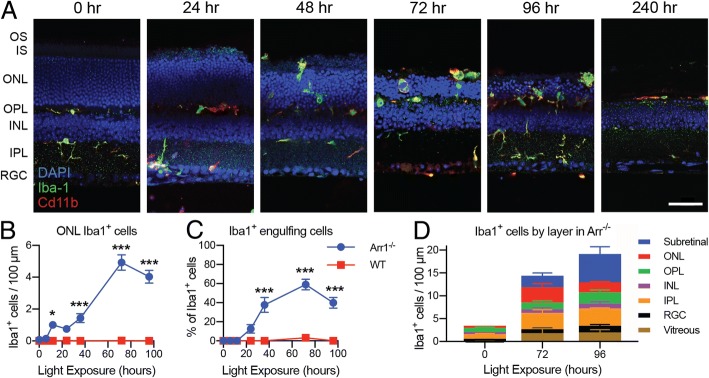
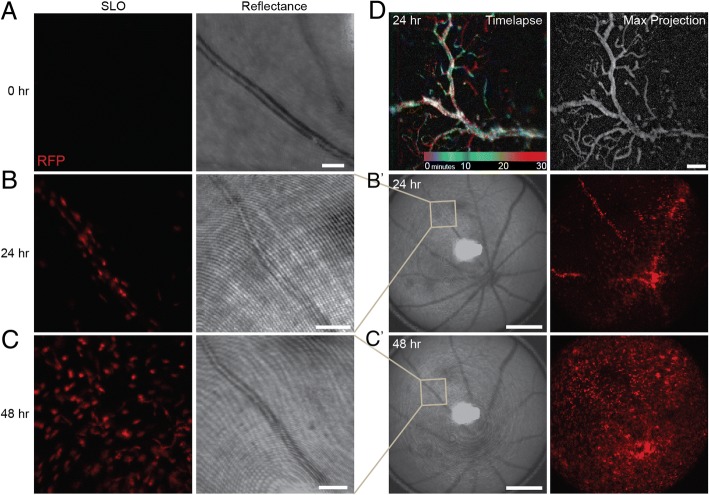
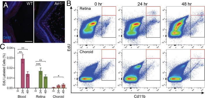
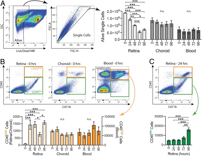
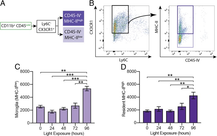
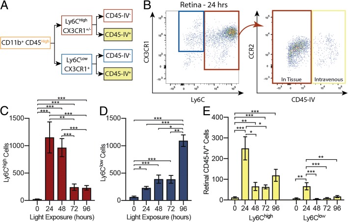
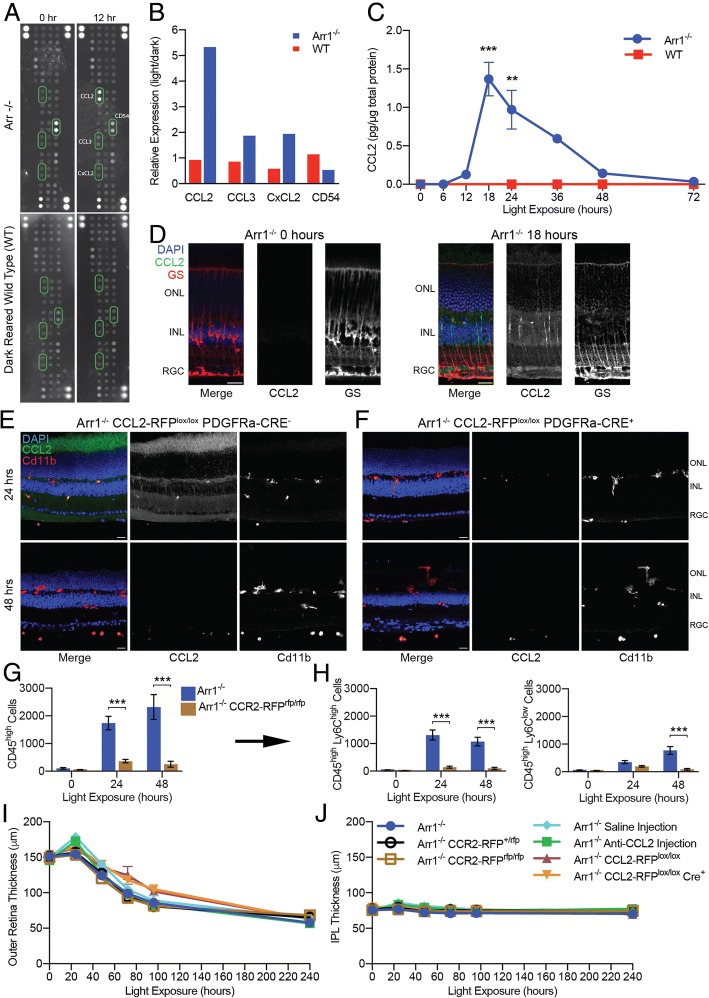
BACKGROUND: Activation of resident microglia accompanies every known form of neurodegeneration, but the involvement of peripheral monocytes that extravasate and rapidly transform into microglia-like macrophages within the central nervous system during degeneration is far less clear. METHODS: Using a combination of in vivo ocular imaging, flow cytometry, and immunohistochemistry, we investigated the response of infiltrating cells in a light-inducible mouse model of photoreceptor degeneration. RESULTS: cell extravasation from the retinal vasculature. Proliferation of microglia and monocytes occurred concurrently; however, there was no indication of proliferation in either population until 72-96 h after neurodegeneration began. Eliminating CCL2-CCR2 signaling blocked monocyte recruitment, but did not alter the extent of retinal degeneration. CONCLUSIONS: These results demonstrate that the immune response to photoreceptor degeneration includes both resident microglia and monocytes, even at very early times. Surprisingly, preventing monocyte infiltration did not block neurodegeneration, suggesting that in this model, degeneration is limited by cell clearance from other phagocytes or by the timing of intrinsic cell death programs. These results show monocyte involvement is not limited to disease states that overwhelm or deplete the resident microglial population and that interventions focused on modulating the peripheral immune system are not universally beneficial for staving off degeneration.
Overview
The claims and statistics of this paper haven’t been checked yet.
Run the check — free, no account needed. It reads the open-access text and checks the sources it cites.
Integrity signals
Registry
Cites
Statistics
Figures
Verification
Claims the paper makes
The claims and statistics of this paper haven’t been checked yet.
Run the check — free, no account needed. It reads the open-access text and checks the sources it cites.
Claims other papers make about this one
Each is checked against this paper’s own text: does it say what the citing paper says it does?
In the citing paper
“Given that peripheral monocytes have been shown to participate in CNS disease via both infiltration and repopulation of neuroglia following microglia depletion (4–10), it is important to determine if CSF1R inhibition causes functional changes in peripheral immune cells that can become part of the CNS (3, 4, 10–14).”
From CSF1R inhibition by a small-molecule inhibitor is not microglia specific; affecting hematopoiesis and the function of macrophages (2020) · cites this paper as [7]
Self-check
Percentages, sums and reported differences re-computed from the paper’s own numbers.
The self-check needs the paper’s full text.
Citations
Cites
Polazzi, E., Mengoni, I., Caprini, M., Peña-Altamira, E., Kurtys, E., & Monti, B. (2013) · Neurosignals · doi:10.1159/000337115 · Cited by 8,994
Monocyte and macrophage heterogeneity
Gordon, S., & Taylor, P. R. (2005) · Nature Reviews Immunology · doi:10.1038/nri1733 · Cited by 5,190
Microglia: Active sensor and versatile effector cells in the normal and pathologic brain
Hanisch, U.-K., & Kettenmann, H. (2007) · Nature Neuroscience · doi:10.1038/nn1997 · Cited by 3,676
From Monocytes to M1/M2 Macrophages: Phenotypical vs. Functional Differentiation
Italiani, P., & Boraschi, D. (2014) · Frontiers in Immunology · doi:10.3389/fimmu.2014.00514 · Cited by 2,067
Müller cells in the healthy and diseased retina
BRINGMANN, A., PANNICKE, T., GROSCHE, J., FRANCKE, M., WIEDEMANN, P., SKATCHKOV, S., OSBORNE, N., & REICHENBACH, A. (2006) · Progress in Retinal and Eye Research · doi:10.1016/j.preteyeres.2006.05.003 · Cited by 1,711
Local self-renewal can sustain CNS microglia maintenance and function throughout adult life
Ajami, B., Bennett, J. L., Krieger, C., Tetzlaff, W., & Rossi, F. M. V. (2007) · Nature Neuroscience · doi:10.1038/nn2014 · Cited by 1,643
Microglia in physiology and disease
Wolf, S. A., Boddeke, H. W. G. M., & Kettenmann, H. (2017) · Annual Review of Physiology · doi:10.1146/annurev-physiol-022516-034406 · Cited by 1,426
Microglia and brain macrophages in the molecular age: from origin to neuropsychiatric disease
Prinz, M., & Priller, J. (2014) · Nature Reviews Neuroscience · doi:10.1038/nrn3722 · Cited by 1,329
Yang, J., Zhang, L., Yu, C., Yang, X.-F., & Wang, H. (2014) · Biomarker Research · doi:10.1186/2050-7771-2-1 · Cited by 1,099
Microglia in the adult brain arise from Ly-6ChiCCR2+ monocytes only under defined host conditions
Mildner, A., Schmidt, H., Nitsche, M., Merkler, D., Hanisch, U.-K., Mack, M., Heikenwalder, M., Brück, W., Priller, J., & Prinz, M. (2007) · Nature Neuroscience · doi:10.1038/nn2015 · Cited by 1,089
Cited by
Lei, F., Cui, N., Zhou, C., Chodosh, J., Vavvas, D. G., & Paschalis, E. I. (2020) · Proceedings of the National Academy of Sciences · doi:10.1073/pnas.1922788117 · Cited by 322
Ronning, K. E., Karlen, S. J., Miller, E. B., & Burns, M. E. (2019) · Scientific Reports · doi:10.1038/s41598-019-41141-0 · Cited by 88
Self-citationWiley, L. A., Boyce, T. M., Meyering, E. E., Ochoa, D., Sheehan, K. M., Stone, E. M., Mullins, R. F., Tucker, B. A., & Han, I. C. (2023) · Human Gene Therapy · doi:10.1089/hum.2022.222 · Cited by 30
Sectoral activation of glia in an inducible mouse model of autosomal dominant retinitis pigmentosa
Massengill, M. T., Ash, N. F., Young, B. M., Ildefonso, C. J., & Lewin, A. S. (2020) · Scientific Reports · doi:10.1038/s41598-020-73749-y · Cited by 13
Metadata
- Authors
- Karlen, S. J., Miller, E. B., Wang, X., Levine, E. S., Zawadzki, R. J., & Burns, M. E.
- Venue
- Journal of Neuroinflammation
- Published
- 2018-12
- Volume
- 15
- Issue
- 1
- ISSN
- 1742-2094
- PMC
- PMC7659426
- Cited by
- 71
- References
- 50
- Language
- en
- Licence
- creativecommons.org/licenses/by/4.0/ (tdm)
- creativecommons.org/licenses/by/4.0/ (vor, 14 d)
- Funders
- National Eye Institute (R01-EY24320, P30-EY012576)
- National Institutes of Health (T32-GM007377, T32-EY105387, NIH S10OD018223)
About these checks
Registry, retraction, Cites and statistics results are computed, not judged, with one exception: a printed reference that no index lists is read once by a model before it counts as a citation error. Claims are extracted by a model and kept only when their quote appears verbatim in the text; each cited source is then checked, with a model’s help, for whether it says what the claim relies on. Those verdicts can be wrong — read the claim’s quote and the cited source before relying on one.
Statistics and arithmetic are recomputed, not judged.
Confidence bands are shown only for fields calibrated on our annotated test set. None is calibrated yet, so no band is shown.
What counts as a claim ›How we measure confidence ›How we check this ›
Are you an author, or do you see a problem with this page? Request a review
Data sources
Bibliographic data: Semantic Scholar, OpenAlex, Crossref.
Contains information from the Semantic Scholar Academic Graph Datasets, made available under the ODC Attribution License (ODC-BY 1.0).
Semantic Scholar’s data is described in: Kinney, R., et al. (2023). The Semantic Scholar Open Data Platform. arXiv:2301.10140.
