

The DOI matches its Crossref record; 84 of the 84 references it cites were found in a registry or an index.
Propagation of Tau aggregates
Open accessAbstract
Since 2009, evidence has accumulated to suggest that Tau aggregates form first in a small number of brain cells, from where they propagate to other regions, resulting in neurodegeneration and disease. Propagation of Tau aggregates is often called prion-like, which refers to the capacity of an assembled protein to induce the same abnormal conformation in a protein of the same kind, initiating a self-amplifying cascade. In addition, prion-like encompasses the release of protein aggregates from brain cells and their uptake by neighbouring cells. In mice, the intracerebral injection of Tau inclusions induced the ordered assembly of monomeric Tau, followed by its spreading to distant brain regions. Short fibrils constituted the major species of seed-competent Tau. The existence of several human Tauopathies with distinct fibril morphologies has led to the suggestion that different molecular conformers (or strains) of aggregated Tau exist.
Overview
The claims and statistics of this paper haven’t been checked yet.
Run the check — free, no account needed. It reads the open-access text and checks the sources it cites.
Integrity signals
Registry
Cites
Statistics
Figures
Verification
Claims the paper makes
The claims and statistics of this paper haven’t been checked yet.
Run the check — free, no account needed. It reads the open-access text and checks the sources it cites.
Claims other papers make about this one
Each is checked against this paper’s own text: does it say what the citing paper says it does?
The source backs part of the claim, not all of it — or backs it for a different population, condition or comparison.
In the citing paper
“A synapse-centred hypothesis also does not preclude the spreading hypothesis [34, 66], which could be either a downstream pathological event or a potential trigger of Tau-related excitotoxicity and synaptic failure.”
From The new genetic landscape of Alzheimer’s disease: from amyloid cascade to genetically driven synaptic failure hypothesis? (2019) · cites this paper as [34]
Experimental — accuracy not yet measuredIn the citing paper
“In animal models that express tau only in certain brain regions, or that are injected with exogenous tau, spreading of tau pathology to anatomically connected regions occurs over time54,64,228.”
From Tau-targeting therapies for Alzheimer disease (2018) · cites this paper as [64]
Self-check
Percentages, sums and reported differences re-computed from the paper’s own numbers.
The self-check needs the paper’s full text.
Citations
Cites
Amyloid oligomers exacerbate tau pathology in a mouse model of tauopathy
Selenica, M.-L. B., Brownlow, M., Jimenez, J. P., Lee, D. C., Pena, G., Dickey, C. A., Gordon, M. N., & Morgan, D. (2013) · Neurodegenerative Diseases · doi:10.1159/000337230 · Cited by 10,359
Corder, E. H., Saunders, A. M., Strittmatter, W. J., Schmechel, D. E., Gaskell, P. C., Small, G. W., Roses, A. D., Haines, J. L., & Pericak-Vance, M. A. (1993) · Science · doi:10.1126/science.8346443 · Cited by 9,429
The amyloid hypothesis of Alzheimer's disease at 25 years
Selkoe, D. J., & Hardy, J. (2016) · EMBO Molecular Medicine · doi:10.15252/emmm.201606210 · Cited by 6,164
Novel Proteinaceous Infectious Particles Cause Scrapie
Prusiner, S. B. (1982) · Science · doi:10.1126/science.6801762 · Cited by 5,221
Association of missense and 5′-splice-site mutations in tau with the inherited dementia FTDP-17
Hutton, M., Lendon, C. L., Rizzu, P., Baker, M., Froelich, S., Houlden, H., Pickering-Brown, S., Chakraverty, S., Isaacs, A., Grover, A., et al. (1998) · Nature · doi:10.1038/31508 · Cited by 3,498
Goedert, M., Spillantini, M. G., Jakes, R., Rutherford, D., & Crowther, R. A. (1989) · Neuron · doi:10.1016/0896-6273(89)90210-9 · Cited by 2,492
Self-citationAtomic structures of amyloid cross-β spines reveal varied steric zippers
Sawaya, M. R., Sambashivan, S., Nelson, R. A., Ivanova, M. I., Sievers, S. A., Apostol, M. I., Thompson, M. J., Balbirnie, M., Wiltzius, J. J. W., McFarlane, H. T., et al. (2007) · Nature · doi:10.1038/nature05695 · Cited by 2,279
Tau in physiology and pathology
Wang, Y., & Mandelkow, E. (2016) · Nature Reviews Neuroscience · doi:10.1038/nrn.2015.1 · Cited by 2,251
Nelson, P. T., Alafuzoff, I., Bigio, E. H., Bouras, C., Braak, H., Cairns, N. J., Castellani, R. J., Crain, B. J., Davies, P., Tredici, K. D., et al. (2012) · Journal of Neuropathology & Experimental Neurology · doi:10.1097/nen.0b013e31825018f7 · Cited by 2,050 · cited 1×
“However, it is also clear that pathological changes in Tau correlate better with nerve cell dysfunction than Aβ deposits [5, 6].”
BackgroundTransmission and spreading of tauopathy in transgenic mouse brain
Clavaguera, F., Bolmont, T., Crowther, R. A., Abramowski, D., Frank, S., Probst, A., Fraser, G., Stalder, A. K., Beibel, M., Staufenbiel, M., et al. (2009) · Nature Cell Biology · doi:10.1038/ncb1901 · Cited by 1,803
Self-citationDepletion of microglia and inhibition of exosome synthesis halt tau propagation
Asai, H., Ikezu, S., Tsunoda, S., Medalla, M., Luebke, J., Haydar, T., Wolozin, B., Butovsky, O., Kügler, S., & Ikezu, T. (2015) · Nature Neuroscience · doi:10.1038/nn.4132 · Cited by 1,591
Mutation in the tau gene in familial multiple system tauopathy with presenile dementia
Spillantini, M. G., Murrell, J. R., Goedert, M., Farlow, M. R., Klug, A., & Ghetti, B. (1998) · Proceedings of the National Academy of Sciences · doi:10.1073/pnas.95.13.7737 · Cited by 1,474
Self-citationPrimary age-related tauopathy (PART): a common pathology associated with human aging
Crary, J. F., Trojanowski, J. Q., Schneider, J. A., Abisambra, J. F., Abner, E. L., Alafuzoff, I., Arnold, S. E., Attems, J., Beach, T. G., Bigio, E. H., et al. (2014) · Acta Neuropathologica · doi:10.1007/s00401-014-1349-0 · Cited by 1,467
Tau is a candidate gene for chromosome 17 frontotemporal dementia
Poorkaj, P., Bird, T. D., Wijsman, E., Nemens, E., Garruto, R. M., Anderson, L., Andreadis, A., Wiederholt, W. C., Raskind, M., & Schellenberg, G. D. (1998) · Annals of Neurology · doi:10.1002/ana.410430617 · Cited by 1,374
Propagation of Tau Misfolding from the Outside to the Inside of a Cell
Frost, B., Jacks, R. L., & Diamond, M. I. (2009) · Journal of Biological Chemistry · doi:10.1074/jbc.m808759200 · Cited by 1,252
Goedert, M., Wischik, C. M., Crowther, R. A., Walker, J. E., & Klug, A. (1988) · Proceedings of the National Academy of Sciences · doi:10.1073/pnas.85.11.4051 · Cited by 1,134
Self-citationGoedert, M., Spillantini, M. G., Cairns, N. J., & Crowther, R. A. (1992) · Neuron · doi:10.1016/0896-6273(92)90117-v · Cited by 1,122
Self-citationGoedert, M., Jakes, R., Spillantini, M. G., Hasegawa, M., Smith, M. J., & Crowther, R. A. (1996) · Nature · doi:10.1038/383550a0 · Cited by 1,036
Self-citationGoedert, M. (2015) · Science · doi:10.1126/science.1255555 · Cited by 1,028
Self-citationvon Bergen, M., Friedhoff, P., Biernat, J., Heberle, J., Mandelkow, E.-M., & Mandelkow, E. (2000) · Proceedings of the National Academy of Sciences · doi:10.1073/pnas.97.10.5129 · Cited by 1,021
Distinct Tau Prion Strains Propagate in Cells and Mice and Define Different Tauopathies
Sanders, D. W., Kaufman, S. K., DeVos, S. L., Sharma, A. M., Mirbaha, H., Li, A., Barker, S. J., Foley, A. C., Thorpe, J. R., Serpell, L. C., et al. (2014) · Neuron · doi:10.1016/j.neuron.2014.04.047 · Cited by 986
Wischik, C. M., Novak, M., Thøgersen, H. C., Edwards, P. C., Runswick, M. J., Jakes, R., Walker, J. E., Milstein, C., Roth, M., & Klug, A. (1988) · Proceedings of the National Academy of Sciences · doi:10.1073/pnas.85.12.4506 · Cited by 939
Goedert, M., & Jakes, R. (1990) · The EMBO Journal · doi:10.1002/j.1460-2075.1990.tb07870.x · Cited by 929 · cited 1×
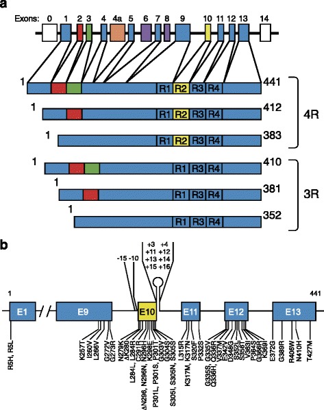
“Similar amounts of 3R and 4R Tau are expressed in the cerebral cortex of adults [16].”
BackgroundSelf-citationHeparan sulfate proteoglycans mediate internalization and propagation of specific proteopathic seeds
Holmes, B. B., DeVos, S. L., Kfoury, N., Li, M., Jacks, R., Yanamandra, K., Ouidja, M. O., Brodsky, F. M., Marasa, J., Bagchi, D. P., et al. (2013) · Proceedings of the National Academy of Sciences · doi:10.1073/pnas.1301440110 · Cited by 859
A common inversion under selection in Europeans
Stefánsson, H., Helgason, A., Þorleifsson, G., Steinthórsdóttir, V., Másson, G., Barnard, J., Baker, A., Jónasdóttir, Á., Ingason, A., Gudnadottir, V. G., et al. (2005) · Nature Genetics · doi:10.1038/ng1508 · Cited by 847
Neuronal activity enhances tau propagation and tau pathology in vivo
Wu, J. W., Hussaini, S. A., Bastille, I. M., Rodriguez, G. A., Mrejeru, A., Rilett, K., Sanders, D. W., Cook, C., Fu, H., Boonen, R. A. C. M., et al. (2016) · Nature Neuroscience · doi:10.1038/nn.4328 · Cited by 820
Tau and neurodegenerative disease: the story so far
Iqbal, K., Liu, F., & Gong, C.-X. (2016) · Nature Reviews Neurology · doi:10.1038/nrneurol.2015.225 · Cited by 806
Brain homogenates from human tauopathies induce tau inclusions in mouse brain
Clavaguera, F., Akatsu, H., Fraser, G. M., Crowther, R. A., Frank, S., Hench, J., Probst, A., Winkler, D. T., Reichwald, J., Staufenbiel, M., et al. (2013) · Proceedings of the National Academy of Sciences · doi:10.1073/pnas.1301175110 · Cited by 792
Self-citationThe genetics of myelodysplastic syndrome: from clonal haematopoiesis to secondary leukaemia
Sperling, A. S., Gibson, C. J., & Ebert, B. L. (2016) · Nature reviews. Cancer · doi:10.1038/nrc.2016.112 · Cited by 706
Iba, M., Guo, J. L., McBride, J. D., Zhang, B., Trojanowski, J. Q., & Lee, V. M.-Y. (2013) · The Journal of Neuroscience · doi:10.1523/jneurosci.2642-12.2013 · Cited by 664
Propagation of Tau aggregates and neurodegeneration
Goedert, M., Eisenberg, D. S., & Crowther, R. A. (2017) · Annual Review of Neuroscience · doi:10.1146/annurev-neuro-072116-031153 · Cited by 632
Self-citationvon Bergen�, M., Barghorn, S., Li, L., Marx, A., Biernat, J., Mandelkow, E., & Mandelkow�, E. (2001) · Journal of Biological Chemistry · doi:10.1074/jbc.m105196200 · Cited by 618
Structural characterization of the core of the paired helical filament of Alzheimer disease.
Wischik, C. M., Novák, M., Edwards, P. C., Klug, A., Tichelaar, W., & Crowther, R. A. (1988) · Proceedings of the National Academy of Sciences · doi:10.1073/pnas.85.13.4884 · Cited by 609
Arendt, T., Stieler, J. T., & Holzer, M. (2016) · Brain Research Bulletin · doi:10.1016/j.brainresbull.2016.08.018 · Cited by 609
Wilcock, G. K., & Esiri, M. M. (1982) · Journal of the Neurological Sciences · doi:10.1016/0022-510x(82)90155-1 · Cited by 608
Identification of common variants influencing risk of the tauopathy progressive supranuclear palsy
PSP Genetics Study Group, Höglinger, G. U., Melhem, N. M., Dickson, D. W., Sleiman, P. M. A., Wang, L.-S., Klei, L., Rademakers, R., de Silva, R., Litvan, I., et al. (2011) · Nature Genetics · doi:10.1038/ng.859 · Cited by 603
Yanamandra, K., Kfoury, N., Jiang, H., Mahan, T. E., Ma, S., Maloney, S. E., Wozniak, D. F., Diamond, M. I., & Holtzman, D. M. (2013) · Neuron · doi:10.1016/j.neuron.2013.07.046 · Cited by 574
DeVos, S. L., Miller, R. L., Schoch, K. M., Holmes, B. B., Kebodeaux, C. S., Wegener, A. J., Chen, G., Shen, T., Tran, H., Nichols, B., et al. (2017) · Science Translational Medicine · doi:10.1126/scitranslmed.aag0481 · Cited by 516
Ahmed, Z., Cooper, J., Murray, T. K., Garn, K., McNaughton, E., Clarke, H., Parhizkar, S., Ward, M. A., Cavallini, A., Jackson, S., et al. (2014) · Acta Neuropathologica · doi:10.1007/s00401-014-1254-6 · Cited by 484
Self-citationMicrotubule-Associated Protein MAP2 Shares a Microtubule Binding Motif with Tau Protein
Lewis, S. A., Wang, D. H., & Cowan, N. J. (1988) · Science · doi:10.1126/science.3142041 · Cited by 483
Morris, M., Knudsen, G. M., Maeda, S., Trinidad, J. C., Ioanoviciu, A., Burlingame, A. L., & Mucke, L. (2015) · Nature Neuroscience · doi:10.1038/nn.4067 · Cited by 472
Ghetti, B. F., Oblak, A. L., Boeve, B. F., Johnson, K. A., Dickerson, B. C., & Goedert, M. (2014) · Neuropathology and Applied Neurobiology · doi:10.1111/nan.12213 · Cited by 458
Self-citationKaufman, S. K., Sanders, D. W., Thomas, T. L., Ruchinskas, A. J., Vaquer-Alicea, J., Sharma, A. M., Miller, T. M., & Diamond, M. I. (2016) · Neuron · doi:10.1016/j.neuron.2016.09.055 · Cited by 450
Guo, J. L., Narasimhan, S., Changolkar, L., He, Z., Stieber, A., Zhang, B., Gathagan, R. J., Iba, M., McBride, J. D., Trojanowski, J. Q., et al. (2016) · Journal of Experimental Medicine · doi:10.1084/jem.20160833 · Cited by 440
Straight and paired helical filaments in Alzheimer disease have a common structural unit.
Crowther, R. A. (1991) · Proceedings of the National Academy of Sciences · doi:10.1073/pnas.88.6.2288 · Cited by 366
Passive Immunization with Anti-Tau Antibodies in Two Transgenic Models
Chai, X., Wu, S., Murray, T. K., Kinley, R. D., Cella, C. V., Sims, H., Buckner, N., Hanmer, J. M., Davies, P., O’Neill, M. J., et al. (2011) · Journal of Biological Chemistry · doi:10.1074/jbc.m111.229633 · Cited by 363
The car☐yl third of tau is tightly bound to paired helical filaments
Kondo, J., Honda, T., Mori, H., Hamada, Y., Miura, R., OGAWARA, M., & Ihara, Y. (1988) · Neuron · doi:10.1016/0896-6273(88)90130-4 · Cited by 357
Arendt, T., Stieler, J., Strijkstra, A. M., Hut, R. A., Rüdiger, J., Van der Zee, E. A., Harkany, T., Holzer, M., & Härtig, W. (2003) · The Journal of Neuroscience · doi:10.1523/jneurosci.23-18-06972.2003 · Cited by 357
Pérez, M., Valpuesta, J., Medina, M., de Garcini, E. M., & Ávila, J. (1996) · Journal of Neurochemistry · doi:10.1046/j.1471-4159.1996.67031183.x · Cited by 355
PART is part of Alzheimer disease
Duyckaerts, C. J., Braak, H., Brion, J., Buée, L., Tredici, K. D., Goedert, M., Halliday, G. M., Neumann, M., Spillantini, M. G., Tolnay, M., et al. (2015) · Acta Neuropathologica · doi:10.1007/s00401-015-1390-7 · Cited by 329
Self-citationBerriman, J. A., Serpell, L. C., Oberg, K. A., Fink, A. L., Goedert, M., & Crowther, R. A. (2003) · Proceedings of the National Academy of Sciences · doi:10.1073/pnas.1530287100 · Cited by 288
Self-citationConformation Determines the Seeding Potencies of Native and Recombinant Tau Aggregates
Falcon, B., Cavallini, A., Angers, R. C., Glover, S. J., Murray, T. K., Barnham, L., Jackson, S., O'Neill, M. J., Isaacs, A. M., Hutton, M. L., et al. (2014) · Journal of Biological Chemistry · doi:10.1074/jbc.m114.589309 · Cited by 265
Self-citationCloning of a big tau microtubule-associated protein characteristic of the peripheral nervous system.
Goedert, M., Spillantini, M. G., & Crowther, R. A. (1992) · Proceedings of the National Academy of Sciences · doi:10.1073/pnas.89.5.1983 · Cited by 255
Self-citationPrimary structure of high molecular weight tau present in the peripheral nervous system.
Couchie, D., Mavilia, C., Georgieff, I. S., Liem, R. K. H., Shelanski, M. L., & Nunez, J. (1992) · Proceedings of the National Academy of Sciences · doi:10.1073/pnas.89.10.4378 · Cited by 241
Coppola, G., Chinnathambi, S., Lee, J. J., Dombroski, B. A., Baker, M. C., Soto-Ortolaza, A. I., Lee, S. E., Klein, E., Huang, A. Y., Sears, R., et al. (2012) · Human Molecular Genetics · doi:10.1093/hmg/dds161 · Cited by 240
Fontaine, S. N., Zheng, D., Sabbagh, J. J., Martin, M. D., Chaput, D., Darling, A. L., Trotter, J. H., Stothert, A. R., Nordhues, B. A., Lussier, A. L., et al. (2016) · The EMBO Journal · doi:10.15252/embj.201593489 · Cited by 214
The fuzzy coat of pathological human Tau fibrils is a two-layered polyelectrolyte brush
Wegmann, S., Medalsy, I. D., Mandelkow�, E., & Müller, D. J. (2012) · Proceedings of the National Academy of Sciences · doi:10.1073/pnas.1212100110 · Cited by 201
Cytosolic Fc receptor TRIM21 inhibits seeded tau aggregation
McEwan, W. A., Falcon, B., Vaysburd, M., Clift, D., Oblak, A. L., Ghetti, B., Goedert, M., & James, L. C. (2017) · Proceedings of the National Academy of Sciences · doi:10.1073/pnas.1607215114 · Cited by 201
Self-citationAbnormal Tau-Containing Filaments in Neurodegenerative Diseases
Crowther, R. A., & Goedert, M. (2000) · Journal of Structural Biology · doi:10.1006/jsbi.2000.4270 · Cited by 188
Self-citationJanning, D., Igaev, M., Sündermann, F., Brühmann, J., Beutel, O., Heinisch, J. J., Bakota, L., Piehler, J., Junge, W., & Brandt, R. (2014) · Molecular Biology of the Cell · doi:10.1091/mbc.e14-06-1099 · Cited by 181
Woerman, A. L., Aoyagi, A., Patel, S. S., Kazmi, S. A., Lobach, I. V., Grinberg, L. T., McKee, A. C., Seeley, W. W., Olson, S. H., & Prusiner, S. B. (2016) · Proceedings of the National Academy of Sciences · doi:10.1073/pnas.1616344113 · Cited by 180
Jackson, S. J., Kerridge, C., Cooper, J., Cavallini, A., Falcon, B., Cella, C. V., Landi, A., Szekeres, P. G., Murray, T. K., Ahmed, Z., et al. (2016) · Journal of Neuroscience · doi:10.1523/jneurosci.3542-15.2016 · Cited by 163
Self-citationIovino, M., Agathou, S., González-Rueda, A., Del Castillo Velasco-Herrera, M., Borroni, B., Alberici, A., Lynch, T., O’Dowd, S., Geti, I., Gaffney, D., et al. (2015) · Brain · doi:10.1093/brain/awv222 · Cited by 155
Self-citationFolding of the Tau Protein on Microtubules
Kadavath, H., Jaremko, M., Jaremko, Ł., Biernat, J., Mandelkow, E., & Zweckstetter, M. (2015) · Angewandte Chemie International Edition · doi:10.1002/anie.201501714 · Cited by 153
Sankaranarayanan, S., Barten, D. M., Vana, L., Devidze, N., Yang, L., Cadelina, G. W., Hoque, N., DeCarr, L. B., Keenan, S., Lin, A., et al. (2015) · PLoS ONE · doi:10.1371/journal.pone.0125614 · Cited by 150
Sposito, T., Preza, E., Mahoney, C. J., Setó-Salvia, N., Ryan, N. S., Morris, H. R., Arber, C., Devine, M. J., Houlden, H., Warner, T. T., et al. (2015) · Human Molecular Genetics · doi:10.1093/hmg/ddv246 · Cited by 148
Peripheral administration of tau aggregates triggers intracerebral tauopathy in transgenic mice
Clavaguera, F., Hench, J., Lavenir, I., Schweighauser, G., Frank, S., Goedert, M., & Tolnay, M. (2013) · Acta Neuropathologica · doi:10.1007/s00401-013-1231-5 · Cited by 143
Self-citation“Prion‐Like” Templated Misfolding in Tauopathies
Clavaguera, F., Lavenir, I., Falcon, B., Frank, S., Goedert, M., & Tolnay, M. (2013) · Brain Pathology · doi:10.1111/bpa.12044 · Cited by 133
Self-citationGoedert, M., Baur, C.-P., Ahringer, J., Jakes, R., Hasegawa, M., Spillantini, M. G., Smith, M. J., & Hill, F. (1996) · Journal of Cell Science · doi:10.1242/jcs.109.11.2661 · Cited by 123
Self-citationMolecular cloning of a ubiquitously distributed microtubule-associated protein with Mr 190,000.
Aizawa, H., Emori, Y., Murofushi, H., Kawasaki, H., Sakai, H., & Suzuki, K. (1990) · Journal of Biological Chemistry · doi:10.1016/s0021-9258(18)77426-7 · Cited by 122
Tau isoform composition influences rate and extent of filament formation
Zhong, Q., Congdon, E. E., Nagaraja, H. N., & Kuret, J. (2012) · Journal of Biological Chemistry · doi:10.1074/jbc.m112.364067 · Cited by 122
Identification and characterization of the Drosophila tau homolog
Heidary, G., & Fortini, M. (2001) · Mechanisms of Development · doi:10.1016/s0925-4773(01)00487-7 · Cited by 120
Neurofibrillary degeneration and neuronal loss in alzheimer's disease
Bondareff, W., Mountjoy, C. Q., Roth, M., & Hauser, D. L. (1989) · Neurobiology of Aging · doi:10.1016/0197-4580(89)90007-9 · Cited by 109
Zhao, Y., Tseng, I.-C., Heyser, C. J., Rockenstein, E., Mante, M., Adame, A., Zheng, Q., Huang, T., Wang, X., Arslan, P. E., et al. (2015) · Neuron · doi:10.1016/j.neuron.2015.08.020 · Cited by 98 · cited 1×
“sults in elevated levels of appoptosin, a protein that activates caspase-3, which can cleave Tau [50].”
BackgroundHaplotype-specific expression of the N-terminal exons 2 and 3 at the human MAPT locus
Caffrey, T. M., Joachim, C., & Wade-Martins, R. (2008) · Neurobiology of Aging · doi:10.1016/j.neurobiolaging.2007.05.002 · Cited by 88
Kara, E., Ling, H., Pittman, A. M., Shaw, K., de Silva, R., Simone, R., Holton, J. L., Warren, J. D., Rohrer, J. D., Xiromerisiou, G., et al. (2012) · Neurobiology of Aging · doi:10.1016/j.neurobiolaging.2012.04.006 · Cited by 81
An evolutionary roadmap to the microtubule-associated protein MAP Tau
Sündermann, F., Fernández, M. P., & Morgan, R. O. (2016) · BMC Genomics · doi:10.1186/s12864-016-2590-9 · Cited by 79
Audouard, E., Houben, S., Masaracchia, C., Yilmaz, Z., Suain, V., Authelet, M., Decker, R. D., Buée, L., Boom, A., Leroy, K., et al. (2016) · American Journal Of Pathology · doi:10.1016/j.ajpath.2016.06.008 · Cited by 73
Niewidok, B., Igaev, M., Sündermann, F., Janning, D., Bakota, L., & Brandt, R. (2016) · Molecular Biology of the Cell · doi:10.1091/mbc.e16-06-0402 · Cited by 62
Yoshida, H., & Goedert, M. (2002) · Biochemistry · doi:10.1021/bi026464m · Cited by 50
Self-citationPástor, P., Moreno, F., Clarimón, J., Ruiz, A., Combarros, O., Calero, M., de Munaín, A. L., Bullido, M. J., de Pancorbo, M. M., Carro, E., et al. (2015) · Journal of Alzheimer s Disease · doi:10.3233/jad-150555 · Cited by 42
Unclassifiable tauopathy associated with an A152T variation in MAPT exon 7
Kovács, G. G., Wöehrer, A., Ströbel, T., Botond, G., Attems, J., & Budka, H. (2011) · Clinical Neuropathology · doi:10.5414/npp30003 · Cited by 40
The Role of MAPT Haplotype H2 and Isoform 1N/4R in Parkinsonism of Older Adults
Valença, G. T., Srivastava, G. P., Filho, J. O., White, C. C., Yu, L., Schneider, J. A., Buchman, A. S., Shulman, J. M., Bennett, D. A., & Jager, P. L. D. (2016) · PLoS ONE · doi:10.1371/journal.pone.0157452 · Cited by 30
Xie, C., Soeda, Y., Shinzaki, Y., In, Y., Tomoo, K., Ihara, Y., & Miyasaka, T. (2015) · Journal of Neurochemistry · doi:10.1111/jnc.13228 · Cited by 27
Cited by
Sorted by citations
Tau-targeting therapies for Alzheimer disease
Congdon, E. E., & Sigurdsson, E. M. (2018) · Nature Reviews Neurology · doi:10.1038/s41582-018-0013-z · Cited by 1,113
Reconsideration of Amyloid Hypothesis and Tau Hypothesis in Alzheimer's Disease
Kametani, F., & Hasegawa, M. (2018) · Frontiers in Neuroscience · doi:10.3389/fnins.2018.00025 · Cited by 836
Ambadipudi, S., Biernat, J., Riedel, D., Mandelkow, E., & Zweckstetter, M. (2017) · Nature Communications · doi:10.1038/s41467-017-00480-0 · Cited by 782
What is the evidence that tau pathology spreads through prion-like propagation?
Mudher, A., Colin, M., Dujardin, S., Medina, M., Dewachter, I., Alavi Naini, S. M., Mandelkow, E.-M., Mandelkow, E., Buée, L., Goedert, M., et al. (2017) · Acta Neuropathologica Communications · doi:10.1186/s40478-017-0488-7 · Cited by 384
Self-citationAlquezar, C., Arya, S., & Kao, A. W. (2021) · Frontiers in Neurology · doi:10.3389/fneur.2020.595532 · Cited by 367
Metadata
- Authors
- Goedert, M., & Spillantini, M. G.
- Affiliations
- MRC Laboratory of Molecular Biology (GB)
- University of Cambridge (GB)
- Corresponding author
- Goedert, Michel
- Venue
- Molecular Brain
- Published
- 2017-05-30
- Volume
- 10
- Issue
- 1
- Pages
- 18-18
- ISSN
- 1756-6606 (ISSN-L)
- PMC
- PMC5450399
- OpenAlex
- W2617946428
- Cited by
- 213
- References
- 84
- Citations by year
- 2026: 15 · 2025: 17 · 2024: 16 · 2023: 13 · 2022: 24 · 2021: 27 · 2020: 25 · 2019: 42 · 2018: 25 · 2017: 9
- FWCI
- 3.39
- Type
- journal article
- Language
- en
- Open access
- gold
- doi.org
- Licence
- cc-by
- Locations
- Molecular Brain (publishedVersion, cc-by)
- Europe PMC (PubMed Central) (publishedVersion, other-oa)
- PubMed Central (publishedVersion, cc-by)
- PubMed (publishedVersion)
- DOAJ (DOAJ: Directory of Open Access Journals) (submittedVersion, cc-by-sa)
- Apollo (University of Cambridge) (submittedVersion, cc-by)
- Apollo
- Funders
- National Centre for the Replacement, Refinement and Reduction of Animals in Research (NC/C013102/1)
- Topics
- Life Sciences › Neuroscience › Cellular and Molecular Neuroscience › Alzheimer's disease research and treatments
- Life Sciences › Biochemistry, Genetics and Molecular Biology › Molecular Biology › Prion Diseases and Protein Misfolding
- Health Sciences › Medicine › Clinical Neurology › Amyotrophic Lateral Sclerosis Research
- Keywords
- tau aggregates, prion-like propagation, tauopathy, neurodegeneration, fibril morphology, intracerebral injection
- MeSH terms
- Animals, Humans, tau Proteins, Protein Isoforms, Protein Aggregates
About these checks
Registry, retraction, Cites and statistics results are computed, not judged, with one exception: a printed reference that no index lists is read once by a model before it counts as a citation error. Claims are extracted by a model and kept only when their quote appears verbatim in the text; each cited source is then checked, with a model’s help, for whether it says what the claim relies on. Those verdicts can be wrong — read the claim’s quote and the cited source before relying on one.
Statistics and arithmetic are recomputed, not judged.
Confidence bands are shown only for fields calibrated on our annotated test set. None is calibrated yet, so no band is shown.
What counts as a claim ›How we measure confidence ›How we check this ›
Are you an author, or do you see a problem with this page? Request a review
