Cited by 155

The DOI matches its Crossref record; 6 of the 6 references it cites were found in a registry or an index.

Universal immunotherapeutic strategy for hepatocellular carcinoma with exosome vaccines that engage adaptive and innate immune responses

Zuo, Bingfeng, Zhang, Yang, Zhao, Kangjie, Wu, Li, Qi, Han, Yang, Rong, Gao, Xianjun, Geng, Mengyuan, Wu, Yingjie, Jing, Renwei, Zhou, Qibing, Seow, Yiqi, Yin, HaiFang·2022·Journal of Hematology & Oncology

Abstract

Background: Personalized immunotherapy utilizing cancer vaccines tailored to the tumors of individual patients holds promise for tumors with high genetic heterogeneity, potentially enabling eradication of the tumor in its entirety. Methods: Here, we demonstrate a general strategy for biological nanovaccines that trigger tailored tumor-specific immune responses for hepatocellular carcinoma (HCC). Dendritic cell (DC)-derived exosomes (DEX) are painted with a HCC-targeting peptide (P47-P), an α-fetoprotein epitope (AFP212-A2) and a functional domain of high mobility group nucleosome-binding protein 1 (N1ND-N), an immunoadjuvant for DC recruitment and activation, via an exosomal anchor peptide to form a “trigger” DEX vaccine (DEX P&A2&N ). Results: DEX P&A2&N specifically promoted recruitment, accumulation and activation of DCs in mice with orthotopic HCC tumor, resulting in enhanced cross-presentation of tumor neoantigens and de novo T cell response. DEX P&A2&N elicited significant tumor retardation and tumor-specific immune responses in HCC mice with large tumor burdens. Importantly, tumor eradication was achieved in orthotopic HCC mice when antigenic AFP peptide was replaced with the full-length AFP (A) to form DEX P&A&N . Supplementation of Fms-related tyrosine kinase 3 ligand greatly augmented the antitumor immunity of DEX P&A&N by increasing immunological memory against tumor re-challenge in orthotopic HCC mice. Depletion of T cells, cross-presenting DCs and other innate immune cells abrogated the functionality of DEX P&A&N . Conclusions: These findings demonstrate the capacity of universal DEX vaccines to induce tumor-specific immune responses by triggering an immune response tailored to the tumors of each individual, thus presenting a generalizable approach for personalized immunotherapy of HCC, by extension of other tumors, without the need to identify tumor antigens.

Overview

The claims and statistics of this paper haven’t been checked yet.

Run the check — free, no account needed. It reads the open-access text and checks the sources it cites.

A few free checks a day · papers already verified open instantly · results are public

Integrity signals

Registry

Crossref · 10.1186/s13045-022-01266-8

Cites

6 of 6 references verified0 citation errors · 0 not checkable

Statistics

Figures

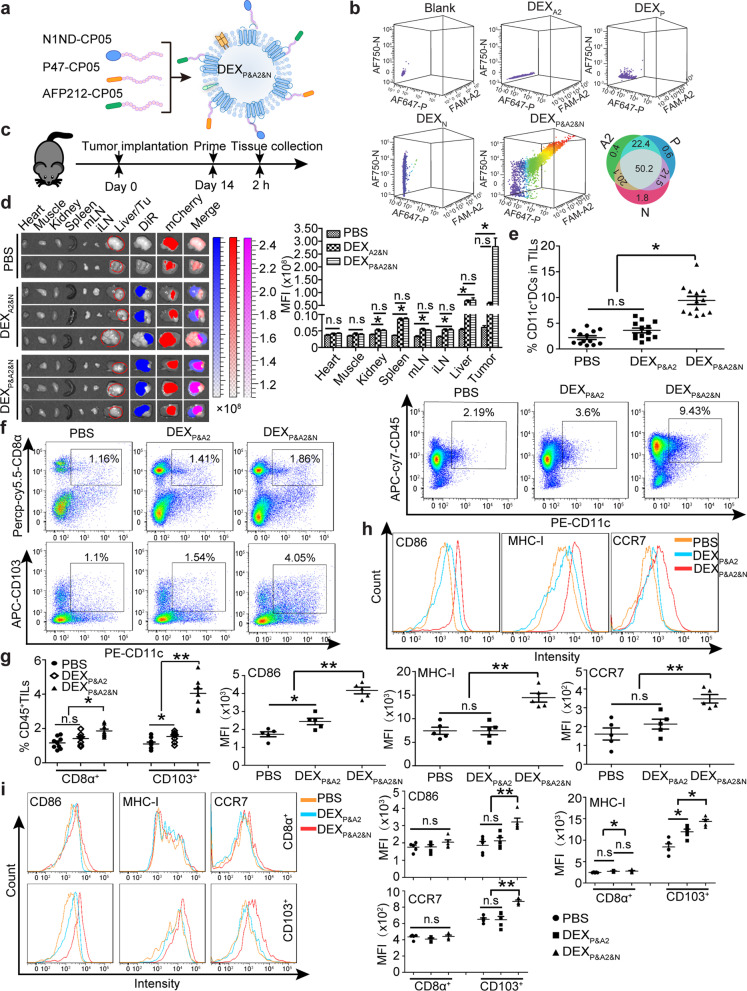
Fig. 1 Evaluation of DEXP&A2&N’s tumor-targeting, DC-recruiting and DC-activating ability in orthotopic mCherry-expressing HCC mice. a Schematic illustration for designer DEX vaccine-DEXP&A2&N. P-P47; A2-AFP212; N-N1ND. DEX refers to DC-derived exosomes. b Flow cytometric analysis to assess the simultaneous binding efficiency of three moieties on DEX (n = 4). Diagram for dosing regimen (c) and tissue distribution and quantitative analysis of labeled DEXP&A2&N (d) in day-14 orthotopic HCC mice bearing mCherry-expressing tumors. DiR-labeled DEXA2&N (n = 9), DEXP&A2&N (n = 9) (80 μg/mouse) or PBS (n = 4) were injected into day-14 orthotopic HCC mice bearing mCherry-expressing tumors intravenously, and tissues were harvested 2 h after injection (one-way ANOVA post hoc Student–Newman–Keuls test was used except for liver and tumor in which one-way ANOVA on ranks was used). mLN-mesenteric lymph node; iLN-inguinal lymph node; Tu-tumor. e Flow cytometric and quantitative analysis of CD11c+ DCs in tumor-infiltrating lymphocytes (TILs) from HCC mice bearing mCherry-expressing tumors (n = 13; one-way ANOVA post hoc Student–Newman–Keuls test). Flow cytometric (f) and quantitative analysis (g) of CD103+CD11c+ (one-way ANOVA on ranks) and CD8α+ CD11c+ DCs (one-way ANOVA post hoc Student–Newman–Keuls test) in TILs from HCC mice (n = 9). h Flow cytometric and quantitative analysis of surface protein markers on CD11c+ DCs from tumors of orthotopic HCC mice treated with DEXP&A2 or DEXP&A2&N (n = 5; one-way ANOVA post hoc Student–Newman–Keuls test). i Flow cytometric and quantitative analysis of surface protein markers on CD103+CD11c+ and CD8α+CD11c+ DCs from tumors of orthotopic HCC mice treated with DEXP&A2 or DEXP&A2&N (n = 5; one-way ANOVA post hoc Student–Newman–Keuls test). *p < 0.05, **p < 0.001; n.s, not significant
Fig. 1 Evaluation of DEXP&A2&N’s tumor-targeting, DC-recruiting and DC-activating ability in orthotopic mCherry-expressing HCC mice. a Schematic illustration for designer DEX vaccine-DEXP&A2&N. P-P47; A2-AFP212; N-N1ND. DEX refers to DC-derived exosomes. b Flow cytometric analysis to assess the simultaneous binding efficiency of three moieties on DEX (n = 4). Diagram for dosing regimen (c) and tissue distribution and quantitative analysis of labeled DEXP&A2&N (d) in day-14 orthotopic HCC mice bearing mCherry-expressing tumors. DiR-labeled DEXA2&N (n = 9), DEXP&A2&N (n = 9) (80 μg/mouse) or PBS (n = 4) were injected into day-14 orthotopic HCC mice bearing mCherry-expressing tumors intravenously, and tissues were harvested 2 h after injection (one-way ANOVA post hoc Student–Newman–Keuls test was used except for liver and tumor in which one-way ANOVA on ranks was used). mLN-mesenteric lymph node; iLN-inguinal lymph node; Tu-tumor. e Flow cytometric and quantitative analysis of CD11c+ DCs in tumor-infiltrating lymphocytes (TILs) from HCC mice bearing mCherry-expressing tumors (n = 13; one-way ANOVA post hoc Student–Newman–Keuls test). Flow cytometric (f) and quantitative analysis (g) of CD103+CD11c+ (one-way ANOVA on ranks) and CD8α+ CD11c+ DCs (one-way ANOVA post hoc Student–Newman–Keuls test) in TILs from HCC mice (n = 9). h Flow cytometric and quantitative analysis of surface protein markers on CD11c+ DCs from tumors of orthotopic HCC mice treated with DEXP&A2 or DEXP&A2&N (n = 5; one-way ANOVA post hoc Student–Newman–Keuls test). i Flow cytometric and quantitative analysis of surface protein markers on CD103+CD11c+ and CD8α+CD11c+ DCs from tumors of orthotopic HCC mice treated with DEXP&A2 or DEXP&A2&N (n = 5; one-way ANOVA post hoc Student–Newman–Keuls test). *p < 0.05, **p < 0.001; n.s, not significantCC BY · PubMed Central
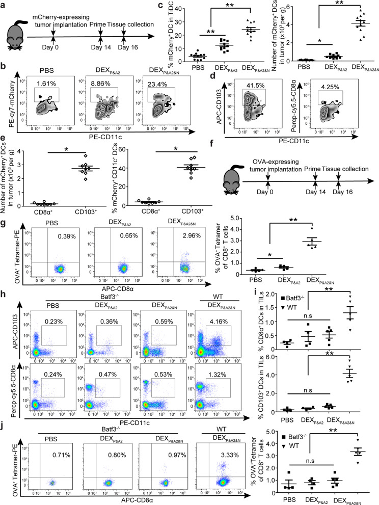
Fig. 2 Investigation of DEXP&A2&N promoting DC uptake and cross-presentation of tumor neoantigens in orthotopic HCC mice. a Diagram for dosing regimen of DEXP&A2&N. DEXP&A2 or DEXP&A2&N (80 μg/mouse) were injected into day-14 orthotopic HCC mice bearing mCherry-expressing tumors intravenously, and tissues were harvested 2 days after injection. Flow cytometric (b) and quantitative analysis (c) of mCherry+CD11c+ DCs from tumor of orthotopic HCC mice treated with DEXP&A2 or DEXP&A2&N (n = 11) (One-way ANOVA on ranks was used for the number and one-way ANOVA post hoc Student–Newman–Keuls test was applied for the percent analysis). TiDC: tumor-infiltrating DC. Flow cytometric (d) and quantitative analysis (e) of mCherry+CD103+CD11c+ or CD8α+CD11c+ DCs from tumor of orthotopic HCC mice treated with DEXP&A2&N (n = 8; Mann–Whitney rank-sum test). f Diagram for dosing regimen of DEXP&A2&N. DEXP&A2 or DEXP&A2&N (80 μg/mouse) were injected into day-14 orthotopic HCC mice bearing OVA-expressing tumors intravenously, and tissues were harvested 2 days after injection. g Flow cytometric and quantitative analysis of OVA+ tetramer T cells from splenocytes of orthotopic HCC mice treated with DEXP&A2 or DEXP&A2&N (n = 5; one-way ANOVA on ranks). Flow cytometric (h) and quantitative analysis (i) of CD103+CD11c+ or CD8α+CD11c+ DCs from TILs of orthotopic Batf3−/− HCC mice treated with PBS or DEXP&A2 (n = 4) or DEXP&A2&N (n = 5) and orthotopic wild-type (WT) HCC mice treated with DEXP&A2&N (n = 5) (one-way ANOVA on ranks). j Flow cytometric and quantitative analysis of OVA+ tetramer T cells from tumor of orthotopic Batf3−/− HCC mice treated with PBS or DEXP&A2 (n = 4) or DEXP&A2&N (n = 5) and orthotopic wild-type (WT) HCC mice treated with DEXP&A2&N (n = 5) (one-way ANOVA post hoc Student–Newman–Keuls test). *p < 0.05, **p < 0.001; n.s, not significant
Fig. 2 Investigation of DEXP&A2&N promoting DC uptake and cross-presentation of tumor neoantigens in orthotopic HCC mice. a Diagram for dosing regimen of DEXP&A2&N. DEXP&A2 or DEXP&A2&N (80 μg/mouse) were injected into day-14 orthotopic HCC mice bearing mCherry-expressing tumors intravenously, and tissues were harvested 2 days after injection. Flow cytometric (b) and quantitative analysis (c) of mCherry+CD11c+ DCs from tumor of orthotopic HCC mice treated with DEXP&A2 or DEXP&A2&N (n = 11) (One-way ANOVA on ranks was used for the number and one-way ANOVA post hoc Student–Newman–Keuls test was applied for the percent analysis). TiDC: tumor-infiltrating DC. Flow cytometric (d) and quantitative analysis (e) of mCherry+CD103+CD11c+ or CD8α+CD11c+ DCs from tumor of orthotopic HCC mice treated with DEXP&A2&N (n = 8; Mann–Whitney rank-sum test). f Diagram for dosing regimen of DEXP&A2&N. DEXP&A2 or DEXP&A2&N (80 μg/mouse) were injected into day-14 orthotopic HCC mice bearing OVA-expressing tumors intravenously, and tissues were harvested 2 days after injection. g Flow cytometric and quantitative analysis of OVA+ tetramer T cells from splenocytes of orthotopic HCC mice treated with DEXP&A2 or DEXP&A2&N (n = 5; one-way ANOVA on ranks). Flow cytometric (h) and quantitative analysis (i) of CD103+CD11c+ or CD8α+CD11c+ DCs from TILs of orthotopic Batf3−/− HCC mice treated with PBS or DEXP&A2 (n = 4) or DEXP&A2&N (n = 5) and orthotopic wild-type (WT) HCC mice treated with DEXP&A2&N (n = 5) (one-way ANOVA on ranks). j Flow cytometric and quantitative analysis of OVA+ tetramer T cells from tumor of orthotopic Batf3−/− HCC mice treated with PBS or DEXP&A2 (n = 4) or DEXP&A2&N (n = 5) and orthotopic wild-type (WT) HCC mice treated with DEXP&A2&N (n = 5) (one-way ANOVA post hoc Student–Newman–Keuls test). *p < 0.05, **p < 0.001; n.s, not significantCC BY · PubMed Central
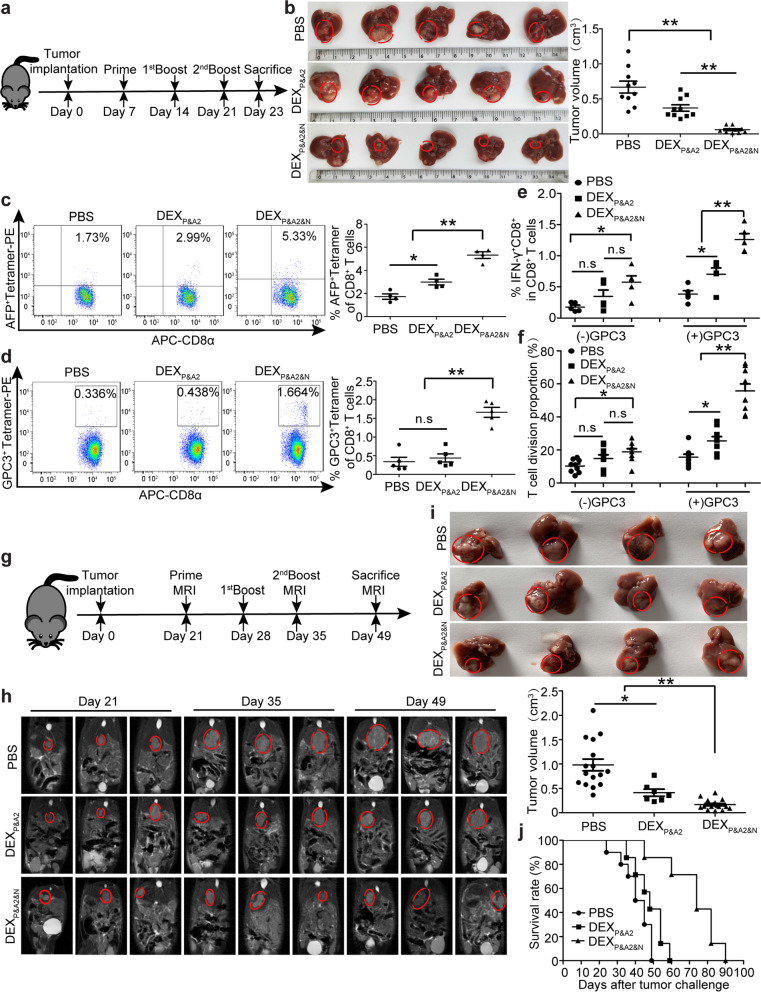
Fig. 3 Antitumor effects of DEXP&A2&N in orthotopic HCC mice. a Diagram for dosing regimen of DEXP&A2&N. DEXP&A2 or DEXP&A2&N (80 μg/mouse) were injected into day-7 orthotopic HCC mice intravenously three times weekly, and tissues were harvested 2 days after last injection. b Measurement of tumor volume in day-7 orthotopic HCC mice treated with DEXP&A2 or DEXP&A2&N (n = 10; one-way ANOVA on ranks). c Flow cytometric and quantitative analysis of AFP+ tetramer T cells from splenocytes of orthotopic HCC mice treated with DEXP&A2 or DEXP&A2&N (n = 4; one-way ANOVA post hoc Student–Newman–Keuls test). d Flow cytometric and quantitative analysis of GPC3+ tetramer T cells from splenocytes of orthotopic HCC mice treated with DEXP&A2 or DEXP&A2&N (n = 5; one-way ANOVA post hoc Student–Newman–Keuls test). Flow cytometric and quantitative analysis of IFN-γ+CD8+ T cells (e) (n = 5; one-way ANOVA post hoc Student–Newman–Keuls test) and T cell division (f) (n = 9; one-way ANOVA on ranks) in splenocytes of orthotopic HCC mice treated with DEXP&A2 or DEXP&A2&N, followed by in vitro stimulation of GPC3 epitopes. g Diagram for dosing regimen of DEXP&A2&N. DEXP&A2 or DEXP&A2&N (120 μg/mouse) were injected into day-21 orthotopic HCC mice bearing large established tumors intravenously three times weekly, and tissues were harvested 2 weeks after last injection. h MRI monitoring of tumor growth in day-21 orthotopic HCC mice bearing large established tumors at different time points. i Assessment of tumor size in orthotopic HCC mice bearing large established tumors treated with PBS (n = 16), DEXP&A2 (n = 7) or DEXP&A2&N (n = 16) at 28 days after initial treatment (one-way ANOVA on ranks). j Survival rate of orthotopic HCC mice treated with PBS (n = 9), DEXP&A2 or DEXP&A2&N (n = 8). *p < 0.05, **p < 0.001; n.s, not significant
Fig. 3 Antitumor effects of DEXP&A2&N in orthotopic HCC mice. a Diagram for dosing regimen of DEXP&A2&N. DEXP&A2 or DEXP&A2&N (80 μg/mouse) were injected into day-7 orthotopic HCC mice intravenously three times weekly, and tissues were harvested 2 days after last injection. b Measurement of tumor volume in day-7 orthotopic HCC mice treated with DEXP&A2 or DEXP&A2&N (n = 10; one-way ANOVA on ranks). c Flow cytometric and quantitative analysis of AFP+ tetramer T cells from splenocytes of orthotopic HCC mice treated with DEXP&A2 or DEXP&A2&N (n = 4; one-way ANOVA post hoc Student–Newman–Keuls test). d Flow cytometric and quantitative analysis of GPC3+ tetramer T cells from splenocytes of orthotopic HCC mice treated with DEXP&A2 or DEXP&A2&N (n = 5; one-way ANOVA post hoc Student–Newman–Keuls test). Flow cytometric and quantitative analysis of IFN-γ+CD8+ T cells (e) (n = 5; one-way ANOVA post hoc Student–Newman–Keuls test) and T cell division (f) (n = 9; one-way ANOVA on ranks) in splenocytes of orthotopic HCC mice treated with DEXP&A2 or DEXP&A2&N, followed by in vitro stimulation of GPC3 epitopes. g Diagram for dosing regimen of DEXP&A2&N. DEXP&A2 or DEXP&A2&N (120 μg/mouse) were injected into day-21 orthotopic HCC mice bearing large established tumors intravenously three times weekly, and tissues were harvested 2 weeks after last injection. h MRI monitoring of tumor growth in day-21 orthotopic HCC mice bearing large established tumors at different time points. i Assessment of tumor size in orthotopic HCC mice bearing large established tumors treated with PBS (n = 16), DEXP&A2 (n = 7) or DEXP&A2&N (n = 16) at 28 days after initial treatment (one-way ANOVA on ranks). j Survival rate of orthotopic HCC mice treated with PBS (n = 9), DEXP&A2 or DEXP&A2&N (n = 8). *p < 0.05, **p < 0.001; n.s, not significantCC BY · PubMed Central
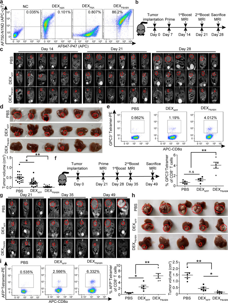
Fig. 4 Antitumor immunity of DEXP&A&N in orthotopic HCC mice. a Flow cytometric analysis to assess the double loading efficiency of two moieties on DEXAFP. DEXP&A&N refer to DEXP47&AFP&N1ND. b Diagram for dosing regimen of DEXP&A&N. DEXAFP or DEXP&A&N (80 μg/mouse) were injected into day-7 orthotopic HCC mice intravenously three times weekly, and tissues were harvested 7 days after last injection. c Dynamic monitoring of tumor growth in orthotopic HCC mice with MRI at different time points. d Measurement of tumor size in orthotopic HCC mice at 21 days after priming (n = 20; one-way ANOVA on ranks). e Flow cytometric and quantitative analysis of GPC3+ tetramer T cells from tumor of orthotopic HCC mice treated with DEXAFP or DEXP&A&N (n = 5; one-way ANOVA post hoc Student–Newman–Keuls test). f Diagram for dosing regimen of DEXP&A&N. DEXAFP or DEXP&A&N (120 μg/mouse) were injected into day-21 orthotopic HCC mice bearing large established tumors intravenously three times weekly, and tissues were harvested 2 weeks after last injection. g Dynamic monitoring of tumor growth in orthotopic HCC mice bearing large established tumors treated with DEXAFP or DEXP&A&N with MRI at different time points. h Measurement of tumor size in orthotopic HCC mice at 28 days after priming (n = 5; one-way ANOVA post hoc Student–Newman–Keuls test). i Flow cytometric and quantitative analysis of AFP+ tetramer T cells from tumor of orthotopic HCC mice treated with DEXAFP or DEXP&A&N (n = 5; one-way ANOVA post hoc Student–Newman–Keuls test). *p < 0.05, **p < 0.001; n.s, not significant
Fig. 4 Antitumor immunity of DEXP&A&N in orthotopic HCC mice. a Flow cytometric analysis to assess the double loading efficiency of two moieties on DEXAFP. DEXP&A&N refer to DEXP47&AFP&N1ND. b Diagram for dosing regimen of DEXP&A&N. DEXAFP or DEXP&A&N (80 μg/mouse) were injected into day-7 orthotopic HCC mice intravenously three times weekly, and tissues were harvested 7 days after last injection. c Dynamic monitoring of tumor growth in orthotopic HCC mice with MRI at different time points. d Measurement of tumor size in orthotopic HCC mice at 21 days after priming (n = 20; one-way ANOVA on ranks). e Flow cytometric and quantitative analysis of GPC3+ tetramer T cells from tumor of orthotopic HCC mice treated with DEXAFP or DEXP&A&N (n = 5; one-way ANOVA post hoc Student–Newman–Keuls test). f Diagram for dosing regimen of DEXP&A&N. DEXAFP or DEXP&A&N (120 μg/mouse) were injected into day-21 orthotopic HCC mice bearing large established tumors intravenously three times weekly, and tissues were harvested 2 weeks after last injection. g Dynamic monitoring of tumor growth in orthotopic HCC mice bearing large established tumors treated with DEXAFP or DEXP&A&N with MRI at different time points. h Measurement of tumor size in orthotopic HCC mice at 28 days after priming (n = 5; one-way ANOVA post hoc Student–Newman–Keuls test). i Flow cytometric and quantitative analysis of AFP+ tetramer T cells from tumor of orthotopic HCC mice treated with DEXAFP or DEXP&A&N (n = 5; one-way ANOVA post hoc Student–Newman–Keuls test). *p < 0.05, **p < 0.001; n.s, not significantCC BY · PubMed Central
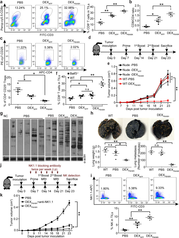
Fig. 5 DEXP&A&N evoke potent antitumor immunity with efficacy dependent on innate and adaptive immune responses. Flow cytometric and quantitative analysis of tumor-infiltrating CD8+ (a) and ratio of tumor-infiltrating CD8+ to CD4+ (b) and flow cytometric and quantitative analysis of tumor CD4+CD25+ (c) T cells of DEXP&A&N -treated orthotopic HCC mice bearing large established tumors (n = 5). d Diagram for dosing regimen of DEXP&A&N. DEXAFP or DEXP&A&N (80 μg) were injected into day-7 thymus-deficient nude mice bearing subcutaneous HCC tumors intravenously three times weekly, and tissues were harvested 2 days after last injection. e Measurement of tumor size in nude mice bearing subcutaneous HCC tumors at different time points (n = 5). One-way ANOVA on ranks test was used for day 7 and 21; one-way ANOVA post hoc Student–Newman–Keuls test was used for day 9, 11, 14, 16, 18 and 23. f Flow cytometric and quantitative analysis of tumor-infiltrating CD8+ T cells of DEXP&A&N-treated orthotopic Batf3−/− HCC mice (n = 5; one-way ANOVA on ranks). g Western blot to examine Hepa1-6 tumor cell lysates with serum from DEXP&A&N- or DEXAFP-treated mice. α-actin was used as a loading control. h Representative images and quantification of tumor nodules in lungs 21 days after serum re-infusion and tumor challenge (n = 4). i Flow cytometric and quantitative analysis of tumor-infiltrating NK cells of DEXP&A&N-treated orthotopic HCC mice bearing large established tumors (n = 5). j Diagram for NK depletion experiments. k Measurement of tumor volume in DEXP&A&N and anti-NK1.1-treated ectopic HCC mice at different time points (n = 5). One-way ANOVA post hoc Student–Newman–Keuls test was used for day 7, 9, 11, 14, 21 and 23; one-way ANOVA on ranks test was applied for day 16 and 18; and Two-tailed t test was applied for DEXP&A&N with or without anti-NK1.1 antibody on day 23. *p < 0.05, **p < 0.001; n.s means not significant. Note: One-way ANOVA post hoc Student–Newman–Keuls test was used for Fig. 5a-c and 5 h, 5i
Fig. 5 DEXP&A&N evoke potent antitumor immunity with efficacy dependent on innate and adaptive immune responses. Flow cytometric and quantitative analysis of tumor-infiltrating CD8+ (a) and ratio of tumor-infiltrating CD8+ to CD4+ (b) and flow cytometric and quantitative analysis of tumor CD4+CD25+ (c) T cells of DEXP&A&N -treated orthotopic HCC mice bearing large established tumors (n = 5). d Diagram for dosing regimen of DEXP&A&N. DEXAFP or DEXP&A&N (80 μg) were injected into day-7 thymus-deficient nude mice bearing subcutaneous HCC tumors intravenously three times weekly, and tissues were harvested 2 days after last injection. e Measurement of tumor size in nude mice bearing subcutaneous HCC tumors at different time points (n = 5). One-way ANOVA on ranks test was used for day 7 and 21; one-way ANOVA post hoc Student–Newman–Keuls test was used for day 9, 11, 14, 16, 18 and 23. f Flow cytometric and quantitative analysis of tumor-infiltrating CD8+ T cells of DEXP&A&N-treated orthotopic Batf3−/− HCC mice (n = 5; one-way ANOVA on ranks). g Western blot to examine Hepa1-6 tumor cell lysates with serum from DEXP&A&N- or DEXAFP-treated mice. α-actin was used as a loading control. h Representative images and quantification of tumor nodules in lungs 21 days after serum re-infusion and tumor challenge (n = 4). i Flow cytometric and quantitative analysis of tumor-infiltrating NK cells of DEXP&A&N-treated orthotopic HCC mice bearing large established tumors (n = 5). j Diagram for NK depletion experiments. k Measurement of tumor volume in DEXP&A&N and anti-NK1.1-treated ectopic HCC mice at different time points (n = 5). One-way ANOVA post hoc Student–Newman–Keuls test was used for day 7, 9, 11, 14, 21 and 23; one-way ANOVA on ranks test was applied for day 16 and 18; and Two-tailed t test was applied for DEXP&A&N with or without anti-NK1.1 antibody on day 23. *p < 0.05, **p < 0.001; n.s means not significant. Note: One-way ANOVA post hoc Student–Newman–Keuls test was used for Fig. 5a-c and 5 h, 5iCC BY · PubMed Central
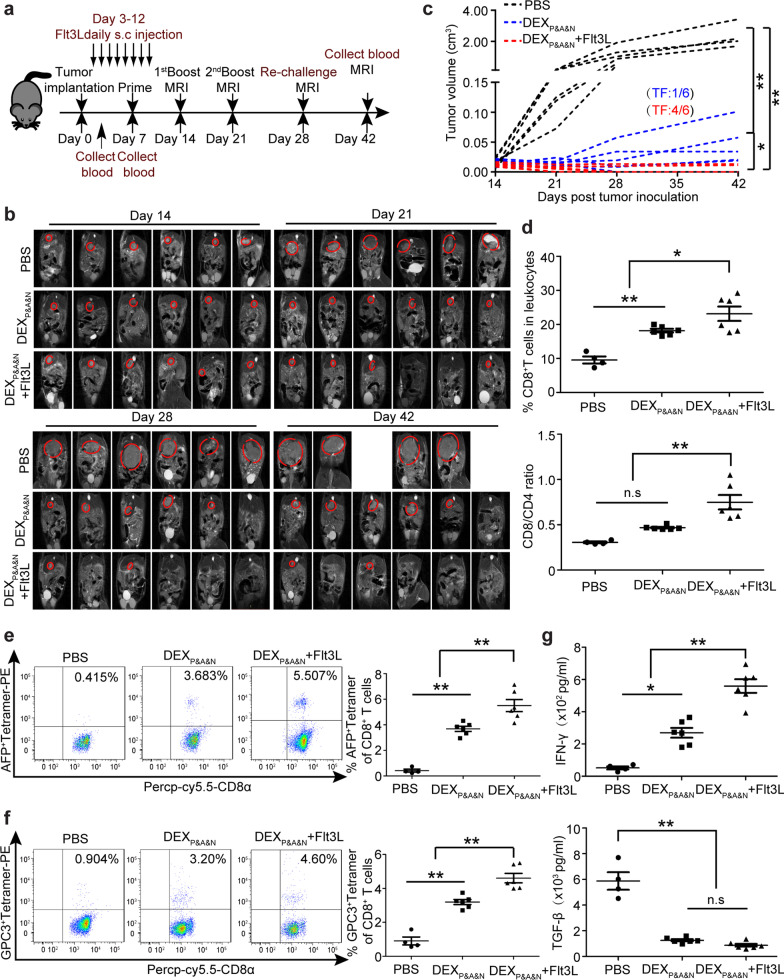
Fig. 6 Flt3L augments DEXP&A&N’s antitumor potency in orthotopic HCC mice. a Diagram for dosing regimen of DEXP&A&N and Flt3L in orthotopic HCC mice. Flt3L was administered subcutaneously to day-3 orthotopic HCC mice at the dose of 800 μg /kg/day for 8 days consecutively and DEXP&A&N (80 μg/mouse) were intravenously administered on day 7 after tumor implantation three times weekly. b Dynamic monitoring of tumor growth in orthotopic HCC mice treated with DEXP&A&N or DEXP&A&N and Flt3L with MRI at different time points. c Measurement of tumor volume for each individual mouse treated with PBS, DEXP&A&N or DEXP&A&N and Flt3L (n = 6). One-way ANOVA on ranks test was used for day 21, 28 and 42; one-way ANOVA post hoc Student–Newman–Keuls test was used for day 14. TF means tumor-free. d Quantitative analysis of CD8+ T cells and ratio of CD8+ to CD4+ T cells in blood of orthotopic HCC mice treated with PBS (n = 4), DEXP&A&N or DEXP&A&N and Flt3L (n = 6) at day 42 after tumor implantation (One-way ANOVA on ranks). Flow cytometric and quantitative analysis of AFP+ tetramer (e) and GPC3+ tetramer (f) T cells in blood of orthotopic HCC mice treated with PBS (n = 4), DEXP&A&N or DEXP&A&N and Flt3L (n = 6) at day 42 after tumor implantation (one-way ANOVA post hoc Student–Newman–Keuls test). g Assessment of IFN-γ and TGF-β in blood of orthotopic HCC mice treated with PBS (n = 4), DEXP&A&N or DEXP&A&N and Flt3L (n = 6) at day 42 after tumor implantation (one-way ANOVA post hoc Student–Newman–Keuls test). *p < 0.05, **p < 0.001; n.s, not significant
Fig. 6 Flt3L augments DEXP&A&N’s antitumor potency in orthotopic HCC mice. a Diagram for dosing regimen of DEXP&A&N and Flt3L in orthotopic HCC mice. Flt3L was administered subcutaneously to day-3 orthotopic HCC mice at the dose of 800 μg /kg/day for 8 days consecutively and DEXP&A&N (80 μg/mouse) were intravenously administered on day 7 after tumor implantation three times weekly. b Dynamic monitoring of tumor growth in orthotopic HCC mice treated with DEXP&A&N or DEXP&A&N and Flt3L with MRI at different time points. c Measurement of tumor volume for each individual mouse treated with PBS, DEXP&A&N or DEXP&A&N and Flt3L (n = 6). One-way ANOVA on ranks test was used for day 21, 28 and 42; one-way ANOVA post hoc Student–Newman–Keuls test was used for day 14. TF means tumor-free. d Quantitative analysis of CD8+ T cells and ratio of CD8+ to CD4+ T cells in blood of orthotopic HCC mice treated with PBS (n = 4), DEXP&A&N or DEXP&A&N and Flt3L (n = 6) at day 42 after tumor implantation (One-way ANOVA on ranks). Flow cytometric and quantitative analysis of AFP+ tetramer (e) and GPC3+ tetramer (f) T cells in blood of orthotopic HCC mice treated with PBS (n = 4), DEXP&A&N or DEXP&A&N and Flt3L (n = 6) at day 42 after tumor implantation (one-way ANOVA post hoc Student–Newman–Keuls test). g Assessment of IFN-γ and TGF-β in blood of orthotopic HCC mice treated with PBS (n = 4), DEXP&A&N or DEXP&A&N and Flt3L (n = 6) at day 42 after tumor implantation (one-way ANOVA post hoc Student–Newman–Keuls test). *p < 0.05, **p < 0.001; n.s, not significantCC BY · PubMed Central
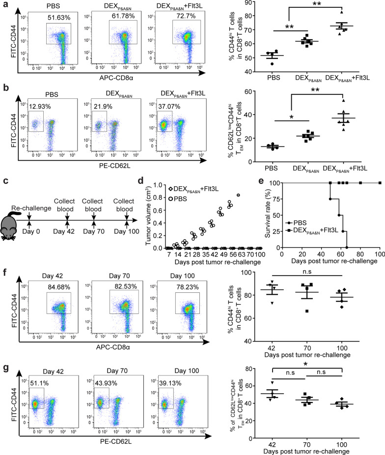
Fig. 7 Long-term protective memory immune responses elicited by DEXP&A&N and Flt3L against tumor re-challenge. Flow cytometric and quantitative analysis of CD44hi CD8+ (a) and CD62LlowCD44hiCD8+ (b) T cells in blood of orthotopic HCC mice treated with PBS (n = 4), DEXP&A&N, DEXP&A&N and Flt3L (n = 6) at day 42 after tumor implantation (one-way ANOVA post hoc Student–Newman–Keuls test). c Diagram for tumor re-challenge in DEXP&A&N and Flt3L-treated tumor-free mice. d Measurement of tumor volume in DEXP&A&N and Flt3L-treated tumor-free mice after re-challenge (n = 4). Mann–Whitney rank-sum test was used for day 7; one-way ANOVA on ranks test was used for day 14, 21, 28, 35 and 42; one-way ANOVA post hoc Student–Newman–Keuls test was used for day 49. e Survival rate of DEXP&A&N and Flt3L-treated tumor-free mice after re-challenge (n = 4). Flow cytometric and quantitative analysis of CD44hiCD8+ (f) and CD62LlowCD44hi CD8+ (g) T cells in blood of DEXP&A&N and Flt3L-treated tumor-free mice at different time points after tumor re-challenge (n = 4; one-way ANOVA post hoc Student–Newman–Keuls test). *p < 0.05, **p < 0.001; n.s, not significant
Fig. 7 Long-term protective memory immune responses elicited by DEXP&A&N and Flt3L against tumor re-challenge. Flow cytometric and quantitative analysis of CD44hi CD8+ (a) and CD62LlowCD44hiCD8+ (b) T cells in blood of orthotopic HCC mice treated with PBS (n = 4), DEXP&A&N, DEXP&A&N and Flt3L (n = 6) at day 42 after tumor implantation (one-way ANOVA post hoc Student–Newman–Keuls test). c Diagram for tumor re-challenge in DEXP&A&N and Flt3L-treated tumor-free mice. d Measurement of tumor volume in DEXP&A&N and Flt3L-treated tumor-free mice after re-challenge (n = 4). Mann–Whitney rank-sum test was used for day 7; one-way ANOVA on ranks test was used for day 14, 21, 28, 35 and 42; one-way ANOVA post hoc Student–Newman–Keuls test was used for day 49. e Survival rate of DEXP&A&N and Flt3L-treated tumor-free mice after re-challenge (n = 4). Flow cytometric and quantitative analysis of CD44hiCD8+ (f) and CD62LlowCD44hi CD8+ (g) T cells in blood of DEXP&A&N and Flt3L-treated tumor-free mice at different time points after tumor re-challenge (n = 4; one-way ANOVA post hoc Student–Newman–Keuls test). *p < 0.05, **p < 0.001; n.s, not significantCC BY · PubMed Central
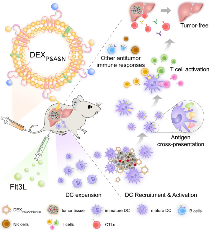
Fig. 8 Graphical abstract for illustrating the mechanism underlying the antitumor immunogenicity of universal DEX vaccines
Fig. 8 Graphical abstract for illustrating the mechanism underlying the antitumor immunogenicity of universal DEX vaccinesCC BY · PubMed Central

Verification

Claims the paper makes

The claims and statistics of this paper haven’t been checked yet.

Run the check — free, no account needed. It reads the open-access text and checks the sources it cites.

A few free checks a day · papers already verified open instantly · results are public

Claims other papers make about this one

Each is checked against this paper’s own text: does it say what the citing paper says it does?

Self-check

Percentages, sums and reported differences re-computed from the paper’s own numbers.

The self-check needs the paper’s full text.

Citations

Cites

Research works printed but not linked: 45

  1. Zhu RX, Seto WK, Lai CL, Yuen MF. Epidemiology of hepatocellular carcinoma in the Asia-Pacific Region. Gut Liver. 2016;10:332–9. · DOI
  2. Xu W, et al. Immunotherapy for hepatocellular carcinoma: recent advances and future perspectives. Ther Adv Med Oncol. 2019;11:1758835919862692. · DOI
  3. Blass E, Ott PA. Advances in the development of personalized neoantigen-based therapeutic cancer vaccines. Nat Rev Clin Oncol. 2021;18:215–29. · DOI · Paper page
  4. Loffler MW, et al. Multi-omics discovery of exome-derived neoantigens in hepatocellular carcinoma. Genome Med. 2019;11:28. · DOI
  5. Lu L, et al. Targeting neoantigens in hepatocellular carcinoma for immunotherapy: a futile strategy? Hepatology. 2021;73:414–21. · DOI
  6. Aslan C, et al. Exosomes for mRNA delivery: a novel biotherapeutic strategy with hurdles and hope. BMC Biotechnol. 2021;21:20. · DOI
  7. Nikfarjam S, Rezaie J, Kashanchi F, Jafari R. Dexosomes as a cell-free vaccine for cancer immunotherapy. J Exp Clin Cancer Res. 2020;39:258. · DOI
  8. Xu Z, Zeng S, Gong Z, Yan Y. Exosome-based immunotherapy: a promising approach for cancer treatment. Mol Cancer. 2020;19:160. · DOI · Paper page
  9. Yang D, et al. High-mobility group nucleosome-binding protein 1 acts as an alarmin and is critical for lipopolysaccharide-induced immune responses. J Exp Med. 2012;209:157–71. · DOI
  10. Zuo B, et al. Alarmin-painted exosomes elicit persistent antitumor immunity in large established tumors in mice. Nat Commun. 2020;11:1790. · DOI
  11. Jing R, et al. Fluorescent peptide highlights micronodules in murine hepatocellular carcinoma models and humans in vitro. Hepatology. 2018;68:1391–411. · DOI
  12. Hong Y, et al. Epitope-optimized alpha-fetoprotein genetic vaccines prevent carcinogen-induced murine autochthonous hepatocellular carcinoma. Hepatology. 2014;59:1448–58. · DOI
  13. Joffre OP, Segura E, Savina A, Amigorena S. Cross-presentation by dendritic cells. Nat Rev Immunol. 2012;12:557–69. · DOI
  14. Lu Z, et al. Dendritic cell-derived exosomes elicit tumor regression in autochthonous hepatocellular carcinoma mouse models. J Hepatol. 2017;67:739–48. · DOI · Paper page
  15. Schmid MA, Kingston D, Boddupalli S, Manz MG. Instructive cytokine signals in dendritic cell lineage commitment. Immunol Rev. 2010;234:32–44. · DOI
  16. Balan S, Bhardwaj N. Cross-presentation of tumor antigens is ruled by synaptic transfer of vesicles among dendritic cell subsets. Cancer Cell. 2020;37:751–3. · DOI
  17. Gardner A, Ruffell B. Dendritic cells and cancer immunity. Trends Immunol. 2016;37:855–65. · DOI
  18. Li AW, et al. A facile approach to enhance antigen response for personalized cancer vaccination. Nat Mater. 2018;17:528–34. · DOI
  19. Weigel BJ, et al. Comparative analysis of murine marrow-derived dendritic cells generated by Flt3L or GM-CSF/IL-4 and matured with immune stimulatory agents on the in vivo induction of antileukemia responses. Blood. 2002;100:4169–76. · DOI
  20. Roberts EW, et al. Critical role for CD103(+)/CD141(+) dendritic cells bearing CCR7 for tumor antigen trafficking and priming of T cell immunity in melanoma. Cancer Cell. 2016;30:324–36. · DOI
  21. Chiang MC, et al. Differential uptake and cross-presentation of soluble and necrotic cell antigen by human DC subsets. Eur J Immunol. 2016;46:329–39. · DOI
  22. Salmon H, et al. Expansion and activation of CD103(+) dendritic cell progenitors at the tumor site enhances tumor responses to therapeutic PD-L1 and BRAF inhibition. Immunity. 2016;44:924–38. · DOI
  23. De Vries IJ, et al. Effective migration of antigen-pulsed dendritic cells to lymph nodes in melanoma patients is determined by their maturation state. Cancer Res. 2003;63:12–7.
  24. Moynihan KD, et al. Eradication of large established tumors in mice by combination immunotherapy that engages innate and adaptive immune responses. Nat Med. 2016;22:1402–10. · DOI
  25. Edelson BT, et al. Peripheral CD103+ dendritic cells form a unified subset developmentally related to CD8alpha+ conventional dendritic cells. J Exp Med. 2010;207:823–36. · DOI
  26. Iwama T, et al. Identification of an H2-Kb or H2-Db restricted and glypican-3-derived cytotoxic T-lymphocyte epitope peptide. Int J Oncol. 2013;42:831–8. · DOI
  27. Dunn GP, Koebel CM, Schreiber RD. Interferons, immunity and cancer immunoediting. Nat Rev Immunol. 2006;6:836–48. · DOI · Paper page
  28. Vitale M, Cantoni C, Pietra G, Mingari MC, Moretta L. Effect of tumor cells and tumor microenvironment on NK-cell function. Eur J Immunol. 2014;44:1582–92. · DOI
  29. Marzagalli M, Ebelt ND, Manuel ER. Unraveling the crosstalk between melanoma and immune cells in the tumor microenvironment. Semin Cancer Biol. 2019;59:236–50. · DOI
  30. Habif G, Crinier A, Andre P, Vivier E, Narni-Mancinelli E. Targeting natural killer cells in solid tumors. Cell Mol Immunol. 2019;16:415–22. · DOI
  31. Vivier E, et al. Innate lymphoid cells: 10 years on. Cell. 2018;174:1054–66. · DOI
  32. Zhu G, et al. Albumin/vaccine nanocomplexes that assemble in vivo for combination cancer immunotherapy. Nat Commun. 2017;8:1954. · DOI
  33. Liu K, Nussenzweig MC. Origin and development of dendritic cells. Immunol Rev. 2010;234:45–54. · DOI
  34. Lai J, et al. Adoptive cellular therapy with T cells expressing the dendritic cell growth factor Flt3L drives epitope spreading and antitumor immunity. Nat Immunol. 2020;21:914–26. · DOI
  35. Anandasabapathy N, et al. Efficacy and safety of CDX-301, recombinant human Flt3L, at expanding dendritic cells and hematopoietic stem cells in healthy human volunteers. Bone Marrow Transplant. 2015;50:924–30. · DOI
  36. Badovinac VP, Messingham KA, Jabbari A, Haring JS, Harty JT. Accelerated CD8+ T-cell memory and prime-boost response after dendritic-cell vaccination. Nat Med. 2005;11:748–56. · DOI
  37. Viaud S, et al. Updated technology to produce highly immunogenic dendritic cell-derived exosomes of clinical grade: a critical role of interferon-gamma. J Immunother. 2011;34:65–75. · DOI
  38. Bhardwaj N, et al. Flt3 ligand augments immune responses to anti-DEC-205-NY-ESO-1 vaccine through expansion of dendritic cell subsets. Nat Cancer. 2020;1:1204–17. · DOI
  39. Hammerich L, et al. Systemic clinical tumor regressions and potentiation of PD1 blockade with in situ vaccination. Nat Med. 2019;25:814–24. · DOI
  40. Cheng AL, Hsu C, Chan SL, Choo SP, Kudo M. Challenges of combination therapy with immune checkpoint inhibitors for hepatocellular carcinoma. J Hepatol. 2020;72:307–19. · DOI
  41. Pinter M, Jain RK, Duda DG. The current landscape of immune checkpoint blockade in hepatocellular carcinoma: a review. JAMA Oncol. 2021;7:113–23. · DOI
  42. Johnston MP, Khakoo SI. Immunotherapy for hepatocellular carcinoma: current and future. World J Gastroenterol. 2019;25:2977–89. · DOI
  43. Shen Z, Reznikoff G, Dranoff G, Rock KL. Cloned dendritic cells can present exogenous antigens on both MHC class I and class II molecules. J Immunol. 1997;158:2723–30. · DOI · Paper page
  44. Rao Q, et al. Tumor-derived exosomes elicit tumor suppression in murine hepatocellular carcinoma models and humans in vitro. Hepatology. 2016;64:456–72. · DOI · Paper page
  45. Kreiter S, et al. Mutant MHC class II epitopes drive therapeutic immune responses to cancer. Nature. 2015;520:692–6. · DOI · Paper page

Cited by

Sorted by citations

See all 155

Metadata

Authors
Zuo, B., Zhang, Y., Zhao, K., Wu, L., Qi, H., Yang, R., Gao, X., Geng, M., Wu, Y., Jing, R., Zhou, Q., Seow, Y., & Yin, H.
Venue
Journal of Hematology & Oncology
Published
2022-04-29
Volume
15
Issue
1
ISSN
1756-8722
Cited by
155
References
6
Language
en

About these checks

Registry, retraction, Cites and statistics results are computed, not judged, with one exception: a printed reference that no index lists is read once by a model before it counts as a citation error. Claims are extracted by a model and kept only when their quote appears verbatim in the text; each cited source is then checked, with a model’s help, for whether it says what the claim relies on. Those verdicts can be wrong — read the claim’s quote and the cited source before relying on one.

Statistics and arithmetic are recomputed, not judged.

Confidence bands are shown only for fields calibrated on our annotated test set. None is calibrated yet, so no band is shown.

What counts as a claim ›How we measure confidence ›How we check this ›

Are you an author, or do you see a problem with this page? Request a review