

The DOI matches its Crossref record; 1 of the 1 references it cites were found in a registry or an index.
The cytoskeleton dynamics-dependent LINC complex in periodontal ligament stem cells transmits mechanical stress to the nuclear envelope and promotes YAP nuclear translocation
Open accessAbstract
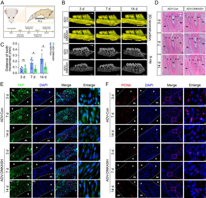
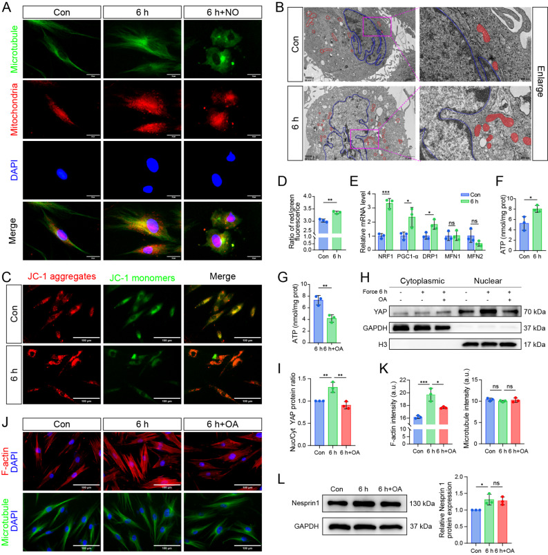
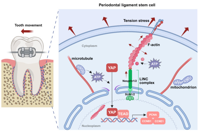
BACKGROUND: Periodontal ligament stem cells (PDLSCs) are important seed cells in tissue engineering and clinical applications. They are the priority receptor cells for sensing various mechanical stresses. Yes-associated protein (YAP) is a recognized mechanically sensitive transcription factor. However, the role of YAP in regulating the fate of PDLSCs under tension stress (TS) and its underlying mechanism is still unclear. METHODS: The effects of TS on the morphology and fate of PDLSCs were investigated using fluorescence staining, transmission electron microscopy, flow cytometry and quantitative real-time polymerase chain reaction (qRT-PCR). Then qRT-PCR, western blotting, immunofluorescence staining and gene knockdown experiments were performed to investigate the expression and distribution of YAP and its correlation with PDLSCs proliferation. The effects of cytoskeleton dynamics on YAP nuclear translocation were subsequently explored by adding cytoskeleton inhibitors. The effect of cytoskeleton dynamics on the expression of the LINC complex was proved through qRT-PCR and western blotting. After destroying the LINC complex by adenovirus, the effects of the LINC complex on YAP nuclear translocation and PDLSCs proliferation were investigated. Mitochondria-related detections were then performed to explore the role of mitochondria in YAP nuclear translocation. Finally, the in vitro results were verified by constructing orthodontic tooth movement models in Sprague-Dawley rats. RESULTS: TS enhanced the polymerization and stretching of F-actin, which upregulated the expression of the LINC complex. This further strengthened the pull on the nuclear envelope, enlarged the nuclear pore, and facilitated YAP's nuclear entry, thus enhancing the expression of proliferation-related genes. In this process, mitochondria were transported to the periphery of the nucleus along the reconstructed microtubules. They generated ATP to aid YAP's nuclear translocation and drove F-actin polymerization to a certain degree. When the LINC complex was destroyed, the nuclear translocation of YAP was inhibited, which limited PDLSCs proliferation, impeded periodontal tissue remodeling, and hindered tooth movement. CONCLUSIONS: Our study confirmed that appropriate TS could promote PDLSCs proliferation and periodontal tissue remodeling through the mechanically driven F-actin/LINC complex/YAP axis, which could provide theoretical guidance for seed cell expansion and for promoting healthy and effective tooth movement in clinical practice.
Overview
The claims and statistics of this paper haven’t been checked yet.
Run the check — free, no account needed. It reads the open-access text and checks the sources it cites.
Integrity signals
Registry
Cites
Statistics
Figures
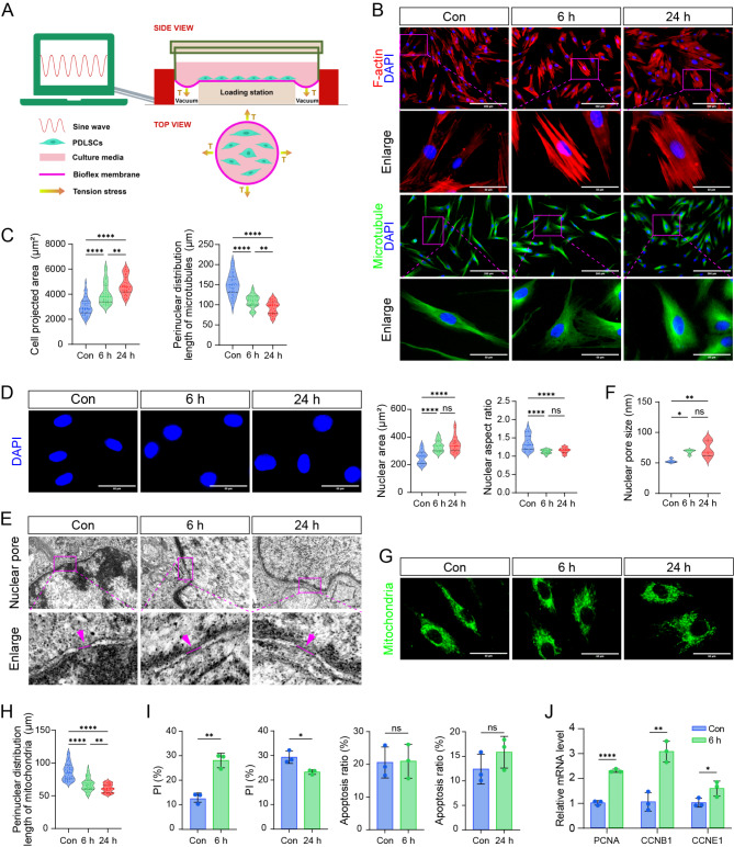
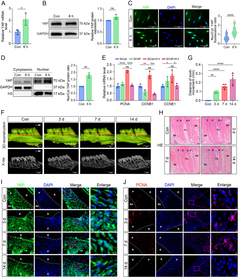
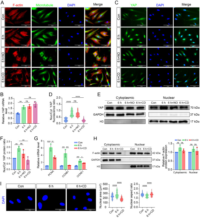
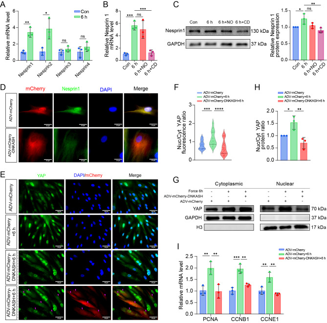
Verification
Claims the paper makes
The claims and statistics of this paper haven’t been checked yet.
Run the check — free, no account needed. It reads the open-access text and checks the sources it cites.
Claims other papers make about this one
Each is checked against this paper’s own text: does it say what the citing paper says it does?
The source backs part of the claim, not all of it — or backs it for a different population, condition or comparison.
In the citing paper
“previous studies have shown that stiff substrates reorganize F‐actin from branched networks into parallel stress fibers and transmit traction forces to the nucleus via the LINC/perinuclear actin cap axis [34, 35]”
From Liver Stiffness Rises Early in MASLD and Drives Inflammation, Lipid Dysmetabolism, and Fibrosis via Piezo1–YAP Mechanotransduction (2026) · cites this paper as [35]
Experimental — accuracy not yet measured
Self-check
Percentages, sums and reported differences re-computed from the paper’s own numbers.
The self-check needs the paper’s full text.
Citations
Cites
The actin cytoskeleton and actin-based motility
Svitkina, T. (2018) · Cold Spring Harbor Perspectives in Biology · doi:10.1101/cshperspect.a018267 · Cited by 761
Research works printed but not linked: 63
- Tomokiyo A, Wada N, Maeda H. Periodontal Ligament Stem cells: regenerative potency in Periodontium. Stem Cells Dev. 2019;28(15):974–85. · DOI
- Wang L, Liang H, Sun B, Mi J, Tong X, Wang Y et al. Role of TRPC6 in periodontal tissue reconstruction mediated by appropriate stress. Stem Cell Res Ther. 2022;13(1). · DOI
- Wang J, Yang H, Ma X, Liu J, Li L, Chen L, et al. LRP6/filamentous-actin signaling facilitates osteogenic commitment in mechanically induced periodontal ligament stem cells. Cell Mol Biol Lett. 2023;28(1):7. · DOI
- Zhang D, Lin W, Jiang S, Deng P, Liu L, Wang Q et al. Lepr-expressing PDLSCs contribute to Periodontal Homeostasis and respond to Mechanical Force by Piezo1. Adv Sci. 2023;10(29). · DOI
- Shi Q, Zheng L, Na J, Li X, Yang Z, Chen X et al. Fluid shear stress promotes periodontal ligament cells proliferation via p38-AMOT-YAP. Cell Mol Life Sci. 2022;79(11). · DOI
- Brockhaus J, Craveiro RB, Azraq I, Niederau C, Schröder SK, Weiskirchen R, et al. In Vitro Compression Model for Orthodontic Tooth Movement Modulates Human Periodontal Ligament Fibroblast Proliferation, apoptosis and cell cycle. Biomolecules. 2021;11(7):932. · DOI
- Li Y, Zhan Q, Bao M, Yi J, Li Y. Biomechanical and biological responses of periodontium in orthodontic tooth movement: up-date in a new decade. Int J Oral Sci. 2021;13(1). · DOI
- Iskratsch T, Wolfenson H, Sheetz MP. Appreciating force and shape — the rise of mechanotransduction in cell biology. Nat Rev Mol Cell Biol. 2014;15(12):825–33. · DOI · Paper page
- Iwata T, Yamato M, Zhang Z, Mukobata S, Washio K, Ando T, et al. Validation of human periodontal ligament-derived cells as a reliable source for cytotherapeutic use. J Clin Periodontol. 2010;37(12):1088–99. · DOI
- Mohebichamkhorami F, Fattahi R, Niknam Z, Aliashrafi M, Khakpour Naeimi S, Gilanchi S et al. Periodontal ligament stem cells as a promising therapeutic target for neural damage. Stem Cell Res Ther. 2022;13(1). · DOI
- Shao X, Hu Z, Su H, Wang Y, Lin Y. Effects of tension on mitochondrial autophagy and osteogenic differentiation of periodontal ligament stem cells. Cell Prolif. 2023. · DOI
- Sugimori T, Yamaguchi M, Shimizu M, Kikuta J, Hikida T, Hikida M, et al. Micro-osteoperforations accelerate orthodontic tooth movement by stimulating periodontal ligament cell cycles. Am J Orthod Dentofac Orthop. 2018;154(6):788–96. · DOI
- Piccolo S, Dupont S, Cordenonsi M. The Biology of YAP/TAZ: Hippo Signaling and Beyond. Physiol Rev. 2014;94(4):1287–312. · DOI
- Dupont S, Morsut L, Aragona M, Enzo E, Giulitti S, Cordenonsi M, et al. Role of YAP/TAZ in mechanotransduction. Nature. 2011;474(7350):179–83. · DOI · Paper page
- Azzolin L, Panciera T, Soligo S, Enzo E, Bicciato S, Dupont S, et al. YAP/TAZ incorporation in the beta-catenin destruction complex orchestrates the wnt response. Cell. 2014;158(1):157–70. · DOI · Paper page
- Romani P, Valcarcel-Jimenez L, Frezza C, Dupont S. Crosstalk between mechanotransduction and metabolism. Nat Rev Mol Cell Biol. 2020;22(1):22–38. · DOI
- Aragona M, Panciera T, Manfrin A, Giulitti S, Michielin F, Elvassore N, et al. A mechanical checkpoint controls multicellular growth through YAP/TAZ regulation by actin-processing factors. Cell. 2013;154(5):1047–59. · DOI · Paper page
- Sansores-Garcia L, Bossuyt W, Wada K, Yonemura S, Tao C, Sasaki H, et al. Modulating F-actin organization induces organ growth by affecting the Hippo pathway. EMBO J. 2011;30(12):2325–35. · DOI
- Crisp M, Liu Q, Roux K, Rattner JB, Shanahan C, Burke B, et al. Coupling of the nucleus and cytoplasm: role of the LINC complex. J Cell Biol. 2006;172(1):41–53. · DOI
- Starr DA. KASH and SUN proteins. Curr Biol. 2011;21(11):R414–5. · DOI
- Razafsky D, Hodzic D. Bringing KASH under the SUN: the many faces of nucleo-cytoskeletal connections. J Cell Biol. 2009;186(4):461–72. · DOI
- Szczesny SE, Mauck RL. The Nuclear option: evidence implicating the cell nucleus in Mechanotransduction. J Biomech Eng. 2017;139(2):0210061–02100616. · DOI
- Lombardi ML, Jaalouk DE, Shanahan CM, Burke B, Roux KJ, Lammerding J. The interaction between nesprins and sun proteins at the nuclear envelope is critical for force transmission between the nucleus and cytoskeleton. J Biol Chem. 2011;286(30):26743–53. · DOI
- Tajik A, Zhang Y, Wei F, Sun J, Jia Q, Zhou W, et al. Transcription upregulation via force-induced direct stretching of chromatin. Nat Mater. 2016;15(12):1287–96. · DOI
- Elosegui-Artola A, Andreu I, Beedle AEM, Lezamiz A, Uroz M, Kosmalska AJ, et al. Force triggers YAP Nuclear Entry by regulating transport across Nuclear pores. Cell. 2017;171(6):1397–e41014. · DOI · Paper page
- Denis KB, Cabe JI, Danielsson BE, Tieu KV, Mayer CR, Conway DE. The LINC complex is required for endothelial cell adhesion and adaptation to shear stress and cyclic stretch. Mol Biol Cell. 2021;32(18):1654–63. · DOI
- Carley E, Stewart RM, Zieman A, Jalilian I, King DE, Zubek A et al. The LINC complex transmits integrin-dependent tension to the nuclear lamina and represses epidermal differentiation. Elife. 2021;10. · DOI
- Shah M, Chacko LA, Joseph JP, Ananthanarayanan V. Mitochondrial dynamics, positioning and function mediated by cytoskeletal interactions. Cell Mol Life Sci. 2021;78(8):3969–86. · DOI
- Huang H, Yang R, Zhou YH. Mechanobiology of Periodontal Ligament Stem Cells in Orthodontic Tooth Movement. Stem Cells Int. 2018;2018:6531216. · DOI
- Zheng W, Wang S, Ma D, Tang L, Duan Y, Jin Y. Loss of proliferation and differentiation capacity of aged human periodontal ligament stem cells and rejuvenation by exposure to the young extrinsic environment. Tissue Eng Part A. 2009;15(9):2363–71. · DOI
- Zhang Q, Narayanan V, Mui KL, O’Bryan CS, Anderson RH, Kc B, et al. Mechanical stabilization of the glandular acinus by Linker of Nucleoskeleton and Cytoskeleton Complex. Curr Biol. 2019;29(17):2826–e394. · DOI
- Li H, Deng Y, Tan M, Feng G, Kuang Y, Li J et al. Low-intensity pulsed ultrasound upregulates osteogenesis under inflammatory conditions in periodontal ligament stem cells through unfolded protein response. Stem Cell Res Ther. 2020;11(1). · DOI
- Huang H, Pan W, Wang Y, Kim HS, Shao D, Huang B, et al. Nanoparticulate cell-free DNA scavenger for treating inflammatory bone loss in periodontitis. Nat Commun. 2022;13(1):5925. · DOI
- Martin-Martinez MD, Stoenoiu M, Verkaeren C, Devuyst O, Delporte C. Recombinant adenovirus administration in rat peritoneum: endothelial expression and safety concerns. Nephrol Dial Transpl. 2004;19(5):1293–7. · DOI
- Seo B-M, Miura M, Gronthos S, Mark Bartold P, Batouli S, Brahim J, et al. Investigation of multipotent postnatal stem cells from human periodontal ligament. Lancet. 2004;364(9429):149–55. · DOI
- Ma S, Meng Z, Chen R, Guan K-L. The Hippo Pathway: Biology and Pathophysiology. Annu Rev Biochem. 2019;88(1):577–604. · DOI · Paper page
- Driskill JH, Pan D. Control of stem cell renewal and fate by YAP and TAZ. Nat Rev Mol Cell Biol. 2023;24(12):895–911. · DOI
- Fletcher DA, Mullins RD. Cell mechanics and the cytoskeleton. Nature. 2010;463(7280):485–92. · DOI
- Saucerman JJ, Tan PM, Buchholz KS, McCulloch AD, Omens JH. Mechanical regulation of gene expression in cardiac myocytes and fibroblasts. Nat Rev Cardiol. 2019;16(6):361–78. · DOI
- Oliver-De La Cruz J, Nardone G, Vrbsky J, Pompeiano A, Perestrelo AR, Capradossi F, et al. Substrate mechanics controls adipogenesis through YAP phosphorylation by dictating cell spreading. Biomaterials. 2019;205:64–80. · DOI
- Kim N-G, Koh E, Chen X, Gumbiner BM. E-cadherin mediates contact inhibition of proliferation through Hippo signaling-pathway components. Proc Natl Acad Sci U S A. 2011;108(29):11930–5. · DOI
- Xue X, Hong X, Li Z, Deng CX, Fu J. Acoustic tweezing cytometry enhances osteogenesis of human mesenchymal stem cells through cytoskeletal contractility and YAP activation. Biomaterials. 2017;134:22–30. · DOI
- Hu JK, Du W, Shelton SJ, Oldham MC, DiPersio CM, Klein OD. An FAK-YAP-mTOR signaling Axis regulates stem cell-based tissue Renewal in mice. Cell Stem Cell. 2017;21(1):91–106. e6. · DOI
- Hasegawa K, Fujii S, Matsumoto S, Tajiri Y, Kikuchi A, Kiyoshima T. YAP signaling induces PIEZO1 to promote oral squamous cell carcinoma cell proliferation. J Pathol. 2021;253(1):80–93. · DOI
- Fan S, Gao Y, Qu A, Jiang Y, Li H, Xie G, et al. YAP-TEAD mediates PPAR alpha-induced hepatomegaly and liver regeneration in mice. Hepatology. 2022;75(1):74–88. · DOI
- Wang C, Yang Q, Han Y, Liu H, Wang Y, Huang Y, et al. A reduced level of the long non-coding RNA SNHG8 activates the NF-kappaB pathway by releasing functional HIF-1alpha in a hypoxic inflammatory microenvironment. Stem Cell Res Ther. 2022;13(1):229. · DOI
- Zhang Z, Cui S, Fu Y, Wang J, Liu J, Wei F. Mechanical force induces mitophagy-mediated anaerobic oxidation in periodontal ligament stem cells. Cell Mol Biol Lett. 2023;28(1). · DOI
- Gao J, He L, Zhou L, Jing Y, Wang F, Shi Y, et al. Mechanical force regulation of YAP by F-actin and GPCR revealed by super-resolution imaging. Nanoscale. 2020;12(4):2703–14. · DOI
- Li S, Li C, Zhang Y, He X, Chen X, Zeng X, et al. Targeting mechanics-Induced Fibroblast activation through CD44-RhoA-YAP pathway ameliorates crystalline silica-Induced Silicosis. Theranostics. 2019;9(17):4993–5008. · DOI
- Plessner M, Melak M, Chinchilla P, Baarlink C, Grosse R. Nuclear F-actin formation and reorganization upon cell spreading. J Biol Chem. 2015;290(18):11209–16. · DOI
- Wen SM, Wen WC, Chao PG. Zyxin and actin structure confer anisotropic YAP mechanotransduction. Acta Biomater. 2022;152:313–20. · DOI
- Zhu R, Antoku S, Gundersen GG. Centrifugal displacement of nuclei reveals multiple LINC complex mechanisms for Homeostatic Nuclear Positioning. Curr Biol. 2017;27(20):3097–110. e5. · DOI
- Heffler J, Shah PP, Robison P, Phyo S, Veliz K, Uchida K, et al. A Balance between Intermediate filaments and microtubules maintains Nuclear Architecture in the cardiomyocyte. Circ Res. 2020;126(3):e10–26. · DOI
- Luxton GW, Gomes ER, Folker ES, Vintinner E, Gundersen GG. Linear arrays of nuclear envelope proteins harness retrograde actin flow for nuclear movement. Science. 2010;329(5994):956–9. · DOI
- Luxton GWG, Gomes ER, Folker ES, Worman H, Gundersen GG. TAN Lines Nucleus. 2014;2(3):173–81. · DOI
- Alam SG, Zhang Q, Prasad N, Li Y, Chamala S, Kuchibhotla R, et al. The mammalian LINC complex regulates genome transcriptional responses to substrate rigidity. Sci Rep. 2016;6:38063. · DOI
- Wang N, Tytell JD, Ingber DE. Mechanotransduction at a distance: mechanically coupling the extracellular matrix with the nucleus. Nat Rev Mol Cell Biol. 2009;10(1):75–82. · DOI
- Mayer CR, Arsenovic PT, Bathula K, Denis KB, Conway DE. Characterization of 3D printed stretching devices for Imaging Force transmission in live-cells. Cell Mol Bioeng. 2019;12(4):289–300. · DOI
- Saporito S, Natale CF, Menna C, Netti PA, Ventre M. A role for nuclear stretching and NPCs changes in the cytoplasmic-nuclear trafficking of YAP: an experimental and numerical modelling approach. Mater Today Bio. 2022;15:100335. · DOI
- Matsuda A, Mofrad MRK. On the nuclear pore complex and its emerging role in cellular mechanotransduction. APL Bioeng. 2022;6(1). · DOI
- Bartolak-Suki E, Imsirovic J, Nishibori Y, Krishnan R, Suki B. Regulation of mitochondrial structure and Dynamics by the Cytoskeleton and mechanical factors. Int J Mol Sci. 2017;18(8). · DOI
- Yang C, Svitkina TM. Ultrastructure and dynamics of the actin-myosin II cytoskeleton during mitochondrial fission. Nat Cell Biol. 2019;21(5):603–13. · DOI
- Schuler M-H, Lewandowska A, Caprio GD, Skillern W, Upadhyayula S, Kirchhausen T, et al. Miro1-mediated mitochondrial positioning shapes intracellular energy gradients required for cell migration. Mol Biol Cell. 2017;28(16):2159–69. · DOI
Cited by
Sorted by citations
Ma, J., Xie, N., Wang, Z., Liang, X., Han, Y., Zhao, Q., Wang, M., Lu, H., Kou, W., Alazawi, W., et al. (2026) · Advanced Science · doi:10.1002/advs.202519109 · Cited by 14
Metadata
- Authors
- Meng, X., Zhu, Y., Tan, H., Daraqel, B., Ming, Y., Li, X., Yang, G., He, X., Song, J., & Zheng, L.
- Affiliations
- Chongqing Medical University (CN)
- Al-Quds University (PS)
- Corresponding author
- Song, Jinlin
- Zheng, Leilei
- Venue
- Stem Cell Research & Therapy
- Published
- 2024-09-06
- Volume
- 15
- Issue
- 1
- Pages
- 284-284
- ISSN
- 1757-6512 (ISSN-L)
- PMC
- PMC11380336
- OpenAlex
- W4402308709
- Cited by
- 26
- References
- 1
- Citations by year
- 2026: 17 · 2025: 8 · 2024: 1
- FWCI
- 5.85
- Type
- journal article
- Language
- en
- Open access
- gold
- doi.org
- Licence
- Locations
- Stem Cell Research & Therapy (publishedVersion, cc-by-nc-nd)
- PubMed Central (publishedVersion, cc-by-nc-nd)
- PubMed (publishedVersion)
- DOAJ (DOAJ: Directory of Open Access Journals) (submittedVersion)
- Funders
- National Natural Science Foundation of China (81470772)
- Chongqing Talent Program: Innovative leading talents (CQYC20210303384)
- Chongqing Medical Scientific Research Project (cstc2020jcyj-msxmX0307, CSTB2023NSCQ-MSX0233)
- Program for Youth Innovation in Future Medicine, Chongqing Medical University (W0033)
- Topics
- Life Sciences › Biochemistry, Genetics and Molecular Biology › Cell Biology › Hippo pathway signaling and YAP/TAZ
- Life Sciences › Biochemistry, Genetics and Molecular Biology › Cell Biology › Cellular Mechanics and Interactions
- Life Sciences › Biochemistry, Genetics and Molecular Biology › Molecular Biology › Nuclear Structure and Function
- Keywords
- periodontal ligament stem cells, LINC complex, YAP nuclear translocation, tensile stress, cytoskeletal dynamics, F-actin polymerization, nuclear envelope, mitochondria, microtubules, cell proliferation, periodontal tissue remodeling, orthodontic tooth movement, mechanotransduction
- MeSH terms
- YAP-Signaling Proteins, Animals, Cells, Cultured, Cytoskeleton, Humans, Male, Nuclear Envelope, Periodontal Ligament, Stem Cells, Stress, Mechanical, Transcription Factors, Adaptor Proteins, Signal Transducing, Cell Proliferation, Rats
About these checks
Registry, retraction, Cites and statistics results are computed, not judged, with one exception: a printed reference that no index lists is read once by a model before it counts as a citation error. Claims are extracted by a model and kept only when their quote appears verbatim in the text; each cited source is then checked, with a model’s help, for whether it says what the claim relies on. Those verdicts can be wrong — read the claim’s quote and the cited source before relying on one.
Statistics and arithmetic are recomputed, not judged.
Confidence bands are shown only for fields calibrated on our annotated test set. None is calibrated yet, so no band is shown.
What counts as a claim ›How we measure confidence ›How we check this ›
Are you an author, or do you see a problem with this page? Request a review
