

The DOI matches its Crossref record; 143 of the 144 references it cites were found in a registry or an index.
A primer on the use of mouse models for identifying direct sex chromosome effects that cause sex differences in non-gonadal tissues
Open accessAbstract
In animals with heteromorphic sex chromosomes, all sex differences originate from the sex chromosomes, which are the only factors that are consistently different in male and female zygotes. In mammals, the imbalance in Y gene expression, specifically the presence vs. absence of Sry , initiates the differentiation of testes in males, setting up lifelong sex differences in the level of gonadal hormones, which in turn cause many sex differences in the phenotype of non-gonadal tissues. The inherent imbalance in the expression of X and Y genes, or in the epigenetic impact of X and Y chromosomes, also has the potential to contribute directly to the sexual differentiation of non-gonadal cells. Here, we review the research strategies to identify the X and Y genes or chromosomal regions that cause direct, sexually differentiating effects on non-gonadal cells. Some mouse models are useful for separating the effects of sex chromosomes from those of gonadal hormones. Once direct “sex chromosome effects” are detected in these models, further studies are required to narrow down the list of candidate X and/or Y genes and then to identify the sexually differentiating genes themselves. Logical approaches to the search for these genes are reviewed here.
Overview
The claims and statistics of this paper haven’t been checked yet.
Run the check — free, no account needed. It reads the open-access text and checks the sources it cites.
Integrity signals
Registry
Cites
Statistics
Figures
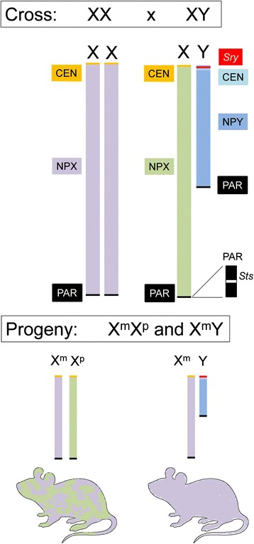
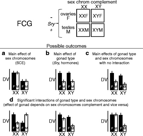
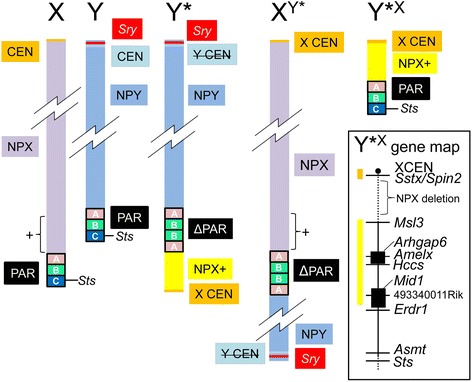
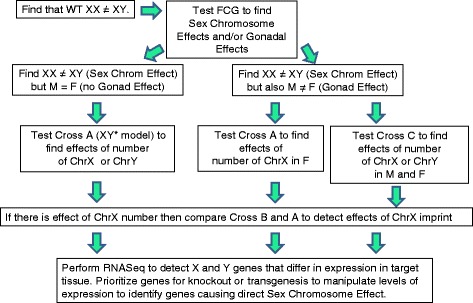
Verification
Claims the paper makes
The claims and statistics of this paper haven’t been checked yet.
Run the check — free, no account needed. It reads the open-access text and checks the sources it cites.
Claims other papers make about this one
Each is checked against this paper’s own text: does it say what the citing paper says it does?
The cited source says what the claim attributes to it.
In the citing paper
“the four core genotypes mouse model which incorporates mice with four different combinations of gonads and sex chromosomes [66, 67], has helped identify the influence of sex chromosomes on physical traits, such as obesity and food intake [68, 69].”
From Integrating Transwomen and Female Athletes with Differences of Sex Development (DSD) into Elite Competition: The FIMS 2021 Consensus Statement (2021) · cites this paper as [67]
Self-check
Percentages, sums and reported differences re-computed from the paper’s own numbers.
The self-check needs the paper’s full text.
Citations
Cites
Analysis of Relative Gene Expression Data Using Real-Time Quantitative PCR and the 2−ΔΔCT Method
Livak, K. J., & Schmittgen, T. D. (2001) · Methods · doi:10.1006/meth.2001.1262 · Cited by 180,860
Mutation of the mouse klotho gene leads to a syndrome resembling ageing
Kuro-o, M., Matsumura, Y., Aizawa, H., Kawaguchi, H., Suga, T., Utsugi, T., Ohyama, Y., Kurabayashi, M., Kaname, T., Kume, E., et al. (1997) · Nature · doi:10.1038/36285 · Cited by 3,837
Wang, H., Yang, H., Shivalila, C. S., Dawlaty, M. M., Cheng, A. W., Zhang, F., & Jaenisch, R. (2013) · Cell · doi:10.1016/j.cell.2013.04.025 · Cited by 3,495
Sinclair, A. H., Berta, P., Palmer, M. S., Hawkins, J. R., Griffiths, B. L., Smith, M. J., Foster, J. W., Frischauf, A., Lovell-Badge, R. H., & Goodfellow, P. N. (1990) · Nature · doi:10.1038/346240a0 · Cited by 3,234
Male development of chromosomally female mice transgenic for Sry
Koopman, P. A., Gubbay, J., Vivian, N., Goodfellow, P. N., & Lovell-Badge, R. H. (1991) · Nature · doi:10.1038/351117a0 · Cited by 2,193
The male-specific region of the human Y chromosome is a mosaic of discrete sequence classes
Skaletsky, H., Kuroda-Kawaguchi, T., Minx, P. J., Cordum, H. S., Hillier, L., Brown, L. G., Repping, S., Pyntikova, T., Ali, J., Bieri, T. A., et al. (2003) · Nature · doi:10.1038/nature01722 · Cited by 2,148
Gubbay, J., Collignon, J., Koopman, P. A., Capel, B., Economou, A., Münsterberg, A., Vivian, N., Goodfellow, P. N., & Lovell-Badge, R. H. (1990) · Nature · doi:10.1038/346245a0 · Cited by 1,675
Myogenin, a factor regulating myogenesis, has a domain homologous to MyoD
Wright, W. E., Sassoon, D. A., & Lin, V. K. (1989) · Cell · doi:10.1016/0092-8674(89)90583-7 · Cited by 1,261
Four Evolutionary Strata on the Human X Chromosome
Lahn, B. T., & Page, D. C. (1999) · Science · doi:10.1126/science.286.5441.964 · Cited by 1,010
The degeneration of Y chromosomes
Charlesworth, B., & Charlesworth, D. (2000) · Philosophical Transactions of the Royal Society B Biological Sciences · doi:10.1098/rstb.2000.0717 · Cited by 931
Cited by
Sorted by citations
Items 101–154 of 154
Aarde, S. M., Bagley, J. R., & Jentsch, J. D. (2022) · bioRxiv (Cold Spring Harbor Laboratory) · doi:10.1101/2022.10.25.513748 · Cited by 3
Zhuang, Q. K.-W., Bauermeister, K., Gálvez, J. H., AlOgayil, N., Batdorj, E., de Villena, F. P., Taketo, T., Bourque, G., & Naumova, A. K. (2025) · Genome biology · doi:10.1186/s13059-025-03547-0 · Cited by 2
Ma, H., Liao, D. J., Zhang, K., Yang, J., Qin, Z., Lu, T., Lou, D., Ran, Q., Huang, H., Cheng, S., et al. (2022) · Current Genomics · doi:10.2174/1389202923666220610121344 · Cited by 2
Régulation du chromosome X et spécificités fonctionnelles des cellules femelles
Chaumeil, J., & Morey, C. (2024) · médecine/sciences · doi:10.1051/medsci/2024179 · Cited by 3
Sneddon, E. A., Rasizer, L. N., Cavalco, N. G., Jaymes, A. H., Ostlie, N. J., Minshall, B. L., Masters, B. M., Hrncir, H., Arnold, A. P., & Radke, A. K. (2021) · bioRxiv (Cold Spring Harbor Laboratory) · doi:10.1101/2021.04.28.441845 · Cited by 2
Self-citationExploring diversity: a review of animal models for investigating variations in sex characteristics
Oliveira, G. D. F., Niepsuj, T., Gupta, A., & Auger, A. P. (2025) · Frontiers in Pediatrics · doi:10.3389/fped.2025.1430983 · Cited by 1
Guedes, M. S., & Yildiz, D. (2026) · Naunyn-Schmiedeberg s Archives of Pharmacology · doi:10.1007/s00210-026-04990-x · Cited by 3
Age-related cardiovascular disease and mosaic hematopoietic loss of the Y chromosome
Chavkin, N. W. (2025) · The Journal of Cardiovascular Aging · doi:10.20517/jca.2024.29 · Cited by 1
KDM5C and KDM5D influence DNA methylation in adult mouse liver
Gibbons, E., Klein, K. O., Inoue, S., Kimura, T., Greenwood, C. M. T., & Naumova, A. K. (2026) · Biology of Sex Differences · doi:10.1186/s13293-026-00844-6 · Cited by 1
Abu-Moch, M., Azab, A. N., Yousef, M., & Agam, G. (2026) · Biomedicine & Pharmacotherapy · doi:10.1016/j.biopha.2026.119356 · Cited by 1
Why males scar more: hormonal and chromosomal clues to idiopathic pulmonary fibrosis
Elliot, S. J., Clark, K. B., Civettini, G., Roos, B. B., Xia, X., Galdikaite, E., Pereira‐Simon, S., Kaboff, A., Catanuto, P., Grimaldo, S., et al. (2026) · Biology of Sex Differences · doi:10.1186/s13293-026-00942-5 · Cited by 1
Self-citationHercus, J. C., Rubio, D. A. S., Nieto, M. E. O., Keum, C., Wang, Q., Macdonald, J. A., Scott, J. S., Lucas, E. R. J., & Christians, J. K. (2025) · Molecular Human Reproduction · doi:10.1093/molehr/gaaf015 · Cited by 1
Sex Bias in Systemic Lupus Erythematosus and Sjögren’s Syndrome
Scofield, R. H., & Harris, V. M. (2020) · Springer eBooks · doi:10.1007/978-981-15-0114-2_3 · Cited by 1
Editorial overview: Daylighting sex differences in physiology
Goodwin, S. F., & Howlett, S. E. (2018) · Current Opinion in Physiology · doi:10.1016/j.cophys.2018.09.002 · Cited by 1
Zhang, Z., Su, V., Wiese, C. B., Cheng, L., Wang, D., Cui, Y., Kallapur, A. S., Kim, J., Wu, X., Tran, P. H., et al. (2024) · bioRxiv (Cold Spring Harbor Laboratory) · doi:10.1101/2024.11.13.623052 · Cited by 1
Sex chromosomes shape the transcriptional landscape of the preimplantation mouse embryoPreprint
Snell, D. M., Varsally, W., Courtois, A., Menchero, S., Munusamy, P., Rietdijk, R., Ojarikre, O. A., Strohbuecker, S., Kaya, H., Sangrithi, M. N., et al. (2024) · bioRxiv (Cold Spring Harbor Laboratory) · doi:10.1101/2024.11.26.625424 · Cited by 1
Shin, J. E., Blencowe, M., McQuillen, C. N., Hrncir, H., Chen, X., Diamante, G., Ahn, I. S., Zhang, G., Arnold, A. P., MacKenzie‐Graham, A. J., et al. (2025) · bioRxiv (Cold Spring Harbor Laboratory) · doi:10.64898/2025.12.08.693050 · Cited by 1
Self-citationDDX3X-mediated translation of structured cardiac mRNAs is essential for female heart developmentPreprint
Mason, K. K., Park, S. K., Emerson, J. I., Lu, Y. W., Wang, D.-Z., Marzluff, W. F., & Conlon, F. L. (2026) · Genes & Development · doi:10.1101/gad.353320.125 · Cited by 1
Gupta, M., Nayyar, N., Shen, J., Lun, S., Chaulagain, S., Mangla, N., Nino-Meza, O. J., Krug, S., Srikrishna, G., Hoffmann, J. P., et al. (2026) · bioRxiv (Cold Spring Harbor Laboratory) · doi:10.64898/2026.06.11.731661 · Cited by 1
Sex as a modulator of lipid metabolism and metabolic disease
Vergnes, L., Riestenberg, C. K., & Reue, K. (2020) · Elsevier eBooks · doi:10.1016/b978-0-12-819404-1.00003-8
Gender Disparities in Bladder Cancer
Zhang, Y., Theodorescu, D., & Li, X. (2021) · IntechOpen eBooks · doi:10.5772/intechopen.98225
Considerations and Challenges for Sex-Aware Drug RepurposingPreprint
Fisher, J., Jones, E., Flanary, V. L., Williams, A. S., Ramsey, E., & Lasseigne, B. N. (2021) · Preprints.org · doi:10.20944/preprints202112.0380.v1
Considerations and Challenges for Sex-Aware Drug RepurposingPreprint
Fisher, J., Jones, E., Flanary, V. L., Williams, A. A., Ramsey, E., & Lasseigne, B. N. (2022) · Preprints.org · doi:10.20944/preprints202112.0380.v2
Sex chromosome mechanisms in cardiac development and diseasePreprint
Conlon, F. L., & Arnold, A. P. (2022) · Research Square · doi:10.21203/rs.3.rs-1933881/v1
Self-citationGabory, A., & Junien, C. (2023) · Elsevier eBooks · doi:10.1016/b978-0-12-823937-7.00012-2
Genetics of Sex Differences in Immunity
Gal-Oz, S. T., & Shay, T. (2023) · Current topics in microbiology and immunology · doi:10.1007/978-3-031-35139-6_1
Sex differences in ischemic stroke: risk factors, pathology, and treatment
Branyan, T. E., & Sohrabji, F. (2024) · Elsevier eBooks · doi:10.1016/b978-0-12-820480-1.00115-7
Women in Cardiovascular Genetics and Systems Medicine
(2024) · Frontiers research topics · doi:10.3389/978-2-8325-4796-0
Zhuang, Q. K.-W., Bauermeister, K., Gálvez, J. H., AlOgayil, N., Batdorj, E., de Villena, F. P., Taketo, T., Bourque, G., & Naumova, A. K. (2024) · bioRxiv (Cold Spring Harbor Laboratory) · doi:10.1101/2024.09.10.612032
Chromosomes X et Y : acteurs clés des inégalités de sexe face aux maladies
Loda, A., Cleland, J., & Heard, É. (2024) · Pour la Science · doi:10.3917/pls.567.0022
Clayton, Z. S., & Moreau, K. L. (2025) · American Journal of Physiology-Heart and Circulatory Physiology · doi:10.1152/ajpheart.00130.2025
Amato-Menker, C. J., Hopen, Q., Pettit, A., Gandhi, J., Hu, G., Schafer, R., & Franko, J. (2024) · Figshare · doi:10.6084/m9.figshare.26693230
Amato-Menker, C. J., Hopen, Q., Pettit, A., Gandhi, J., Hu, G., Schafer, R., & Franko, J. (2024) · Figshare · doi:10.6084/m9.figshare.26693236
Amato-Menker, C. J., Hopen, Q., Pettit, A., Gandhi, J., Hu, G., Schafer, R., & Franko, J. (2024) · Figshare · doi:10.6084/m9.figshare.26693248
Amato-Menker, C. J., Hopen, Q., Pettit, A., Gandhi, J., Hu, G., Schafer, R., & Franko, J. (2024) · Figshare · doi:10.6084/m9.figshare.25414905
Sakamuri, A., Visniauskas, B., Kilanowski‐Doroh, I., McNally, A. B., Sugi, A. I., Kamau, A. N., Sengottaian, D., McLachlan, J. A., Anguera, M. C., Mauvais‐Jarvis, F., et al. (2024) · Figshare · doi:10.6084/m9.figshare.26730379
Sakamuri, A., Visniauskas, B., Kilanowski‐Doroh, I., McNally, A. B., Sugi, A. I., Kamau, A. N., Sengottaian, D., McLachlan, J. A., Anguera, M. C., Mauvais‐Jarvis, F., et al. (2024) · Figshare · doi:10.6084/m9.figshare.26730382
Sakamuri, A., Visniauskas, B., Kilanowski‐Doroh, I., McNally, A. B., Sugi, A. I., Kamau, A. N., Sengottaian, D., McLachlan, J. A., Anguera, M. C., Mauvais‐Jarvis, F., et al. (2024) · Figshare · doi:10.6084/m9.figshare.26730376
Sakamuri, A., Visniauskas, B., Kilanowski‐Doroh, I., McNally, A. B., Sugi, A. I., Kamau, A. N., Sengottaian, D., McLachlan, J. A., Anguera, M. C., Mauvais‐Jarvis, F., et al. (2024) · Figshare · doi:10.6084/m9.figshare.26730385
Amato-Menker, C. J., Hopen, Q., Pettit, A., Gandhi, J., Hu, G., Schafer, R., & Franko, J. (2024) · Figshare · doi:10.6084/m9.figshare.26693239
Amato-Menker, C. J., Hopen, Q., Pettit, A., Gandhi, J., Hu, G., Schafer, R., & Franko, J. (2024) · Figshare · doi:10.6084/m9.figshare.26693245
Amato-Menker, C. J., Hopen, Q., Pettit, A., Gandhi, J., Hu, G., Schafer, R., & Franko, J. (2024) · Figshare · doi:10.6084/m9.figshare.26693233
Amato-Menker, C. J., Hopen, Q., Pettit, A., Gandhi, J., Hu, G., Schafer, R., & Franko, J. (2024) · Figshare · doi:10.6084/m9.figshare.26693242
Qi, S., Al Mamun, A., Ngwa, C., Romana, S., Ritzel, R., Arnold, A. P., McCullough, L. D., & Liu, F. (2021) · Figshare · doi:10.6084/m9.figshare.14211386
Self-citationAmato-Menker, C. J., Hopen, Q., Pettit, A., Gandhi, J., Hu, G., Schafer, R., & Franko, J. (2024) · Figshare · doi:10.6084/m9.figshare.25414981
Deegan, D. F., Calendo, G., Nigam, P., Naqvi, R. A., Arnold, A. P., & Engel, N. (2026) · bioRxiv (Cold Spring Harbor Laboratory) · doi:10.64898/2026.01.09.696197 · Cited by 1
Self-citationPinto‐Benito, D., Paradela‐Leal, C., Cano-Adamuz, N., Grassi, D., Azcoitia, Í., Grade, S., & Arévalo, M. Á. (2026) · bioRxiv (Cold Spring Harbor Laboratory) · doi:10.64898/2026.01.28.702273
The Role of Testosterone in Atherosclerosis: View From Cell Cultures and Animal Models
Dabravolski, S. A., Ekta, M. B., Utkina, A. S., Asoyan, A., Maltseva, O. N., & Orekhov, A. N. (2026) · Journal of Cardiovascular Translational Research · doi:10.1007/s12265-025-10722-y
Zhang, Z., Su, V., Wiese, C. B., Cheng, L., Wang, D., Cui, Y., Kallapur, A. S., Kim, J., Wu, X., Tran, P. H., et al. (2026) · PLoS Biology · doi:10.1371/journal.pbio.3003601 · Cited by 1
Sexual Dimorphism in the Development of Cardiometabolic Syndrome
Velasco, M., Albarado-Ibáñez, A., Larqué, C., Pánico, P., Torres-Jácome, J., & Hiriart, M. (2026) · Handbook of experimental pharmacology · doi:10.1007/164_2026_790
Chaulagain, S., Creisher, P. S., Park, H.-S., Liu, J. A., Gay, L. C., Seibert, B., Wang, A., Jiménez, M., Cáceres, C. J., Perez, D. R., et al. (2026) · Research Square · doi:10.21203/rs.3.rs-9066058/v1
Sexual dimorphism in the liver
Jain, A., & Monga, S. P. (2026) · Nature Reviews Gastroenterology & Hepatology · doi:10.1038/s41575-026-01244-2
Neuroepithelial regulation across development, regeneration, aging, and sex
Zheng, Y., Mueller, K., Pangilinan, G. R., & Shwartz, Y. (2026) · Seminars in Cell and Developmental Biology · doi:10.1016/j.semcdb.2026.103700
X and Y chromosomes as determinants of aging and disease
Dubal, D. B., Ambrosio, F., Stout, M. B., & Theodorescu, D. (2026) · Science · doi:10.1126/science.aeh0145 · Cited by 1
Metadata
- Authors
- Burgoyne, P. S., & Arnold, A. P.
- Affiliations
- National Institute for Medical Research (GB)
- University of California, Los Angeles (US)
- Corresponding author
- Arnold, Arthur P.
- Venue
- Biology of Sex Differences
- Published
- 2016-12
- Volume
- 7
- Issue
- 1
- Pages
- 68-68
- ISSN
- 2042-6410 (ISSN-L)
- PMC
- PMC5154145
- OpenAlex
- W2563939294
- Cited by
- 154
- References
- 144
- Citations by year
- 2026: 17 · 2025: 11 · 2024: 34 · 2023: 14 · 2022: 23 · 2021: 17 · 2020: 14 · 2019: 7 · 2018: 9 · 2017: 8
- FWCI
- 1.07
- Type
- journal article
- Language
- en
- Open access
- gold
- doi.org
- Licence
- Locations
- Biology of Sex Differences (publishedVersion, cc-by)
- PubMed Central (publishedVersion, cc-by)
- PubMed (publishedVersion)
- Figshare (submittedVersion, cc-by)
- The Francis Crick Institute (cc-by)
- Funders
- UK Medical Research Council (U117532009)
- NIMH (MH59268)
- National Institute on Deafness and Other Communication Disorders (DC000217)
- National Institute of Neurological Disorders and Stroke (NS045966, NS043196,)
- NIDDK (DK083561)
- National Institute of Child Health and Human Development (HD076125)
- National Heart, Lung, and Blood Institute (HL119886, HL131182)
- Topics
- Life Sciences › Biochemistry, Genetics and Molecular Biology › Genetics › Genetic and Clinical Aspects of Sex Determination and Chromosomal Abnormalities
- Health Sciences › Medicine › Endocrinology, Diabetes and Metabolism › Sexual Differentiation and Disorders
- Health Sciences › Medicine › Public Health, Environmental and Occupational Health › Sex and Gender in Healthcare
- Keywords
- sex differences, mouse model, SRY, Y chromosome, X chromosome, gonadal hormones, sexual differentiation, candidate gene identification, sex chromosome inactivation
About these checks
Registry, retraction, Cites and statistics results are computed, not judged, with one exception: a printed reference that no index lists is read once by a model before it counts as a citation error. Claims are extracted by a model and kept only when their quote appears verbatim in the text; each cited source is then checked, with a model’s help, for whether it says what the claim relies on. Those verdicts can be wrong — read the claim’s quote and the cited source before relying on one.
Statistics and arithmetic are recomputed, not judged.
Confidence bands are shown only for fields calibrated on our annotated test set. None is calibrated yet, so no band is shown.
What counts as a claim ›How we measure confidence ›How we check this ›
Are you an author, or do you see a problem with this page? Request a review
