

The DOI matches its Crossref record; 274 of the 274 references it cites were found in a registry or an index.
Oligodendrocyte progenitor cells in Alzheimer's disease: from physiology to pathology
Open accessAbstract
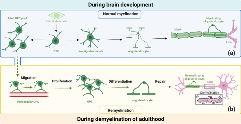
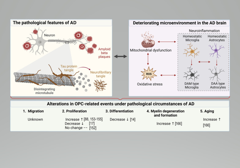
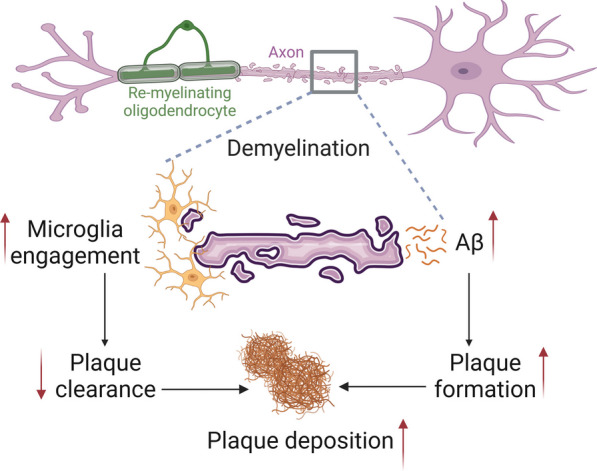
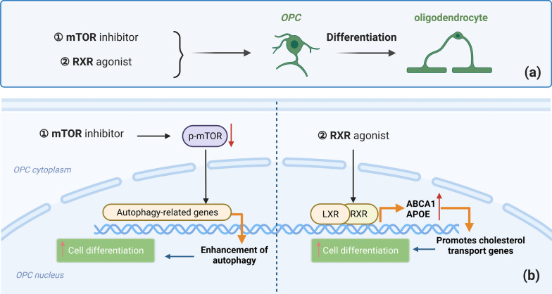
Oligodendrocyte progenitor cells (OPCs) play pivotal roles in myelin formation and phagocytosis, communicating with neighboring cells and contributing to the integrity of the blood–brain barrier (BBB). However, under the pathological circumstances of Alzheimer’s disease (AD), the brain’s microenvironment undergoes detrimental changes that significantly impact OPCs and their functions. Starting with OPC functions, we delve into the transformation of OPCs to myelin-producing oligodendrocytes, the intricate signaling interactions with other cells in the central nervous system (CNS), and the fascinating process of phagocytosis, which influences the function of OPCs and affects CNS homeostasis. Moreover, we discuss the essential role of OPCs in BBB formation and highlight the critical contribution of OPCs in forming CNS-protective barriers. In the context of AD, the deterioration of the local microenvironment in the brain is discussed, mainly focusing on neuroinflammation, oxidative stress, and the accumulation of toxic proteins. The detrimental changes disturb the delicate balance in the brain, impacting the regenerative capacity of OPCs and compromising myelin integrity. Under pathological conditions, OPCs experience significant alterations in migration and proliferation, leading to impaired differentiation and a reduced ability to produce mature oligodendrocytes. Moreover, myelin degeneration and formation become increasingly active in AD, contributing to progressive neurodegeneration. Finally, we summarize the current therapeutic approaches targeting OPCs in AD. Strategies to revitalize OPC senescence, modulate signaling pathways to enhance OPC differentiation, and explore other potential therapeutic avenues are promising in alleviating the impact of AD on OPCs and CNS function. In conclusion, this review highlights the indispensable role of OPCs in CNS function and their involvement in the pathogenesis of AD. The intricate interplay between OPCs and the AD brain microenvironment underscores the complexity of neurodegenerative diseases. Insights from studying OPCs under pathological conditions provide a foundation for innovative therapeutic strategies targeting OPCs and fostering neurodegeneration. Future research will advance our understanding and management of neurodegenerative diseases, ultimately offering hope for effective treatments and improved quality of life for those affected by AD and related disorders.
Overview
The claims and statistics of this paper haven’t been checked yet.
Run the check — free, no account needed. It reads the open-access text and checks the sources it cites.
Integrity signals
Registry
Cites
Statistics
Figures
Verification
Claims the paper makes
The claims and statistics of this paper haven’t been checked yet.
Run the check — free, no account needed. It reads the open-access text and checks the sources it cites.
Claims other papers make about this one
Each is checked against this paper’s own text: does it say what the citing paper says it does?
The cited source says what the claim attributes to it.
In the citing paper
“Despite the elevated rate of myelin formation in AD mice, the overall myelin levels were reduced, suggesting the increased rate of myelin regeneration is insufficient to fully counterbalance the accelerated myelin degeneration”
From Serum/glucocorticoid regulated kinase 1 (SGK1) in neurological disorders: pain or gain (2024) · cites this paper as Zou et al., 2023
Self-check
Percentages, sums and reported differences re-computed from the paper’s own numbers.
The self-check needs the paper’s full text.
Citations
Cites
Lecanemab in early Alzheimer's disease
van Dyck, C. H., Swanson, C. J., Aisen, P., Bateman, R. J., Chen, C., Gee, M., Kanekiyo, M., Li, D., Reyderman, L., Cohen, S., et al. (2023) · New England Journal of Medicine · doi:10.1056/nejmoa2212948 · Cited by 5,678
A Unique Microglia Type Associated with Restricting Development of Alzheimer’s Disease
Keren-Shaul, H., Spinrad, A., Weiner, A., Matcovitch-Natan, O., Dvir-Szternfeld, R., Ulland, T. K., David, E., Baruch, K., Lara-Astaiso, D., Toth, B., et al. (2017) · Cell · doi:10.1016/j.cell.2017.05.018 · Cited by 5,493
Microglia Sculpt Postnatal Neural Circuits in an Activity and Complement-Dependent Manner
Schafer, D. P., Lehrman, E. K., Kautzman, A. G., Koyama, R., Mardinly, A. R., Yamasaki, R., Ransohoff, R. M., Greenberg, M. E., Barres, B. A., & Stevens, B. (2012) · Neuron · doi:10.1016/j.neuron.2012.03.026 · Cited by 4,089
Neuroinflammation and microglial activation in Alzheimer disease: where do we go from here?
Leng, F., & Edison, P. (2021) · Nature Reviews Neurology · doi:10.1038/s41582-020-00435-y · Cited by 3,179
Single-cell transcriptomic analysis of Alzheimer’s disease
Mathys, H., Davila-Velderrain, J., Peng, Z., Gao, F., Mohammadi, S., Young, J. Z., Menon, M., He, L., Abdurrob, F., Jiang, X., et al. (2019) · Nature · doi:10.1038/s41586-019-1195-2 · Cited by 2,568
Senolytics improve physical function and increase lifespan in old age
Xu, M., Pirtskhalava, T., Farr, J. N., Weigand, B. M., Palmer, A. K., Weivoda, M. M., Inman, C. L., Ogrodnik, M. B., Hachfeld, C. M., Fraser, D. G., et al. (2018) · Nature Medicine · doi:10.1038/s41591-018-0092-9 · Cited by 2,287 · cited 1×
“Interestingly, an increasing body of research indicates that OPCs undergo functional dysregulation in the pathological state of AD, suggesting OPCs as a potential therapeutic target for AD [18, 148, 203].”
BackgroundCellular senescence in ageing: from mechanisms to therapeutic opportunities
Di Micco, R., Krizhanovsky, V., Baker, D., & d’Adda di Fagagna, F. (2021) · Nature Reviews Molecular Cell Biology · doi:10.1038/s41580-020-00314-w · Cited by 2,277 · cited 1×
“Cellular senescence refers to a state in which cells are blocked in the G1 phase, unable to proliferate and unresponsive to external stimuli, resulting in the inability to perform normal functions [170].”
BackgroundHammond, T. R., Dufort, C., Dissing-Olesen, L., Giera, S., Young, A., Wysoker, A., Walker, A. J., Segel, M., Nemesh, J., Saunders, A., et al. (2018) · doi:10.1101/406140 · Cited by 2,225
Microglia states and nomenclature: A field at its crossroads
Paolicelli, R. C., Sierra, A., Stevens, B., Tremblay, M.-E., Aguzzi, A., Ajami, B., Amit, I., Audinat, E., Bechmann, I., Bennett, M., et al. (2022) · Neuron · doi:10.1016/j.neuron.2022.10.020 · Cited by 1,973
Two Phase 3 Trials of Bapineuzumab in Mild-to-Moderate Alzheimer's Disease
Salloway, S., Sperling, R., Fox, N. C., Blennow, K., Klunk, W., Raskind, M., Sabbagh, M., Honig, L. S., Porsteinsson, A. P., Ferris, S., et al. (2014) · New England Journal of Medicine · doi:10.1056/nejmoa1304839 · Cited by 1,960
Cited by
Sorted by citations
Cellular senescence in Alzheimer’s disease: from physiology to pathology
Zhu, J., Wu, C., & Yang, L. (2024) · Translational Neurodegeneration · doi:10.1186/s40035-024-00447-4 · Cited by 74
Self-citationDias, D., Portugal, C. C., Relvas, J. B., & Socodato, R. (2025) · Cells · doi:10.3390/cells14040243 · Cited by 71
Shokr, M. M. (2025) · Metabolic Brain Disease · doi:10.1007/s11011-025-01739-y · Cited by 64
The impact of physical exercise on neuroinflammation mechanism in Alzheimer’s disease
Hu, J., Huang, B., & Chen, K. (2024) · Frontiers in Aging Neuroscience · doi:10.3389/fnagi.2024.1444716 · Cited by 55
Myelin dysfunction in aging and brain disorders: mechanisms and therapeutic opportunities
Huang, Z., Zhang, Y., Zou, P., Zong, X., & Zhang, Q. (2025) · Molecular Neurodegeneration · doi:10.1186/s13024-025-00861-w · Cited by 49
Self-citation
Metadata
- Authors
- Zou, P., Wu, C., Liu, T. C.-Y., Duan, R., & Yang, L.
- Affiliations
- Louisiana State University (US)
- South China Normal University (CN)
- Corresponding author
- Yang, Luodan
- Venue
- Translational Neurodegeneration
- Published
- 2023-11-14
- Volume
- 12
- Issue
- 1
- Pages
- 52-52
- ISSN
- 2047-9158 (ISSN-L)
- PMC
- PMC10644503
- OpenAlex
- W4388667039
- Cited by
- 97
- References
- 274
- Citations by year
- 2026: 34 · 2025: 49 · 2024: 14
- FWCI
- 3.72
- Type
- journal article
- Language
- en
- Open access
- gold
- doi.org
- Locations
- Translational Neurodegeneration (publishedVersion, cc-by)
- PubMed Central (publishedVersion, cc-by)
- PubMed (publishedVersion)
- DOAJ (DOAJ: Directory of Open Access Journals) (submittedVersion, cc-by-sa)
- Funders
- National Natural Science Foundation of China (31971096, 32100918)
- China Postdoctoral Science Foundation (2021M690060, 2022T150227)
- Sigma Xi Grants in Aid of Research (GIAR) program (G03152021115804390)
- Guangzhou Scientific Research Grant (SL2022B04J00013, SL2024A04J00578)
- SCNU Young Faculty Development Program (22KJ04)
- Topics
- Life Sciences › Neuroscience › Developmental Neuroscience › Neurogenesis and neuroplasticity mechanisms
- Life Sciences › Neuroscience › Cellular and Molecular Neuroscience › Alzheimer's disease research and treatments
- Life Sciences › Neuroscience › Neurology › Neuroinflammation and Neurodegeneration Mechanisms
- Keywords
- oligodendrocyte precursor cells, Alzheimer's disease, myelination, phagocytosis, blood-brain barrier, neuroinflammation, oxidative stress, amyloid-beta, tau protein, OPC differentiation, myelin degeneration, neurodegeneration, cellular senescence
- MeSH terms
- Oligodendrocyte Precursor Cells, Alzheimer Disease, Cell Differentiation, Humans, Oligodendroglia, Quality of Life
About these checks
Registry, retraction, Cites and statistics results are computed, not judged, with one exception: a printed reference that no index lists is read once by a model before it counts as a citation error. Claims are extracted by a model and kept only when their quote appears verbatim in the text; each cited source is then checked, with a model’s help, for whether it says what the claim relies on. Those verdicts can be wrong — read the claim’s quote and the cited source before relying on one.
Statistics and arithmetic are recomputed, not judged.
Confidence bands are shown only for fields calibrated on our annotated test set. None is calibrated yet, so no band is shown.
What counts as a claim ›How we measure confidence ›How we check this ›
Are you an author, or do you see a problem with this page? Request a review
