

The DOI matches its Crossref record; 274 of the 274 references it cites were found in a registry or an index.
Oligodendrocyte progenitor cells in Alzheimer's disease: from physiology to pathology
Open accessAbstract
Oligodendrocyte progenitor cells (OPCs) play pivotal roles in myelin formation and phagocytosis, communicating with neighboring cells and contributing to the integrity of the blood–brain barrier (BBB). However, under the pathological circumstances of Alzheimer’s disease (AD), the brain’s microenvironment undergoes detrimental changes that significantly impact OPCs and their functions. Starting with OPC functions, we delve into the transformation of OPCs to myelin-producing oligodendrocytes, the intricate signaling interactions with other cells in the central nervous system (CNS), and the fascinating process of phagocytosis, which influences the function of OPCs and affects CNS homeostasis. Moreover, we discuss the essential role of OPCs in BBB formation and highlight the critical contribution of OPCs in forming CNS-protective barriers. In the context of AD, the deterioration of the local microenvironment in the brain is discussed, mainly focusing on neuroinflammation, oxidative stress, and the accumulation of toxic proteins. The detrimental changes disturb the delicate balance in the brain, impacting the regenerative capacity of OPCs and compromising myelin integrity. Under pathological conditions, OPCs experience significant alterations in migration and proliferation, leading to impaired differentiation and a reduced ability to produce mature oligodendrocytes. Moreover, myelin degeneration and formation become increasingly active in AD, contributing to progressive neurodegeneration. Finally, we summarize the current therapeutic approaches targeting OPCs in AD. Strategies to revitalize OPC senescence, modulate signaling pathways to enhance OPC differentiation, and explore other potential therapeutic avenues are promising in alleviating the impact of AD on OPCs and CNS function. In conclusion, this review highlights the indispensable role of OPCs in CNS function and their involvement in the pathogenesis of AD. The intricate interplay between OPCs and the AD brain microenvironment underscores the complexity of neurodegenerative diseases. Insights from studying OPCs under pathological conditions provide a foundation for innovative therapeutic strategies targeting OPCs and fostering neurodegeneration. Future research will advance our understanding and management of neurodegenerative diseases, ultimately offering hope for effective treatments and improved quality of life for those affected by AD and related disorders.
Overview
The claims and statistics of this paper haven’t been checked yet.
Run the check — free, no account needed. It reads the open-access text and checks the sources it cites.
Integrity signals
Registry
Cites
Statistics
Figures
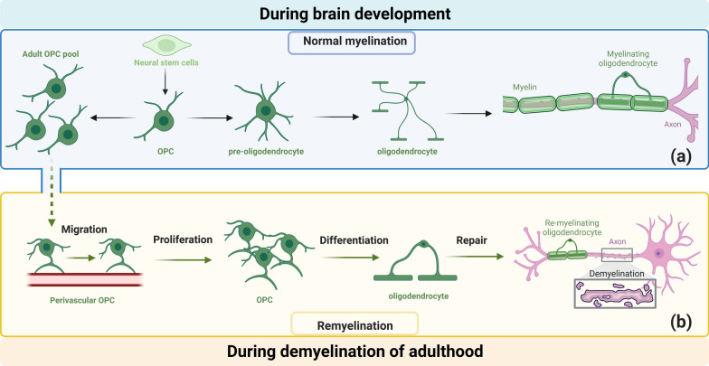
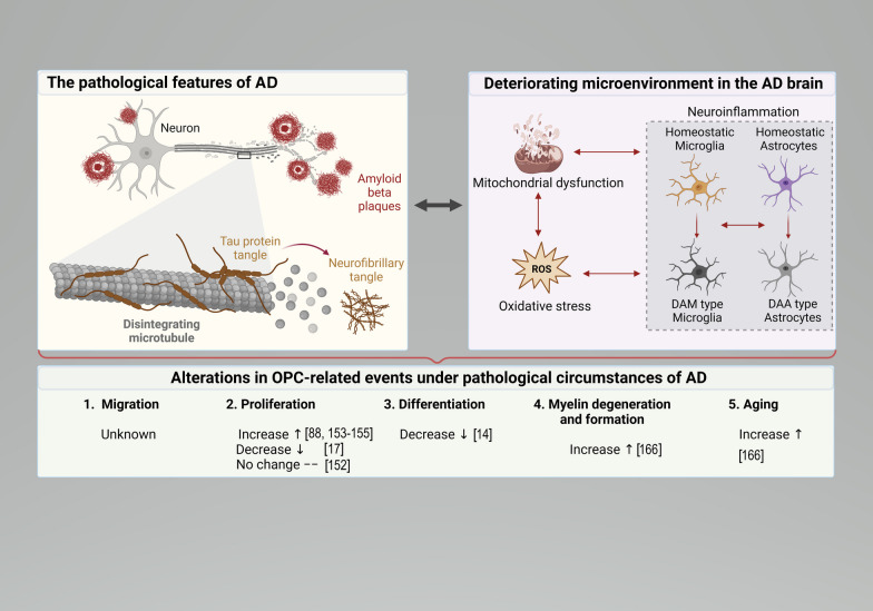
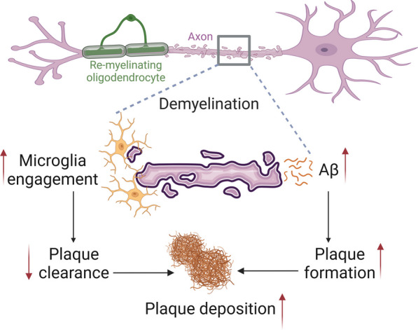
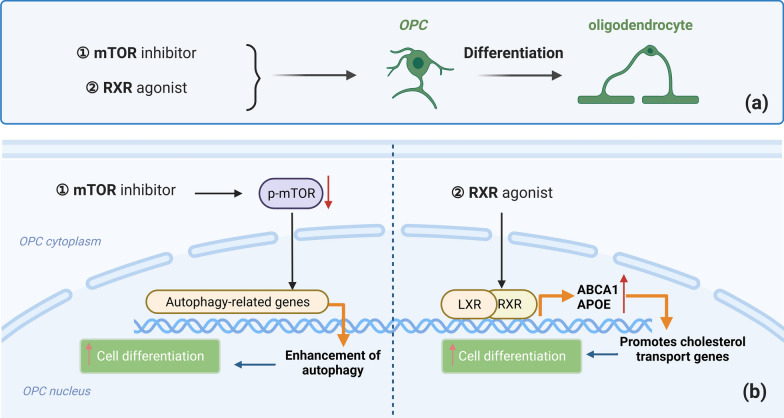
Verification
Claims the paper makes
The claims and statistics of this paper haven’t been checked yet.
Run the check — free, no account needed. It reads the open-access text and checks the sources it cites.
Claims other papers make about this one
Each is checked against this paper’s own text: does it say what the citing paper says it does?
The cited source says what the claim attributes to it.
In the citing paper
“Despite the elevated rate of myelin formation in AD mice, the overall myelin levels were reduced, suggesting the increased rate of myelin regeneration is insufficient to fully counterbalance the accelerated myelin degeneration”
From Serum/glucocorticoid regulated kinase 1 (SGK1) in neurological disorders: pain or gain (2024) · cites this paper as Zou et al., 2023
Self-check
Percentages, sums and reported differences re-computed from the paper’s own numbers.
The self-check needs the paper’s full text.
Citations
Cites
Lecanemab in early Alzheimer's disease
van Dyck, C. H., Swanson, C. J., Aisen, P., Bateman, R. J., Chen, C., Gee, M., Kanekiyo, M., Li, D., Reyderman, L., Cohen, S., et al. (2023) · New England Journal of Medicine · doi:10.1056/nejmoa2212948 · Cited by 5,678
A Unique Microglia Type Associated with Restricting Development of Alzheimer’s Disease
Keren-Shaul, H., Spinrad, A., Weiner, A., Matcovitch-Natan, O., Dvir-Szternfeld, R., Ulland, T. K., David, E., Baruch, K., Lara-Astaiso, D., Toth, B., et al. (2017) · Cell · doi:10.1016/j.cell.2017.05.018 · Cited by 5,493
Microglia Sculpt Postnatal Neural Circuits in an Activity and Complement-Dependent Manner
Schafer, D. P., Lehrman, E. K., Kautzman, A. G., Koyama, R., Mardinly, A. R., Yamasaki, R., Ransohoff, R. M., Greenberg, M. E., Barres, B. A., & Stevens, B. (2012) · Neuron · doi:10.1016/j.neuron.2012.03.026 · Cited by 4,089
Neuroinflammation and microglial activation in Alzheimer disease: where do we go from here?
Leng, F., & Edison, P. (2021) · Nature Reviews Neurology · doi:10.1038/s41582-020-00435-y · Cited by 3,179
Single-cell transcriptomic analysis of Alzheimer’s disease
Mathys, H., Davila-Velderrain, J., Peng, Z., Gao, F., Mohammadi, S., Young, J. Z., Menon, M., He, L., Abdurrob, F., Jiang, X., et al. (2019) · Nature · doi:10.1038/s41586-019-1195-2 · Cited by 2,568
Senolytics improve physical function and increase lifespan in old age
Xu, M., Pirtskhalava, T., Farr, J. N., Weigand, B. M., Palmer, A. K., Weivoda, M. M., Inman, C. L., Ogrodnik, M. B., Hachfeld, C. M., Fraser, D. G., et al. (2018) · Nature Medicine · doi:10.1038/s41591-018-0092-9 · Cited by 2,287 · cited 1×
“Interestingly, an increasing body of research indicates that OPCs undergo functional dysregulation in the pathological state of AD, suggesting OPCs as a potential therapeutic target for AD [18, 148, 203].”
BackgroundCellular senescence in ageing: from mechanisms to therapeutic opportunities
Di Micco, R., Krizhanovsky, V., Baker, D., & d’Adda di Fagagna, F. (2021) · Nature Reviews Molecular Cell Biology · doi:10.1038/s41580-020-00314-w · Cited by 2,277 · cited 1×
“Cellular senescence refers to a state in which cells are blocked in the G1 phase, unable to proliferate and unresponsive to external stimuli, resulting in the inability to perform normal functions [170].”
BackgroundHammond, T. R., Dufort, C., Dissing-Olesen, L., Giera, S., Young, A., Wysoker, A., Walker, A. J., Segel, M., Nemesh, J., Saunders, A., et al. (2018) · doi:10.1101/406140 · Cited by 2,225
Microglia states and nomenclature: A field at its crossroads
Paolicelli, R. C., Sierra, A., Stevens, B., Tremblay, M.-E., Aguzzi, A., Ajami, B., Amit, I., Audinat, E., Bechmann, I., Bennett, M., et al. (2022) · Neuron · doi:10.1016/j.neuron.2022.10.020 · Cited by 1,973
Two Phase 3 Trials of Bapineuzumab in Mild-to-Moderate Alzheimer's Disease
Salloway, S., Sperling, R., Fox, N. C., Blennow, K., Klunk, W., Raskind, M., Sabbagh, M., Honig, L. S., Porsteinsson, A. P., Ferris, S., et al. (2014) · New England Journal of Medicine · doi:10.1056/nejmoa1304839 · Cited by 1,960
Concise review: MSC-derived exosomes for cell-free therapy
Phinney, D. G., & Pittenger, M. F. (2017) · Stem Cells · doi:10.1002/stem.2575 · Cited by 1,834
Biology of Oligodendrocyte and Myelin in the Mammalian Central Nervous System
Baumann, N., & Pham-Dinh, D. (2001) · Physiological Reviews · doi:10.1152/physrev.2001.81.2.871 · Cited by 1,828
Integrated Systems Approach Identifies Genetic Nodes and Networks in Late-Onset Alzheimer’s Disease
Zhang, B., Gaiteri, C., Bodea, L.-G., Wang, Z., McElwee, J., Podtelezhnikov, A. A., Zhang, C., Xie, T., Tran, L., Dobrin, R., et al. (2013) · Cell · doi:10.1016/j.cell.2013.03.030 · Cited by 1,657
Remyelination in the CNS: from biology to therapy
Franklin, R. J. M., & ffrench-Constant, C. (2008) · Nature Reviews Neuroscience · doi:10.1038/nrn2480 · Cited by 1,544
Microglial interactions with synapses are modulated by visual experience
Tremblay, M.-È., Lowery, R. L., & Majewska, A. K. (2010) · PLoS Biology · doi:10.1371/journal.pbio.1000527 · Cited by 1,542
Neuronal activity promotes oligodendrogenesis and adaptive myelination in the mammalian brain
Gibson, E. M., Purger, D., Mount, C. W., Goldstein, A. K., Lin, G. L., Wood, L. S., Inema, I., Miller, S. E., Bieri, G., Zuchero, J. B., et al. (2014) · Science · doi:10.1126/science.1252304 · Cited by 1,472
Apolipoprotein E and Alzheimer disease: pathobiology and targeting strategies
Yamazaki, Y., Zhao, N., Caulfield, T. R., Liu, C.-C., & Bu, G. (2019) · Nature Reviews Neurology · doi:10.1038/s41582-019-0228-7 · Cited by 1,425
Normal development of brain circuits
Tau, G. Z., & Peterson, B. S. (2010) · Neuropsychopharmacology · doi:10.1038/npp.2009.115 · Cited by 1,391
Clearance of senescent glial cells prevents tau-dependent pathology and cognitive decline
Bussian, T. J., Aziz, A., Meyer, C. F., Swenson, B. L., van Deursen, J. M., & Baker, D. J. (2018) · Nature · doi:10.1038/s41586-018-0543-y · Cited by 1,289
Mitochondria dysfunction in the pathogenesis of Alzheimer’s disease: Recent advances.
Wang, W., Zhao, F., Ma, X., Perry, G., & Zhu, X. (2020) · Molecular Neurodegeneration · doi:10.1186/s13024-020-00376-6 · Cited by 1,274
Cellular Senescence: Aging, Cancer, and Injury
Calcinotto, A., Kohli, J., Zagato, E., Pellegrini, L., Demaria, M., & Alimonti, A. (2019) · Physiological Reviews · doi:10.1152/physrev.00020.2018 · Cited by 1,270
Normal aging induces A1-like astrocyte reactivity
Clarke, L. E., Liddelow, S. A., Chakraborty, C., Münch, A. E., Heiman, M., & Barres, B. A. (2018) · Proceedings of the National Academy of Sciences · doi:10.1073/pnas.1800165115 · Cited by 1,237
Signal transduction via the MAP kinases: proceed at your own RSK.
Blenis, J. (1993) · Proceedings of the National Academy of Sciences · doi:10.1073/pnas.90.13.5889 · Cited by 1,233
Senolytic drugs: from discovery to translation
Kirkland, J. L., & Tchkonia, T. (2020) · Journal of Internal Medicine · doi:10.1111/joim.13141 · Cited by 1,227
Motor skill learning requires active central myelination
McKenzie, I. A., Ohayon, D., Li, H., Paes de Faria, J., Emery, B., Tohyama, K., & Richardson, W. D. (2014) · Science · doi:10.1126/science.1254960 · Cited by 1,177
Glutamatergic synapses on oligodendrocyte precursor cells in the hippocampus
Bergles, D. E., Roberts, J. D. B., Somogyi, P., & Jahr, C. E. (2000) · Nature · doi:10.1038/35012083 · Cited by 1,062
Trial of Solanezumab for Mild Dementia Due to Alzheimer’s Disease
Honig, L. S., Vellas, B., Woodward, M., Boada, M., Bullock, R., Borrie, M., Hager, K., Andreasen, N., Scarpini, E., Liu-Seifert, H., et al. (2018) · New England Journal of Medicine · doi:10.1056/nejmoa1705971 · Cited by 1,049 · cited 1×
“…to date, nearly all of the interventions targeting Aβ and tau protein hyper-phosphorylation in AD have failed in phase III clinical trials [193–195], including immunotherapies, drugs reducing Aβ production or enhancing Aβ clearance, as well as drugs inhibiting tau phosphorylation or…”
BackgroundDisease-associated astrocytes in Alzheimer’s disease and aging
Habib, N., McCabe, C., Medina, S., Varshavsky, M., Kitsberg, D., Dvir-Szternfeld, R., Green, G., Dionne, D., Nguyen, L., Marshall, J. L., et al. (2020) · Nature Neuroscience · doi:10.1038/s41593-020-0624-8 · Cited by 1,009
Zhang, P., Kishimoto, Y., Grammatikakis, I., Gottimukkala, K., Cutler, R. G., Zhang, S., Abdelmohsen, K., Bohr, V. A., Misra Sen, J., Gorospe, M., et al. (2019) · Nature Neuroscience · doi:10.1038/s41593-019-0372-9 · Cited by 977
PDGFRA/NG2 glia generate myelinating oligodendrocytes and piriform projection neurons in adult mice
Rivers, L. E., Young, K. M., Rizzi, M., Jamen, F., Psachoulia, K., Wade, A., Kessaris, N., & Richardson, W. D. (2008) · Nature Neuroscience · doi:10.1038/nn.2220 · Cited by 961
Grubman, A., Chew, G., Ouyang, J. F., Sun, G., Choo, X. Y., McLean, C., Simmons, R. K., Buckberry, S., Vargas-Landin, D. B., Poppe, D., et al. (2019) · Nature Neuroscience · doi:10.1038/s41593-019-0539-4 · Cited by 956
Polydendrocytes (NG2 cells): multifunctional cells with lineage plasticity
Nishiyama, A., Komitova, M., Suzuki, R., & Zhu, X. (2009) · Nature Reviews Neuroscience · doi:10.1038/nrn2495 · Cited by 865
A Review: Inflammatory Process in Alzheimer's Disease, Role of Cytokines
Rubio-Perez, J. M., & Morillas-Ruiz, J. M. (2012) · The Scientific World JOURNAL · doi:10.1100/2012/756357 · Cited by 864
Kuhlmann, T., Miron, V., Cuo, Q., Wegner, C., Antel, J., & Bruck, W. (2008) · Brain · doi:10.1093/brain/awn096 · Cited by 864
Oxidative Stress and the Pathogenesis of Alzheimer's Disease
Zhao, Y., & Zhao, B. (2013) · Oxidative Medicine and Cellular Longevity · doi:10.1155/2013/316523 · Cited by 855
Noble, M., Murray, K., Stroobant, P., Waterfield, M. D., & Riddle, P. (1988) · Nature · doi:10.1038/333560a0 · Cited by 830
Myelin Impairs CNS Remyelination by Inhibiting Oligodendrocyte Precursor Cell Differentiation
Kotter, M. R., Li, W.-W., Zhao, C., & Franklin, R. J. M. (2006) · The Journal of Neuroscience · doi:10.1523/jneurosci.2615-05.2006 · Cited by 768
The blood-brain barrier in Alzheimer’s disease
Zenaro, E., Piacentino, G., & Constantin, G. (2017) · Neurobiology of Disease · doi:10.1016/j.nbd.2016.07.007 · Cited by 767
White matter changes in Alzheimer’s disease: A focus on myelin and oligodendrocytes
Nasrabady, S. E., Rizvi, B., Goldman, J. E., & Brickman, A. M. (2018) · Acta Neuropathologica Communications · doi:10.1186/s40478-018-0515-3 · Cited by 723
NG2 cells generate both oligodendrocytes and gray matter astrocytes
Zhu, X., Bergles, D. E., & Nishiyama, A. (2007) · Development · doi:10.1242/dev.004895 · Cited by 699
Regulation of oligodendrocyte differentiation and myelination
Emery, B. (2010) · Science · doi:10.1126/science.1190927 · Cited by 653
Rejuvenation of Regeneration in the Aging Central Nervous System
Ruckh, J. M., Zhao, J.-W., Shadrach, J. L., van Wijngaarden, P., Rao, T. N., Wagers, A. J., & Franklin, R. J. M. (2012) · Cell Stem Cell · doi:10.1016/j.stem.2011.11.019 · Cited by 649
Regenerating CNS myelin — from mechanisms to experimental medicines
Franklin, R. J. M., & ffrench-Constant, C. (2017) · Nature Reviews Neuroscience · doi:10.1038/nrn.2017.136 · Cited by 647 · cited 2×
“However, the nOPCs generate new oligodendrocytes for myelination, and the aOPCs generate that for remyelination [59, 60].”
BackgroundSim, F. J., Zhao, C., Penderis, J., & Franklin, R. J. M. (2002) · The Journal of Neuroscience · doi:10.1523/jneurosci.22-07-02451.2002 · Cited by 625
Oligodendrocytes in development, myelin generation and beyond
Kuhn, S., Gritti, L., Crooks, D., & Dombrowski, Y. (2019) · Cells · doi:10.3390/cells8111424 · Cited by 625
Pathogenic protein seeding in Alzheimer disease and other neurodegenerative disorders
Jucker, M., & Walker, L. C. (2011) · Annals of Neurology · doi:10.1002/ana.22615 · Cited by 622 · cited 1×
“Neurotoxicity-related neuroinflammation, oxidative stress, and cytokine release are pathological factors in AD [104, 105].”
BackgroundNistor, G. I., Totoiu, M. O., Haque, N. S. K., Carpenter, M. K., & Keirstead, H. S. (2004) · Glia · doi:10.1002/glia.20127 · Cited by 582
Disease-specific oligodendrocyte lineage cells arise in multiple sclerosis
Falcão, A. M., van Bruggen, D., Marques, S., Meijer, M., Jäkel, S., Agirre, E., Samudyata, Floriddia, E. M., Vanichkina, D. P., ffrench-Constant, C., et al. (2018) · Nature Medicine · doi:10.1038/s41591-018-0236-y · Cited by 579
Amyloid cascade hypothesis: Pathogenesis and therapeutic strategies in Alzheimer's disease
Barage, S. H., & Sonawane, K. D. (2015) · Neuropeptides · doi:10.1016/j.npep.2015.06.008 · Cited by 579
Retinoid X receptor gamma signaling accelerates CNS remyelination
Huang, J. K., Jarjour, A. A., Nait Oumesmar, B., Kerninon, C., Williams, A., Krezel, W., Kagechika, H., Bauer, J., Zhao, C., Evercooren, A. B.-V., et al. (2011) · Nature Neuroscience · doi:10.1038/nn.2702 · Cited by 547
Niche stiffness underlies the ageing of central nervous system progenitor cells
Segel, M., Neumann, B., Hill, M. F. E., Weber, I. P., Viscomi, C., Zhao, C., Young, A., Agley, C. C., Thompson, A. J., Gonzalez, G. A., et al. (2019) · Nature · doi:10.1038/s41586-019-1484-9 · Cited by 532
Oligodendrocyte Population Dynamics and the Role of PDGF In Vivo
Calver, A. R., Hall, A. C., Yu, W., Walsh, F. S., Heath, J. K., Betsholtz, C., & Richardson, W. D. (1998) · Neuron · doi:10.1016/s0896-6273(00)80469-9 · Cited by 516
Pringle, N. P., & Richardson, W. D. (1993) · Development · doi:10.1242/dev.117.2.525 · Cited by 510
Rapid production of new oligodendrocytes is required in the earliest stages of motor-skill learning
Xiao, L., Ohayon, D., McKenzie, I. A., Sinclair-Wilson, A., Wright, J. L., Fudge, A. D., Emery, B., Li, H., & Richardson, W. D. (2016) · Nature Neuroscience · doi:10.1038/nn.4351 · Cited by 507
Exosomes — beyond stem cells for restorative therapy in stroke and neurological injury
Zhang, Z. G., Buller, B., & Chopp, M. (2019) · Nature Reviews Neurology · doi:10.1038/s41582-018-0126-4 · Cited by 503
Shen, S., Sandoval, J., Swiss, V. A., Li, J., Dupree, J. L., Franklin, R. J. M., & Casaccia‐Bonnefil, P. (2008) · Nature Neuroscience · doi:10.1038/nn.2172 · Cited by 499
Myelin dysfunction drives amyloid-β deposition in models of Alzheimer’s disease
Depp, C., Sun, T., Sasmita, A. O., Spieth, L., Berghoff, S. A., Nazarenko, T., Overhoff, K., Steixner-Kumar, A. A., Subramanian, S., Arinrad, S., et al. (2023) · Nature · doi:10.1038/s41586-023-06120-6 · Cited by 493 · cited 3×
“3) [168].”
BackgroundInfluentialOligodendrocyte precursors migrate along vasculature in the developing nervous system
Tsai, H.-H., Niu, J., Munji, R., Davalos, D., Chang, J., Zhang, H., Tien, A.-C., Kuo, C. J., Chan, J. R., Daneman, R., et al. (2016) · Science · doi:10.1126/science.aad3839 · Cited by 483
Pant, S., Hilton, H., & Burczynski, M. E. (2011) · Biochemical Pharmacology · doi:10.1016/j.bcp.2011.12.037 · Cited by 480
Myelin Membrane Wrapping of CNS Axons by PI(3,4,5)P3-Dependent Polarized Growth at the Inner Tongue
Snaidero, N., Möbius, W., Czopka, T., Hekking, L. H. P., Mathisen, C., Verkleij, D., Goebbels, S., Edgar, J. M., Merkler, D., Lyons, D. A., et al. (2014) · Cell · doi:10.1016/j.cell.2013.11.044 · Cited by 477
Chomiak, T., & Hu, B. (2009) · PLoS ONE · doi:10.1371/journal.pone.0007754 · Cited by 465
Diversity in function and regulation of MAP kinase pathways
BLUMER, K. J., & Johnson, G. L. (1994) · Trends in Biochemical Sciences · doi:10.1016/0968-0004(94)90147-3 · Cited by 465
Oligodendrocyte, Astrocyte, and Microglia Crosstalk in Myelin Development, Damage, and Repair
Domingues, H. S., Portugal, C. C., Socodato, R., & Relvas, J. B. (2016) · Frontiers in Cell and Developmental Biology · doi:10.3389/fcell.2016.00071 · Cited by 460
Metformin restores CNS remyelination capacity by rejuvenating aged stem cells
Neumann, B., Baror, R., Zhao, C., Segel, M., Dietmann, S., Rawji, K. S., Foerster, S., McClain, C. R., Chalut, K., van Wijngaarden, P., et al. (2019) · Cell Stem Cell · doi:10.1016/j.stem.2019.08.015 · Cited by 449
Kirby, L., Jin, J., Cardona, J. G., Smith, M. D., Martin, K. A., Wang, J., Strasburger, H., Herbst, L., Alexis, M., Karnell, J., et al. (2019) · Nature Communications · doi:10.1038/s41467-019-11638-3 · Cited by 436 · cited 1×
“These changes cause antigen cross-presentation by OPCs to cytotoxic CD8 + T cells, resulting in OPC death [83].”
BackgroundRangaraju, S., Dammer, E. B., Raza, S. A., Rathakrishnan, P., Xiao, H., Gao, T., Duong, D. M., Pennington, M. W., Lah, J. J., Seyfried, N. T., et al. (2018) · Molecular Neurodegeneration · doi:10.1186/s13024-018-0254-8 · Cited by 430
Disruption of oligodendrogenesis impairs memory consolidation in adult mice
Steadman, P. E., Xia, F., Ahmed, M., Mocle, A. J., Penning, A. R. A., Geraghty, A. C., Steenland, H. W., Monje, M., Josselyn, S. A., & Frankland, P. W. (2020) · Neuron · doi:10.1016/j.neuron.2019.10.013 · Cited by 426
Proinflammatory cytokines IL-6 and TNF-α and the development of inflammation in obese subjects
Popko, K., Górska, E., Stelmaszczyk‐Emmel, A., Pływaczewski, R., Stokłosa, A., Górecka, D., Pyrżak, B., & Demkow, U. A. (2010) · European journal of medical research · doi:10.1186/2047-783x-15-s2-120 · Cited by 423
Motor skill learning induces changes in white matter microstructure and myelination
Sampaio-Baptista, C., Khrapitchev, A. A., Foxley, S., Schlagheck, T., Scholz, J., Jbabdi, S., DeLuca, G. C., Miller, K. L., Taylor, A., Thomas, N., et al. (2013) · The Journal of Neuroscience · doi:10.1523/jneurosci.3048-13.2013 · Cited by 417
Ogrodnik, M., Evans, S. A., Fielder, E., Victorelli, S., Kruger, P., Salmonowicz, H., Weigand, B. M., Patel, A. D., Pirtskhalava, T., Inman, C. L., et al. (2021) · Aging Cell · doi:10.1111/acel.13296 · Cited by 405
Involvement of PGC-1α in the formation and maintenance of neuronal dendritic spines
Cheng, A., Wan, R., Yang, J.-L., Kamimura, N., Son, T. G., Ouyang, X., Luo, Y., Okun, E., & Mattson, M. P. (2012) · Nature Communications · doi:10.1038/ncomms2238 · Cited by 401
Tsai, H., Frost, E. E., To, V., Robinson, S., ffrench‐Constant, C., Geertman, R. T., Ransohoff, R. M., & Miller, R. H. (2002) · Cell · doi:10.1016/s0092-8674(02)00838-3 · Cited by 398
Microglia Priming with Aging and Stress
Niraula, A., Sheridan, J. F., & Godbout, J. P. (2017) · Neuropsychopharmacology · doi:10.1038/npp.2016.185 · Cited by 398
Antioxidant Therapies for Alzheimer's Disease
Feng, Y., & Wang, X. (2012) · Oxidative Medicine and Cellular Longevity · doi:10.1155/2012/472932 · Cited by 393
Synaptic pathology in Alzheimer's disease: a review of ultrastructural studies
Scheff, S. (2003) · Neurobiology of Aging · doi:10.1016/j.neurobiolaging.2003.08.002 · Cited by 391
Richardson, W. D., Kessaris, N., & Pringle, N. (2006) · Nature reviews. Neuroscience · doi:10.1038/nrn1826 · Cited by 385
Preservation of a remote fear memory requires new myelin formation
Pan, S., Mayoral, S. R., Choi, H. S., Chan, J. R., & Kheirbek, M. A. (2020) · Nature Neuroscience · doi:10.1038/s41593-019-0582-1 · Cited by 371
PDGF A-chain gene is expressed by mammalian neurons during development and in maturity
Yeh, H.-J., Ruit, K. G., Wang, Y.-X., Parks, W. C., Snider, W. D., & Deuel, T. F. (1991) · Cell · doi:10.1016/0092-8674(91)90222-k · Cited by 363
Pharmacogenetic stimulation of neuronal activity increases myelination in an axon-specific manner
Mitew, S., Gobius, I., Fenlon, L. R., McDougall, S. J., Hawkes, D. J., Xing, Y. L., Bujalka, H., Gundlach, A. L., Richards, L. J., Kilpatrick, T. J., et al. (2018) · Nature Communications · doi:10.1038/s41467-017-02719-2 · Cited by 353
Tau and neuroinflammation: what impact for Alzheimer's disease and tauopathies?
Laurent, C., Buée, L., & Blum, D. (2018) · Biomedical Journal · doi:10.1016/j.bj.2018.01.003 · Cited by 348
Nanoparticles as carriers for drug delivery in cancer
Dadwal, A., Baldi, A., & Narang, R. K. (2018) · Artificial Cells Nanomedicine and Biotechnology · doi:10.1080/21691401.2018.1457039 · Cited by 347
Enhancing myelin renewal reverses cognitive dysfunction in a murine model of Alzheimer’s disease
Chen, J.-F., Liu, K., Hu, B., Li, R.-R., Xin, W., Chen, H., Wang, F., Chen, L., Li, R.-X., Ren, S.-Y., et al. (2021) · Neuron · doi:10.1016/j.neuron.2021.05.012 · Cited by 346
Song, T., Song, X., Zhu, C., Patrick, R., Skurla, M., Santangelo, I., Green, M., Harper, D., Ren, B., Forester, B. P., et al. (2021) · Ageing Research Reviews · doi:10.1016/j.arr.2021.101503 · Cited by 346
Microglia phagocytose myelin sheaths to modify developmental myelination
Hughes, A. N., & Appel, B. (2020) · Nature Neuroscience · doi:10.1038/s41593-020-0654-2 · Cited by 344
IGF-I instructs multipotent adult neural progenitor cells to become oligodendrocytes
Hsieh, J., Aimone, J. B., Kaspar, B. K., Kuwabara, T., Nakashima, K., & Gage, F. H. (2004) · The Journal of Cell Biology · doi:10.1083/jcb.200308101 · Cited by 334
Cheng, K. K., Yeung, C. F., Ho, S. W., Chow, S. F., Chow, A. H. L., & Baum, L. (2012) · The AAPS Journal · doi:10.1208/s12248-012-9444-4 · Cited by 329
The clinical promise of biomarkers of synapse damage or loss in Alzheimer’s disease
the Synaptic Health Endpoints Working Group, Colom-Cadena, M., Spires-Jones, T., Zetterberg, H., Blennow, K., Caggiano, A., DeKosky, S. T., Fillit, H., Harrison, J. E., Schneider, L. S., et al. (2020) · Alzheimer's Research & Therapy · doi:10.1186/s13195-020-00588-4 · Cited by 328
Motor learning promotes remyelination via new and surviving oligodendrocytes
Bacmeister, C. M., Barr, H. J., McClain, C. R., Thornton, M. A., Nettles, D., Welle, C. G., & Hughes, E. G. (2020) · Nature Neuroscience · doi:10.1038/s41593-020-0637-3 · Cited by 322
Neuronal activity regulates remyelination via glutamate signalling to oligodendrocyte progenitors
Gautier, H. O. B., Evans, K. A., Volbracht, K., James, R., Sitnikov, S., Lundgaard, I., James, F., Lao‐Peregrín, C., Reynolds, R., Franklin, R. J. M., et al. (2015) · Nature Communications · doi:10.1038/ncomms9518 · Cited by 316
The identity and function of microglia in neurodegeneration
Song, W. M., & Colonna, M. (2018) · Nature Immunology · doi:10.1038/s41590-018-0212-1 · Cited by 308
Brück, W. (2005) · Journal of Neurology · doi:10.1007/s00415-005-5002-7 · Cited by 307
Bapineuzumab for mild to moderate Alzheimer’s disease in two global, randomized, phase 3 trials
Vandenberghe, R., Rinne, J. O., Boada, M., Katayama, S., Scheltens, P., Vellas, B., Tuchman, M., Gass, A., Fiebach, J. B., Hill, D., et al. (2016) · Alzheimer's Research & Therapy · doi:10.1186/s13195-016-0189-7 · Cited by 304
Self-citationBoyd, A. L., Zhang, H., & Williams, A. (2013) · Acta Neuropathologica · doi:10.1007/s00401-013-1112-y · Cited by 298
Moyon, S., Dubessy, A., Aigrot, M. S., Trotter, M. W. B., Huang, J. K., Dauphinot, L., Potier, M., Kerninon, C., Parsadaniantz, S. M., Franklin, R. J. M., et al. (2015) · Journal of Neuroscience · doi:10.1523/jneurosci.0849-14.2015 · Cited by 295
Transcriptional Convergence of Oligodendrocyte Lineage Progenitors during Development
Marques, S., van Bruggen, D., Vanichkina, D. P., Floriddia, E. M., Munguba, H., Väremo, L., Giacomello, S., Falcão, A. M., Meijer, M., Björklund, Å. K., et al. (2018) · Developmental Cell · doi:10.1016/j.devcel.2018.07.005 · Cited by 290
Tau Activation of Microglial cGAS–IFN Reduces MEF2C‐mediated Cognitive Resilience
Udeochu, J. C., Amin, S., Huang, Y., Fan, L., Torres, E. R. S., Carling, G. K., Liu, B., McGurran, H., Coronas-Samano, G., Kauwe, G., et al. (2023) · Nature Neuroscience · doi:10.1038/s41593-023-01315-6 · Cited by 289
Loss of adaptive myelination contributes to methotrexate chemotherapy-related cognitive impairment
Geraghty, A. C., Gibson, E. M., Ghanem, R. A., Greene, J. J., Ocampo, A., Goldstein, A. K., Ni, L., Yang, T., Marton, R. M., Paşca, S. P., et al. (2019) · Neuron · doi:10.1016/j.neuron.2019.04.032 · Cited by 288
Lee, J.-T., Xu, J., Lee, J.-M., Ku, G., Han, X., Yang, D.-I., Chen, S., & Hsu, C. Y. (2004) · The Journal of Cell Biology · doi:10.1083/jcb.200307017 · Cited by 280
Glia: the fulcrum of brain diseases
Giaume, C., Kirchhoff, F., Matute, C., Reichenbach, A., & Verkhratsky, A. (2007) · Cell Death and Differentiation · doi:10.1038/sj.cdd.4402144 · Cited by 260
The adult oligodendrocyte can participate in remyelination
Duncan, I. D., Radcliff, A. B., Heidari, M., Kidd, G., August, B. K., & Wierenga, L. A. (2018) · Proceedings of the National Academy of Sciences · doi:10.1073/pnas.1808064115 · Cited by 259
Cellular senescence and Alzheimer disease: the egg and the chicken scenario
Saez-Atienzar, S., & Masliah, E. (2020) · Nature Reviews Neuroscience · doi:10.1038/s41583-020-0325-z · Cited by 257 · cited 4×
“Previous studies have identified various forms of cellular senescence in neurodegenerative diseases, and these senescent cells have a profound impact on the transmission of signals between neurons and the maintenance of neuronal structures [176, 177].”
BackgroundInfluentialNiu, J., Tsai, H., Hoi, K. K., Huang, N., Yu, G., Kim, K., Baranzini, S. E., Xiao, L., Chan, J. R., & Fancy, S. P. J. (2019) · Nature Neuroscience · doi:10.1038/s41593-019-0369-4 · Cited by 247
Arai, K., & Lo, E. H. (2009) · Journal of Neuroscience · doi:10.1523/jneurosci.0035-09.2009 · Cited by 246
Barthès, J., Özçelik, H., Hindié, M., Halili, A. N., Hasan, A., & Vrana, N. E. (2014) · BioMed Research International · doi:10.1155/2014/921905 · Cited by 238
Multiple Cerebral Hemorrhages in a Patient Receiving Lecanemab and Treated with t-PA for Stroke
Reish, N. J., Jamshidi, P., Stamm, B. J., Flanagan, M. E., Sugg, E., Tang, M., Donohue, K. L., McCord, M., Krumpelman, C. S., Mesulam, M. M., et al. (2023) · New England Journal of Medicine · doi:10.1056/nejmc2215148 · Cited by 234
Robinson, S., Tani, M., Strieter, R. M., Ransohoff, R. M., & Miller, R. H. (1998) · Journal of Neuroscience · doi:10.1523/jneurosci.18-24-10457.1998 · Cited by 228
Nicaise, A. M., Wagstaff, L. J., Willis, C. M., Paisie, C. A., Chandok, H., Robson, P., Fossati, V., Williams, A., & Crocker, S. J. (2019) · Proceedings of the National Academy of Sciences · doi:10.1073/pnas.1818348116 · Cited by 228
Spaas, J., van Veggel, L., Schepers, M., Tiane, A., van Horssen, J., Wilson, D. M., Moya, P. R., Piccart, E., Hellings, N., Eijnde, B. O., et al. (2021) · Cellular and Molecular Life Sciences · doi:10.1007/s00018-021-03802-0 · Cited by 224
Behrendt, G., Baer, K., Buffo, A., Curtis, M. A., Faull, R. L., Rees, M. I., Götz, M., & Dimou, L. (2013) · Glia · doi:10.1002/glia.22432 · Cited by 219
Motor Function in Subjects with Mild Cognitive Impairment and Early Alzheimer’s Disease
Pettersson, A. F., Olsson, E., & Wahlund, L. (2005) · Dementia and Geriatric Cognitive Disorders · doi:10.1159/000084555 · Cited by 218
Act1 mediates IL-17-induced EAE pathogenesis selectively in NG2+ glial cells
Kang, Z., Wang, C., Zepp, J., Wu, L., Sun, K., Zhao, J., Chandrasekharan, U., DiCorleto, P. E., Trapp, B. D., Ransohoff, R. M., et al. (2013) · Nature Neuroscience · doi:10.1038/nn.3505 · Cited by 218
Targeting the Synapse in Alzheimer’s Disease
Jackson, J., Jambrina, E., Li, J., Marston, H., Menzies, F., Phillips, K., & Gilmour, G. (2019) · Frontiers in Neuroscience · doi:10.3389/fnins.2019.00735 · Cited by 215
Long-Term Potentiation of Neuron-Glia Synapses Mediated by Ca 2+ -Permeable AMPA Receptors
Ge, W., Yang, X.-J., Zhang, Z., Wang, H.-K., Shen, W., Deng, Q.-D., & Duan, S. (2006) · Science · doi:10.1126/science.1124669 · Cited by 208
Plemel, J. R., Stratton, J. A., Michaels, N. J., Rawji, K. S., Zhang, E., Sinha, S., Baaklini, C. S., Dong, Y., Ho, M., Thorburn, K., et al. (2020) · Science Advances · doi:10.1126/sciadv.aay6324 · Cited by 208
Ong, W., & Levine, J. M. (1999) · Neuroscience · doi:10.1016/s0306-4522(98)00751-9 · Cited by 206
Weng, Q., Wang, J., Wang, J., He, D., Cheng, Z., Zhang, F., Verma, R., Xu, L., Dong, X., Liao, Y., et al. (2019) · Cell stem cell · doi:10.1016/j.stem.2019.03.006 · Cited by 206
Safety of Mesenchymal Stem Cells for Clinical Application
Wang, Y., Han, Z., Song, Y., & Han, Z. (2012) · Stem Cells International · doi:10.1155/2012/652034 · Cited by 206
Periods of synchronized myelin changes shape brain function and plasticity
de Faria, O., Pivoňková, H., Varga, B. V., Timmler, S., Evans, K. A., & Káradóttir, R. T. (2021) · Nature Neuroscience · doi:10.1038/s41593-021-00917-2 · Cited by 205
Mitochondrial dynamics and transport in Alzheimer's disease
Flannery, P. J., & Trushina, E. V. (2019) · Molecular and Cellular Neuroscience · doi:10.1016/j.mcn.2019.06.009 · Cited by 203
Young CSF restores oligodendrogenesis and memory in aged mice via Fgf17
Iram, T., Kern, F., Kaur, A., Myneni, S., Morningstar, A. R., Shin, H., García, M. Á., Yerra, L. P., Pálovics, R., Yang, A. C., et al. (2022) · Nature · doi:10.1038/s41586-022-04722-0 · Cited by 201
Cellular Senescence and Inflammaging in Age-Related Diseases
Olivieri, F., Prattichizzo, F., Grillari, J., & Balistreri, C. R. (2018) · Mediators of Inflammation · doi:10.1155/2018/9076485 · Cited by 200
Life Extension Factor Klotho Prevents Mortality and Enhances Cognition in hAPP Transgenic Mice
Dubal, D. B., Zhu, L., Sanchez, P. E., Worden, K., Broestl, L., Johnson, E. C. B., Ho, K., Yu, G.-Q., Kim, D., Bétourné, A., et al. (2015) · Journal of Neuroscience · doi:10.1523/jneurosci.5791-12.2015 · Cited by 196
Zhang, J., Buller, B., Zhang, Z. G., Zhang, Y., Lü, M., Rosene, D. L., Medalla, M., Moore, T. L., & Chopp, M. (2021) · Experimental Neurology · doi:10.1016/j.expneurol.2021.113895 · Cited by 196
Mechanisms of Pathogenic Tau and Aβ Protein Spreading in Alzheimer’s Disease
d’Errico, P., & Meyer‐Luehmann, M. (2020) · Frontiers in Aging Neuroscience · doi:10.3389/fnagi.2020.00265 · Cited by 191
Short-term learning induces white matter plasticity in the fornix
Hofstetter, S., Tavor, I., Tzur Moryosef, S., & Assaf, Y. (2013) · Journal of Neuroscience · doi:10.1523/jneurosci.4520-12.2013 · Cited by 190
Cummings, J. L., Zhong, K., Kinney, J. W., Heaney, C., Moll-Tudla, J., Joshi, A., Pontecorvo, M., Devous, M., Tang, A., & Bena, J. (2016) · Alzheimer's Research & Therapy · doi:10.1186/s13195-016-0173-2 · Cited by 190
The role of myelin damage in Alzheimer’s disease pathology
Papuć, E., & Rejdak, K. (2020) · Archives of Medical Science · doi:10.5114/aoms.2018.76863 · Cited by 188
Focal demyelination in Alzheimer’s disease and transgenic mouse models
Mitew, S., Kirkcaldie, M. T. K., Halliday, G. M., Shepherd, C. E., Vickers, J. C., & Dickson, T. C. (2010) · Acta Neuropathologica · doi:10.1007/s00401-010-0657-2 · Cited by 186
Oligodendrocyte Precursor Cells Support Blood-Brain Barrier Integrity via TGF-β Signaling
Seo, J. H., Maki, T., Maeda, M., Miyamoto, N., Liang, A. C., Hayakawa, K., Pham, L. D., Suwa, F., Taguchi, A., Matsuyama, T., et al. (2014) · PLoS ONE · doi:10.1371/journal.pone.0103174 · Cited by 185
Quantitative profiling brain proteomes revealed mitochondrial dysfunction in Alzheimer’s disease
Adav, S. S., Park, J. E., & Sze, S. K. (2019) · Molecular Brain · doi:10.1186/s13041-019-0430-y · Cited by 183
Pattern of remyelination in the CNS
BLAKEMORE, W. F. (1974) · Nature · doi:10.1038/249577a0 · Cited by 182
Lau, V. Z., Ramer, L. M., & Tremblay, M. (2023) · Nature Communications · doi:10.1038/s41467-023-37304-3 · Cited by 175
Feng, S., Wu, C., Zou, P., Deng, Q., Chen, Z., Li, M., Zhu, L., Li, F., Liu, T. C., Duan, R., et al. (2023) · Theranostics · doi:10.7150/thno.81951 · Cited by 174
Self-citationOligodendrocyte precursor cells engulf synapses during circuit remodeling in mice
Auguste, Y. S. S., Ferro, A., Kahng, J. A., Xavier, A. M., Dixon, J. R., Vrudhula, U., Nichitiu, A.-S., Rosado, D., Wee, T.-L., Pedmale, U. V., et al. (2022) · Nature Neuroscience · doi:10.1038/s41593-022-01170-x · Cited by 173
Potential Effects of MSC-Derived Exosomes in Neuroplasticity in Alzheimer’s Disease
Reza-Zaldívar, E. E., Hernández-Sapiéns, M. A., Minjarez, B. D., Gutiérrez-Mercado, Y. K., Márquez-Aguirre, A. L., & Canales-Aguirre, A. A. (2018) · Frontiers in Cellular Neuroscience · doi:10.3389/fncel.2018.00317 · Cited by 169
Yang, L., Tucker, D., Dong, Y., Wu, C., Lu, Y., Li, Y., Zhang, J., Liu, T. C., & Zhang, Q. (2017) · Experimental Neurology · doi:10.1016/j.expneurol.2017.10.013 · Cited by 168
Self-citationPolydendrocytes: NG2 Cells with Many Roles in Development and Repair of the CNS
Nishiyama, A. (2007) · The Neuroscientist · doi:10.1177/1073858406295586 · Cited by 166
Innovative nano-carriers in anticancer drug delivery-a comprehensive review
Dong, P. W., Rakesh, K. P., Manukumar, H. M. G., Mohammed, Y. H. I., Karthik, C. S., Sumathi, S., Mallu, P., & Qin, H. (2019) · Bioorganic Chemistry · doi:10.1016/j.bioorg.2019.01.019 · Cited by 166
The Antiaging Protein Klotho Enhances Oligodendrocyte Maturation and Myelination of the CNS
Chen, C.-D., Sloane, J. A., Li, H., Aytan, N., Giannaris, E. L., Zeldich, E., Hinman, J. D., Dedeoglu, A., Rosene, D. L., Bansal, R., et al. (2013) · Journal of Neuroscience · doi:10.1523/jneurosci.2080-12.2013 · Cited by 165
Oligodendrocyte precursor cells ingest axons in the mouse neocortex
Buchanan, J., Elabbady, L., Collman, F., Jorstad, N. L., Bakken, T. E., Ott, C. M., Glatzer, J. C., Bleckert, A., Bodor, Á. L., Brittain, D., et al. (2022) · Proceedings of the National Academy of Sciences · doi:10.1073/pnas.2202580119 · Cited by 162
Effects of Exercise Training on Anxious–Depressive-like Behavior in Alzheimer Rat
Wu, C., Yang, L., Li, Y., Dong, Y., YANG, B., Tucker, L. D., Zong, X., & Zhang, Q. (2020) · Medicine & Science in Sports & Exercise · doi:10.1249/mss.0000000000002294 · Cited by 161
Self-citationZou, L., Loprinzi, P. D., Yeung, A., Zeng, N., & Huang, T. (2019) · Archives of Physical Medicine and Rehabilitation · doi:10.1016/j.apmr.2019.03.009 · Cited by 160
Oxidative Stress and Metabolic Syndrome: Cause or Consequence of Alzheimer's Disease?
Contreras, D. L., Carvajal, K., Toral-Ríos, D., Franco-Bocanegra, D., & Campos‐Peña, V. (2014) · Oxidative Medicine and Cellular Longevity · doi:10.1155/2014/497802 · Cited by 155
A small-molecule PI3Kα activator for cardioprotection and neuroregeneration
Gong, G. Q., Bilanges, B., Allsop, B., Masson, G. R., Roberton, V. H., Askwith, T., Oxenford, S. J., Madsen, R. R., Conduit, S. E., Bellini, D., et al. (2023) · Nature · doi:10.1038/s41586-023-05972-2 · Cited by 155
Marx, M., Perlmutter, R. A., & Madri, J. A. (1994) · Journal of Clinical Investigation · doi:10.1172/jci116936 · Cited by 152
The role of glial cells in multiple sclerosis disease progression
Healy, L. M., Stratton, J. A., Kuhlmann, T., & Antel, J. (2022) · Nature Reviews Neurology · doi:10.1038/s41582-022-00624-x · Cited by 152 · cited 1×
“However, increasing evidence has shown the essential role of glial cells in the sophisticated structure and dynamics of neural networks [3, 4], including oligodendrocyte progenitor cells (OPCs), one of the precursor cells of glial cells [5].”
BackgroundCell of all trades: oligodendrocyte precursor cells in synaptic, vascular, and immune functionPreprint
Akay, L. A., Effenberger, A. H., & Tsai, L. (2021) · Genes & Development · doi:10.1101/gad.344218.120 · Cited by 150
Current and emerging avenues for Alzheimer's disease drug targets
Loera‐Valencia, R., Cedazo-Mínguez, Á., Kenigsberg, P.-A., Page, G., Duarte, A. I., Giusti, P., Zusso, M., Robert, P. H., Frisoni, G. B., Cattaneo, A., et al. (2019) · Journal of Internal Medicine · doi:10.1111/joim.12959 · Cited by 142
NG2 glia regulate brain innate immunity via TGF-β2/TGFBR2 axis
Zhang, S.-Z., Wang, Q., Yang, Q., Gu, H., Yin, Y., Li, Y., Hou, J., Chen, R., Sun, Q., Sun, Y., et al. (2019) · BMC Medicine · doi:10.1186/s12916-019-1439-x · Cited by 141
Non-invasive photobiomodulation treatment in an Alzheimer Disease-like transgenic rat model
Yang, L., Wu, C., Parker, E., Li, Y., Dong, Y., Tucker, L. D., Brann, D. W., Lin, H. W., & Zhang, Q. (2022) · Theranostics · doi:10.7150/thno.70756 · Cited by 141
Self-citationDumont, M., Stack, C., Elipenahli, C., Jainuddin, S., Gerges, M., Starkova, N. N., Yang, L., Starkov, A. A., & Beal, F. M. (2011) · The FASEB Journal · doi:10.1096/fj.11-186650 · Cited by 139
Self-citationDifferential cell proliferation in the cortex of the appsweps1de9 alzheimer's disease mouse model
Kamphuis, W., Orre, M., Kooijman, L., Dahmen, M., & Hol, E. M. (2012) · Glia · doi:10.1002/glia.22295 · Cited by 137
Rittchen, S., Boyd, A. L., Burns, A., Park, J. Y., Fahmy, T. M., Metcalfe, S. M., & Williams, A. (2015) · Biomaterials · doi:10.1016/j.biomaterials.2015.03.044 · Cited by 135
Clemente, D., Ortega, M. C., Melero‐Jerez, C., & de Castro, F. (2013) · Frontiers in Cellular Neuroscience · doi:10.3389/fncel.2013.00268 · Cited by 131
Vanzulli, I., Papanikolaou, M., De-La-Rocha, I. C., Pieropan, F., Rivera, A. D., Gomez-Nicola, D., Verkhratsky, A., Rodríguez, J. J., & Butt, A. M. (2020) · Neurobiology of Aging · doi:10.1016/j.neurobiolaging.2020.05.016 · Cited by 130
Rhodes, K. E., Raivich, G., & Fawcett, J. W. (2006) · Neuroscience · doi:10.1016/j.neuroscience.2006.01.055 · Cited by 128
From OPC to Oligodendrocyte: An Epigenetic Journey
Tiane, A., Schepers, M., Rombaut, B., Hupperts, R., Prickaerts, J., Hellings, N., van den Hove, D., & Vanmierlo, T. (2019) · Cells · doi:10.3390/cells8101236 · Cited by 128
Miyamoto, Y., Yamauchi, J., & Tanoue, A. (2008) · The Journal of Neuroscience · doi:10.1523/jneurosci.1482-08.2008 · Cited by 126
Integrin‐growth factor interactions as regulators of oligodendroglial development and function
Baron, W., Colognato, H., & ffrench‐Constant, C. (2004) · Glia · doi:10.1002/glia.20132 · Cited by 126
Musella, A., Gentile, A., Rizzo, F. R., Vito, F. D., Fresegna, D., Bullitta, S., Vanni, V., Guadalupi, L., Bassi, M. S., Buttari, F., et al. (2018) · Frontiers in Aging Neuroscience · doi:10.3389/fnagi.2018.00238 · Cited by 126
Acetylation changes tau interactome to degrade tau in Alzheimer’s disease animal and organoid models
Choi, H., Kim, H. J., Yang, J., Chae, S., Lee, W., Chung, S., Kim, J., Choi, H., Song, H., Lee, C. K., et al. (2019) · Aging Cell · doi:10.1111/acel.13081 · Cited by 124
Gorabi, A. M., Kiaie, N., Barreto, G. E., Read, M. I., Tafti, S. H. A., & Sahebkar, A. (2019) · Molecular Neurobiology · doi:10.1007/s12035-019-01663-0 · Cited by 121
Alzheimer’s Disease, Inflammation, and the Role of Antioxidants
Sinyor, B., Mineo, J., & Ochner, C. (2020) · Journal of Alzheimer s Disease Reports · doi:10.3233/adr-200171 · Cited by 120 · cited 1×
“However, prolonged activation of brain immune cells leads to the release of pro-inflammatory cytokines, initiating an inflammatory cascade and ultimately exacerbating neurodegeneration and neuronal death [111].”
BackgroundMicrovesicles and intercellular communication in the context of parasitism
Barteneva, N. S., Maltsev, N., & Vorobjev, I. A. (2013) · Frontiers in Cellular and Infection Microbiology · doi:10.3389/fcimb.2013.00049 · Cited by 120
Ahmed, M. E., Iyer, S. S., Thangavel, R., Kempuraj, D., Selvakumar, G. P., Raikwar, S. P., Zaheer, S. A., & Zaheer, A. (2017) · Journal of Alzheimer s Disease · doi:10.3233/jad-170634 · Cited by 120
Beneficial Effects of Exercise Pretreatment in a Sporadic Alzheimer’s Rat Model
Wu, C., Yang, L., Tucker, D., Dong, Y., Zhu, L., Duan, R., Liu, T. C., & Zhang, Q. (2017) · Medicine & Science in Sports & Exercise · doi:10.1249/mss.0000000000001519 · Cited by 115
Self-citationNG2 glia-derived GABA release tunes inhibitory synapses and contributes to stress-induced anxiety
Zhang, X., Liu, Y., Hong, X., Li, X., Meshul, C. K., Moore, C., Yang, Y., Han, Y., Li, W., Qi, X., et al. (2021) · Nature Communications · doi:10.1038/s41467-021-25956-y · Cited by 115
Tepavčević, V., & Lubetzki, C. (2022) · Brain · doi:10.1093/brain/awac307 · Cited by 115
Maki, T., Maeda, M., Uemura, M. T., Lo, E. K., Terasaki, Y., Liang, A. C., Shindo, A., Choi, Y. K., Taguchi, A., Matsuyama, T., et al. (2015) · Neuroscience Letters · doi:10.1016/j.neulet.2015.04.047 · Cited by 113
The Role of Mitochondrial Damage-Associated Molecular Patterns in Chronic Neuroinflammation
Bajwa, E., Pointer, C. B., & Klegeris, A. (2019) · Mediators of Inflammation · doi:10.1155/2019/4050796 · Cited by 111
Transcriptional control of myelination and remyelination
Sock, E., & Wegner, M. (2019) · Glia · doi:10.1002/glia.23636 · Cited by 109
Cellular Senescence and Iron Dyshomeostasis in Alzheimer’s Disease
Masaldan, S., Belaidi, A. A., Ayton, S., & Bush, A. I. (2019) · Pharmaceuticals · doi:10.3390/ph12020093 · Cited by 106
Zhang, X., Pearsall, V. M., Carver, C. M., Atkinson, E. J., Clarkson, B. D. S., Grund, E. M., Baez-Faria, M., Pavelko, K. D., Kachergus, J. M., White, T. A., et al. (2022) · Nature Communications · doi:10.1038/s41467-022-33226-8 · Cited by 102
Astrocytes protect oligodendrocyte precursor cells via MEK/ERK and PI3K/Akt signaling
Arai, K., & Lo, E. H. (2010) · Journal of Neuroscience Research · doi:10.1002/jnr.22256 · Cited by 101
Monaghan, T. K., MacKenzie, C., Plevin, R. J., & Lutz, E. M. (2007) · Journal of Neurochemistry · doi:10.1111/j.1471-4159.2007.05018.x · Cited by 101
Yang, L., Wu, C., Li, Y., Dong, Y., Wu, C. Y., Lee, R. H., Brann, D. W., Lin, H. W., & Zhang, Q. (2022) · GeroScience · doi:10.1007/s11357-022-00534-2 · Cited by 100
Self-citationNeuronally-directed effects of RXR activation in a mouse model of Alzheimer’s disease
Mariani, M. M., Malm, T., Lamb, R., Jay, T. R., Neilson, L., Casali, B., Medarametla, L., & Landreth, G. E. (2017) · Scientific Reports · doi:10.1038/srep42270 · Cited by 98 · cited 1×
“Moreover, RXR activation promotes Aβ clearance, inhibits Aβ generation, modulates neuronal function, and exerts anti-inflammatory actions [219, 220].”
BackgroundAdaptive and maladaptive myelination in health and disease
Knowles, J. K., Batra, A., Xu, H., & Monje, M. (2022) · Nature Reviews Neurology · doi:10.1038/s41582-022-00737-3 · Cited by 97
Iron Accumulation during Cellular Senescence
KILLILEA, D. W., WONG, S. L., CAHAYA, H. S., ATAMNA, H., & AMES, B. N. (2004) · Annals of the New York Academy of Sciences · doi:10.1196/annals.1297.063 · Cited by 97
NG2 glial cells regulate neuroimmunological responses to maintain neuronal function and survival
Nakano, M., Tamura, Y., Yamato, M., Kume, S., Eguchi, A., Takata, K., Watanabe, Y., & Kataoka, Y. (2017) · Scientific Reports · doi:10.1038/srep42041 · Cited by 93
Nissim, N. R., Pham, D. V., Poddar, T., Blutt, E., & Hamilton, R. H. (2023) · Brain stimulation · doi:10.1016/j.brs.2023.04.001 · Cited by 93
Raffaele, S., Boccazzi, M., & Fumagalli, M. (2021) · Cells · doi:10.3390/cells10030565 · Cited by 90
Song, S., Yu, L., Hasan, M. N., Paruchuri, S. S., Mullett, S. J., Sullivan, M. L. G., Fiesler, V. M., Young, C. B., Stolz, D. B., Wendell, S. G., et al. (2022) · Communications Biology · doi:10.1038/s42003-021-02984-4 · Cited by 90
Zhang, X., Ashcraft, K. A., Warner, A. B., Nair, S. K., & Dewhirst, M. W. (2019) · Cancer Research · doi:10.1158/0008-5472.can-18-2468 · Cited by 89
The active contribution of OPCs to neuroinflammation is mediated by LRP1
Fernández-Castañeda, A., Chappell, M. S., Rosen, D. A., Seki, S. M., Beiter, R. M., Johanson, D. M., Liskey, D., Farber, E., Önengüt-Gümüşcü, S., Overall, C. C., et al. (2019) · Acta Neuropathologica · doi:10.1007/s00401-019-02073-1 · Cited by 89
Li, Y., Luo, Y., Xie, L., Tart, D. S., Sheehy, R. N., Zhang, L., Coleman, L. G., Chen, X., & Song, J. (2023) · Cell stem cell · doi:10.1016/j.stem.2023.02.006 · Cited by 88
Microglial NMDA receptors drive pro‐inflammatory responses via PARP‐1/TRMP2 signaling
Raghunatha, P., Vosoughi, A. R., Kauppinen, T. M., & Jackson, M. F. (2020) · Glia · doi:10.1002/glia.23790 · Cited by 87
α-Synuclein impairs oligodendrocyte progenitor maturation in multiple system atrophy
May, V. E. L., Ettle, B., Poehler, A.-M., Nuber, S., Ubhi, K. K., Rockenstein, E. M., Winner, B., Wegner, M., Masliah, E., & Winkler, J. (2014) · Neurobiology of Aging · doi:10.1016/j.neurobiolaging.2014.02.028 · Cited by 85
Su, Y., Wang, X., Yang, Y., Chen, L., Xia, W., Hoi, K. K., Li, H. Z., Wang, Q., Yu, G., Chen, X., et al. (2022) · Neuron · doi:10.1016/j.neuron.2022.10.032 · Cited by 85
Wang, L., Geng, J., Qu, M., Yuan, F., Wang, Y., Pan, J., Li, Y., Ma, Y., Zhou, P., Zhang, Z., et al. (2020) · Cell Death and Disease · doi:10.1038/s41419-019-2206-9 · Cited by 85
Glial cells as integrators of peripheral and central signals in the regulation of energy homeostasis
Nampoothiri, S. S., Nogueiras, R., Schwaninger, M., & Prévot, V. (2022) · Nature Metabolism · doi:10.1038/s42255-022-00610-z · Cited by 82
Cross-Talk of the CNS With Immune Cells and Functions in Health and Disease
Matejuk, A., Vandenbark, A. A., & Offner, H. (2021) · Frontiers in Neurology · doi:10.3389/fneur.2021.672455 · Cited by 81
Novel Treatment Strategies Targeting Myelin and Oligodendrocyte Dysfunction in Schizophrenia
Junqueira, D. G., Falvella, A. C. B., Antunes, A. S. L. M., Seabra, G., Brandão‐Teles, C., Martins‐de‐Souza, D., & Crunfli, F. (2020) · Frontiers in Psychiatry · doi:10.3389/fpsyt.2020.00379 · Cited by 80
Aging, Cellular Senescence, and Progressive Multiple Sclerosis
Papadopoulos, D., Magliozzi, R., Mitsikostas, D. D., Gorgoulis, V. G., & Nicholas, R. S. (2020) · Frontiers in Cellular Neuroscience · doi:10.3389/fncel.2020.00178 · Cited by 80
Ehrnhoefer, D. E., Wong, B. K. Y., & Hayden, M. R. (2011) · Nature Reviews Drug Discovery · doi:10.1038/nrd3556 · Cited by 78
NG2 glia are required for maintaining microglia homeostatic state
Liu, Y., & Aguzzi, A. (2019) · Glia · doi:10.1002/glia.23721 · Cited by 78
Han, M., Cao, Y., Guo, X., Chu, X., Li, T., Xue, H., Xin, D., Yuan, L., Ke, H., Li, G., et al. (2020) · Biomedicine & Pharmacotherapy · doi:10.1016/j.biopha.2020.111048 · Cited by 78
Sanadgol, N., Barati, M., Houshmand, F., Hassani, S., Clarner, T., Shahlaei, M., & Golab, F. (2019) · Pharmacological Reports · doi:10.1007/s43440-019-00019-8 · Cited by 74
Dimovasili, C., Fair, A. E., Garza, I., Batterman, K. V., Mortazavi, F., Moore, T. L., & Rosene, D. L. (2022) · GeroScience · doi:10.1007/s11357-022-00621-4 · Cited by 74
Oligodendrocyte precursor cells as a therapeutic target for demyelinating diseases
Skaper, S. D. (2019) · Progress in brain research · doi:10.1016/bs.pbr.2019.03.013 · Cited by 73
Cited by
Sorted by citations
Cellular senescence in Alzheimer’s disease: from physiology to pathology
Zhu, J., Wu, C., & Yang, L. (2024) · Translational Neurodegeneration · doi:10.1186/s40035-024-00447-4 · Cited by 74
Self-citationDias, D., Portugal, C. C., Relvas, J. B., & Socodato, R. (2025) · Cells · doi:10.3390/cells14040243 · Cited by 71
Shokr, M. M. (2025) · Metabolic Brain Disease · doi:10.1007/s11011-025-01739-y · Cited by 64
The impact of physical exercise on neuroinflammation mechanism in Alzheimer’s disease
Hu, J., Huang, B., & Chen, K. (2024) · Frontiers in Aging Neuroscience · doi:10.3389/fnagi.2024.1444716 · Cited by 55
Myelin dysfunction in aging and brain disorders: mechanisms and therapeutic opportunities
Huang, Z., Zhang, Y., Zou, P., Zong, X., & Zhang, Q. (2025) · Molecular Neurodegeneration · doi:10.1186/s13024-025-00861-w · Cited by 49
Self-citation
Metadata
- Authors
- Zou, P., Wu, C., Liu, T. C.-Y., Duan, R., & Yang, L.
- Affiliations
- Louisiana State University (US)
- South China Normal University (CN)
- Corresponding author
- Yang, Luodan
- Venue
- Translational Neurodegeneration
- Published
- 2023-11-14
- Volume
- 12
- Issue
- 1
- Pages
- 52-52
- ISSN
- 2047-9158 (ISSN-L)
- PMC
- PMC10644503
- OpenAlex
- W4388667039
- Cited by
- 97
- References
- 274
- Citations by year
- 2026: 34 · 2025: 49 · 2024: 14
- FWCI
- 3.72
- Type
- journal article
- Language
- en
- Open access
- gold
- doi.org
- Locations
- Translational Neurodegeneration (publishedVersion, cc-by)
- PubMed Central (publishedVersion, cc-by)
- PubMed (publishedVersion)
- DOAJ (DOAJ: Directory of Open Access Journals) (submittedVersion, cc-by-sa)
- Funders
- National Natural Science Foundation of China (31971096, 32100918)
- China Postdoctoral Science Foundation (2021M690060, 2022T150227)
- Sigma Xi Grants in Aid of Research (GIAR) program (G03152021115804390)
- Guangzhou Scientific Research Grant (SL2022B04J00013, SL2024A04J00578)
- SCNU Young Faculty Development Program (22KJ04)
- Topics
- Life Sciences › Neuroscience › Developmental Neuroscience › Neurogenesis and neuroplasticity mechanisms
- Life Sciences › Neuroscience › Cellular and Molecular Neuroscience › Alzheimer's disease research and treatments
- Life Sciences › Neuroscience › Neurology › Neuroinflammation and Neurodegeneration Mechanisms
- Keywords
- oligodendrocyte precursor cells, Alzheimer's disease, myelination, phagocytosis, blood-brain barrier, neuroinflammation, oxidative stress, amyloid-beta, tau protein, OPC differentiation, myelin degeneration, neurodegeneration, cellular senescence
- MeSH terms
- Oligodendrocyte Precursor Cells, Alzheimer Disease, Cell Differentiation, Humans, Oligodendroglia, Quality of Life
About these checks
Registry, retraction, Cites and statistics results are computed, not judged, with one exception: a printed reference that no index lists is read once by a model before it counts as a citation error. Claims are extracted by a model and kept only when their quote appears verbatim in the text; each cited source is then checked, with a model’s help, for whether it says what the claim relies on. Those verdicts can be wrong — read the claim’s quote and the cited source before relying on one.
Statistics and arithmetic are recomputed, not judged.
Confidence bands are shown only for fields calibrated on our annotated test set. None is calibrated yet, so no band is shown.
What counts as a claim ›How we measure confidence ›How we check this ›
Are you an author, or do you see a problem with this page? Request a review
