

Baseline T cell dysfunction by single cell network profiling in metastatic breast cancer patients
Open accessThe DOI matches its Crossref record; 23 of the 23 references it cites were found in a registry or an index.
Abstract
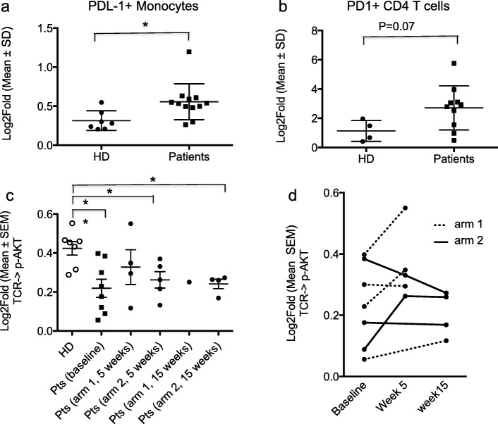
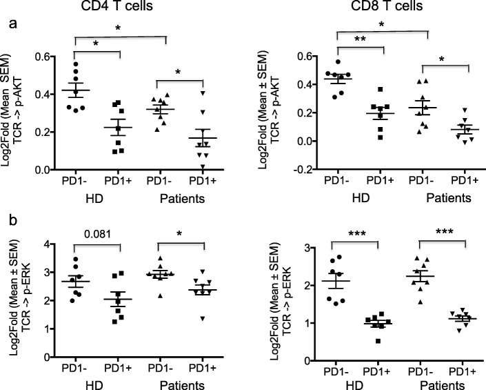
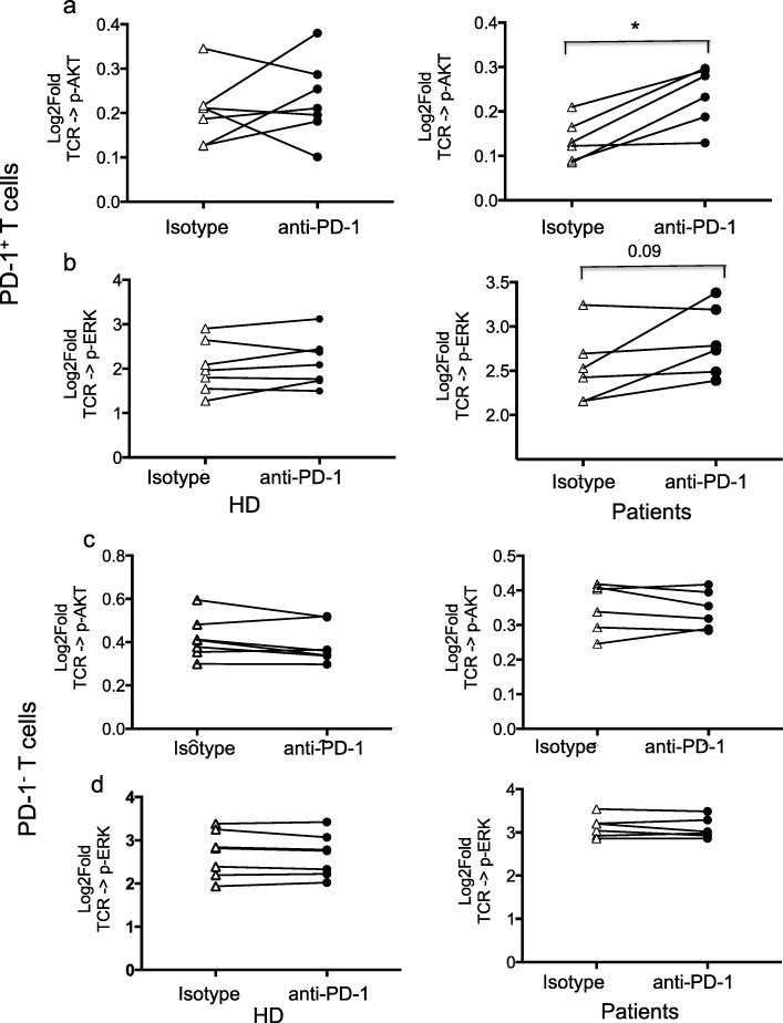
Background We previously reported the results of a multicentric prospective randomized trial of chemo-refractory metastatic breast cancer patients testing the efficacy of two doses of TGFβ blockade during radiotherapy. Despite a lack of objective responses to the combination, patients who received a higher dose of TGFβ blocking antibody fresolimumab had a better overall survival when compared to those assigned to lower dose (hazard ratio of 2.73, p = 0.039). They also demonstrated an improved peripheral blood mononuclear cell (PBMC) counts and increase in the CD8 central memory pool. We performed additional analysis on residual PBMC using single cell network profiling (SCNP). Methods The original trial randomized metastatic breast cancer patients to either 1 or 10 mg/kg of fresolimumab, every 3 weeks for 5 cycles, combined with radiotherapy to a metastatic site at week 1 and 7 (22.5 Gy given in 3 doses of 7.5 Gy). Trial immune monitoring results were previously reported. In 15 patients with available residual blood samples, additional functional studies were performed, and compared with data obtained in parallel from seven healthy female donors (HD): SCNP was applied to analyze T cell receptor (TCR) modulated signaling via CD3 and CD28 crosslinking and measurement of evoked phosphorylation of AKT and ERK in CD4 and CD8 T cell subsets defined by PD-1 expression. Results At baseline, a significantly higher level of expression (p < 0.05) of PD-L1 was identified in patient monocytes compared to HD. TCR modulation revealed dysfunction of circulating T-cells in patient baseline samples as compared to HD, and this was more pronounced in PD-1+ cells. Treatment with radiotherapy and fresolimumab did not resolve this dyfunctional signaling. However, in vitro PD-1 blockade enhanced TCR signaling in patient PD-1+ T cells and not in PD-1- T cells or in PD-1+ T cells from HD. Conclusions Functional T cell analysis suggests that baseline T cell functionality is hampered in metastatic breast cancer patients, at least in part mediated by the PD-1 signaling pathway. These preliminary data support the rationale for investigating the possible beneficial effects of adding PD-1 blockade to improve responses to TGFβ blockade and radiotherapy. Trial registration NCT01401062.
Overview
The claims and statistics of this paper haven’t been checked yet.
Free: it reads the open-access text and checks the first 5 claims that cite a source we can check. Create a free account to check every claim.
Integrity signals
Registry
Cites
Statistics
Figures
Verification
Claims the paper makes
The claims and statistics of this paper haven’t been checked yet.
Free: it reads the open-access text and checks the first 5 claims that cite a source we can check. Create a free account to check every claim.
Claims other papers make about this one
Each is checked against this paper’s own text: does it say what the citing paper says it does?
The cited source says what the claim attributes to it.
In the citing paper
“at least in part reflecting PD-1dependent functional defects of the T-cell compartment at baseline”
From Immunomodulation by targeted anticancer agents (2021) · cites this paper as Formenti et al., 2019
In the citing paper
“clinical responses in this study were limited, most likely as a consequence of PD-1-dependent functional defects of the T-cell compartment at baseline”
From Immunomodulation by targeted anticancer agents (2021) · cites this paper as Formenti et al., 2019
Self-check
Percentages, sums and reported differences re-computed from the paper’s own numbers.
The self-check needs the paper’s full text.
Citations
Cites
PD-1 and PD-L1 Immune Checkpoint Blockade to Treat Breast Cancer
Hartkopf, A. D., Taran, F.-A., Wallwiener, M., Walter, C. B., Krämer, B., Grischke, E.-M., & Brucker, S. Y. (2016) · Breast Care · doi:10.1159/000453569 · Cited by 30,845
TGFβ attenuates tumour response to PD-L1 blockade by contributing to exclusion of T cells
Mariathasan, S., Turley, S. J., Nickles, D., Castiglioni, A., Yuen, K., Wang, Y., Kadel III, E. E., Koeppen, H., Astarita, J. L., Cubas, R., et al. (2018) · Nature · doi:10.1038/nature25501 · Cited by 5,400
Atezolizumab and nab-paclitaxel in advanced triple-negative breast cancer
Schmid, P., Adams, S., Rugo, H. S., Schneeweiss, A., Barrios, C. H., Iwata, H., Diéras, V., Hegg, R., Im, S.-A., Shaw Wright, G., et al. (2018) · New England Journal of Medicine · doi:10.1056/nejmoa1809615 · Cited by 4,350
Wolchok, J. D., Hoos, A., O'Day, S., Weber, J. S., Hamid, O., Lebbé, C., Maio, M., Binder, M., Bohnsack, O., Nichol, G., et al. (2009) · Clinical Cancer Research · doi:10.1158/1078-0432.ccr-09-1624 · Cited by 3,222
TGFβ drives immune evasion in genetically reconstituted colon cancer metastasis
Tauriello, D. V. F., Palomo-Ponce, S., Stork, D., Berenguer-Llergo, A., Badia-Ramentol, J., Iglesias, M., Sevillano, M., Ibiza, S., Cañellas, A., Hernando-Momblona, X., et al. (2018) · Nature · doi:10.1038/nature25492 · Cited by 1,851
Breast Cancer Immunotherapy: Facts and Hopes
Emens, L. A. (2018) · Clinical Cancer Research · doi:10.1158/1078-0432.ccr-16-3001 · Cited by 863
Gorelik, L., & Flavell, R. A. (2001) · Nature Medicine · doi:10.1038/nm1001-1118 · Cited by 770
TGF-β inhibition enhances chemotherapy action against triple-negative breast cancer
Bhola, N. E., Balko, J. M., Dugger, T. C., Kuba, M. G., Sánchez, V., Sanders, M., Stanford, J., Cook, R. S., & Arteaga, C. L. (2013) · Journal of Clinical Investigation · doi:10.1172/jci65416 · Cited by 590
TGFβ is a master regulator of radiation therapy-induced antitumor immunity
Vanpouille-Box, C., Diamond, J. M., Pilones, K. A., Zavadil, J., Babb, J. S., Formenti, S. C., Barcellos-Hoff, M. H., & Demaria, S. (2015) · Cancer Research · doi:10.1158/0008-5472.can-14-3511 · Cited by 514
Self-citationRadiation and the microenvironment – tumorigenesis and therapy
Barcellos-Hoff, M. H., Park, C., & Wright, E. G. (2005) · Nature Reviews Cancer · doi:10.1038/nrc1735 · Cited by 513
Cited by
Immunostimulation with chemotherapy in the era of immune checkpoint inhibitors
Galluzzi, L., Humeau, J., Buqué, A., Zitvogel, L., & Kroemer, G. (2020) · Nature Reviews Clinical Oncology · doi:10.1038/s41571-020-0413-z · Cited by 1,349
TGFβ biology in cancer progression and immunotherapy
Derynck, R., Turley, S. J., & Akhurst, R. J. (2021) · Nature Reviews Clinical Oncology · doi:10.1038/s41571-020-0403-1 · Cited by 944
Cold and hot tumors: from molecular mechanisms to targeted therapy
Wu, B., Zhang, B., Li, B., Wu, H., & Jiang, M. (2024) · Signal Transduction and Targeted Therapy · doi:10.1038/s41392-024-01979-x · Cited by 682
Targeting TGFβ signal transduction for cancer therapy
Liu, S., Ren, J., & ten Dijke, P. (2021) · Signal Transduction and Targeted Therapy · doi:10.1038/s41392-020-00436-9 · Cited by 413
“In a phase 2 clinical trial, the TGFβ-blocking antibody fresolimumab (GC1008) was combined with focal irradiation for the treatment of metastatic breast cancer, and it was found to prolong median overall survival and demonstrated a favorable systemic immune response.(268,269) In addition, fresolimumab (GC1008) combine…”
BackgroundTargeting the tumor stroma for cancer therapy
Xu, M., Zhang, T., Xia, R., Wei, Y., & Wei, X. (2022) · Molecular Cancer · doi:10.1186/s12943-022-01670-1 · Cited by 319
“Several TGF-β-targeted drugs have been actively assessed in clinic to potentiate antitumor effects [338, 339] (NCT01401062, NCT02581787).”
Background
Metadata
- Authors
- Formenti, S. C., Hawtin, R. E., Dixit, N., Evensen, E., Lee, P., Goldberg, J. D., Li, X., Vanpouille-Box, C., Schaue, D., McBride, W. H., & Demaria, S.
- Affiliations
- Weill Cornell Medicine (US)
- Gilead Sciences (United States) (US)
- Nodality (United States) (US)
- University of California, Los Angeles (US)
- New York University (US)
- Corresponding author
- Formenti, Silvia C.
- Demaria, Sandra
- ORCID
- Venue
- Journal for ImmunoTherapy of Cancer
- Publisher
- BMJ
- Published
- 2019-07-11
- Volume
- 7
- Issue
- 1
- Pages
- 177-177
- ISSN
- 2051-1426 (ISSN-L)
- PMC
- PMC6624899
- OpenAlex
- W2959936824
- Cited by
- 53
- References
- 23
- Citations by year
- 2026: 4 · 2025: 7 · 2024: 9 · 2023: 7 · 2022: 5 · 2021: 13 · 2020: 7 · 2019: 1
- FWCI
- 2.72
- Type
- journal article
- Language
- en
- Open access
- gold
- doi.org
- Licence
- cc-by
- creativecommons.org/licenses/by/4.0/ (tdm)
- Locations
- Journal for ImmunoTherapy of Cancer (publishedVersion, cc-by)
- PubMed Central (publishedVersion, cc-by)
- PubMed (publishedVersion)
- DOAJ (DOAJ: Directory of Open Access Journals) (submittedVersion, cc-by-sa)
- eScholarship (California Digital Library) (submittedVersion)
- eScholarship (California Digital Library) (submittedVersion, cc-by)
- Funders
- U.S. Department of Defense (W81XWH-11-1-0530, W81XWH-11-1-0531)
- Breast Cancer Research Foundation
- National Cancer Institute (5P30CA16087)
- Topics
- Health Sciences › Medicine › Oncology › Cancer Immunotherapy and Biomarkers
- Life Sciences › Immunology and Microbiology › Immunology › T-cell and B-cell Immunology
- Life Sciences › Biochemistry, Genetics and Molecular Biology › Molecular Biology › TGF-β signaling in diseases
- Keywords
- metastatic breast cancer, T cell dysfunction, TGF-beta blockade, radiotherapy, PD-1 signaling, PD-L1, T cell receptor signaling, anti-PD-1 therapy, immune monitoring
- MeSH terms
- Adult, Aged, Antineoplastic Agents, Breast Neoplasms, Combined Modality Therapy, Female, Humans, Middle Aged, Receptors, Antigen, T-Cell, CD4-Positive T-Lymphocytes, Transforming Growth Factor beta, CD8-Positive T-Lymphocytes, Young Adult, B7-H1 Antigen, Programmed Cell Death 1 Receptor, Antibodies, Monoclonal, Humanized
About these checks
Registry, retraction, Cites and statistics results are computed, not judged, with one exception: a printed reference that no index lists is read once by a model before it counts as a citation error. Claims are extracted by a model and kept only when their quote appears verbatim in the text; each cited source is then checked, with a model’s help, for whether it says what the claim relies on. Those verdicts can be wrong — read the claim’s quote and the cited source before relying on one.
Statistics and arithmetic are recomputed, not judged.
Confidence bands are shown only for fields calibrated on our annotated test set. None is calibrated yet, so no band is shown.
What counts as a claim ›How we measure confidence ›How we check this ›
Are you an author, or do you see a problem with this page? Request a review
How to cite
Cite CoChat for the checks on this page, and the paper for its own findings.
- This page
- CoChat. (2026). Citation check: Baseline T cell dysfunction by single cell network profiling in metastatic breast cancer patients. https://app.cochat.ai/papers/10.1186/s40425-019-0633-x
- The paper
- Formenti, S. C., Hawtin, R. E., Dixit, N., Evensen, E., Lee, P., Goldberg, J. D., Li, X., Vanpouille-Box, C., Schaue, D., McBride, W. H., & Demaria, S. (2019). Baseline T cell dysfunction by single cell network profiling in metastatic breast cancer patients. Journal for ImmunoTherapy of Cancer. https://doi.org/10.1186/s40425-019-0633-x
Data sources
Bibliographic data: Semantic Scholar, OpenAlex, Crossref.
Contains information from the Semantic Scholar Academic Graph Datasets, made available under the ODC Attribution License (ODC-BY 1.0).
Semantic Scholar’s data is described in: Kinney, R., et al. (2023). The Semantic Scholar Open Data Platform. arXiv:2301.10140.
