

The DOI matches its Crossref record; 171 of the 171 references it cites were found in a registry or an index.
Microglial pathology
Open accessAbstract
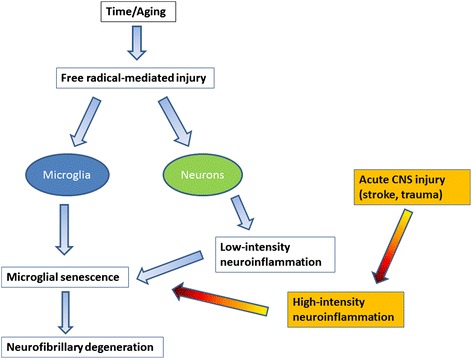
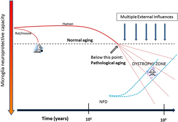
This paper summarizes pathological changes that affect microglial cells in the human brain during aging and in aging-related neurodegenerative diseases, primarily Alzheimer's disease (AD). It also provides examples of microglial changes that have been observed in laboratory animals during aging and in some experimentally induced lesions and disease models. Dissimilarities and similarities between humans and rodents are discussed in an attempt to generate a current understanding of microglial pathology and its significance during aging and in the pathogenesis of Alzheimer dementia (AD). The identification of dystrophic (senescent) microglia has created an ostensible conflict with prior work claiming a role for activated microglia and neuroinflammation during normal aging and in AD, and this has raised a basic question: does the brain's immune system become hyperactive (inflamed) or does it become weakened (senescent) in elderly and demented people, and what is the impact on neuronal function and cognition? Here we strive to reconcile these seemingly contradictory notions by arguing that both low-grade neuroinflammation and microglial senescence are the result of aging-associated free radical injury. Both processes are damaging for microglia as they synergistically exhaust this essential cell population to the point where the brain's immune system is effete and unable to support neuronal function.
Overview
The claims and statistics of this paper haven’t been checked yet.
Free: it reads the open-access text and checks the first 5 claims that cite a source we can check. Create a free account to check every claim.
Integrity signals
Registry
Cites
Statistics
Figures
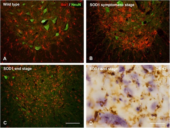

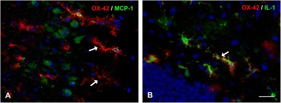
Verification
Claims the paper makes
The claims and statistics of this paper haven’t been checked yet.
Free: it reads the open-access text and checks the first 5 claims that cite a source we can check. Create a free account to check every claim.
Claims other papers make about this one
Each is checked against this paper’s own text: does it say what the citing paper says it does?
In the citing paper
“This striking difference between human and mouse supports the idea that rodent microglia may not experience pathological factors associated with aging such as senescence or dystrophy (70).”
From Microglia in Alzheimer's disease (2017) · cites this paper as [70]
Self-check
Percentages, sums and reported differences re-computed from the paper’s own numbers.
The self-check needs the paper’s full text.
Citations
Cites
Yaşlı Kadınlarda Üreme Sağlığı
Kokanalı, D., & Engin Üstün, Y. (n.d.) · DergiPark (Istanbul University) · Cited by 170,090
Yaşlı Kadınlarda Üreme Sağlığı
Kokanalı, D., & Üstün, Y. E. (2019) · Jinekoloji-Obstetrik ve Neonatoloji Tıp Dergisi · Cited by 169,581
Neuropathological stageing of Alzheimer-related changes.
Braak, H., & Braak, E. (1991) · Acta Neuropathologica · doi:10.1007/bf00308809 · Cited by 16,123 · cited 1×
“Neurofibrillary degeneration (NFD), or tau pathology, and synapse loss offer the best neuropathological correlates of cognitive impairment and dementia in AD [81-85], and it will be important to elucidate the role that microglial cells play in the development of AD-type neurodegeneration.”
BackgroundThe Amyloid Hypothesis of Alzheimer’s Disease: Progress and Problems on the Road to Therapeutics
Hardy, J., & Selkoe, D. J. (2002) · Science · doi:10.1126/science.1072994 · Cited by 13,864
Aging: a theory based on free radical and radiation chemistry
Harman, D. (1956) · Journal of Gerontology · doi:10.1093/geronj/11.3.298 · Cited by 8,567 · cited 2×
“creased metabolic activity and concomitantly decreased free radical production [50].”
BackgroundResting microglial cells are highly dynamic surveillants of brain parenchyma in vivo
Nimmerjahn, A., Kirchhoff, F., & Helmchen, F. (2005) · Science · doi:10.1126/science.1110647 · Cited by 5,827
(2016) · Repository@Hull (Worktribe) (University of Hull) · doi:10.1111/j.1365 · Cited by 5,304
The senescence-associated secretory phenotype: the dark side of tumor suppression
Coppé, J.-P., Desprez, P.-Y., Krtolica, A., & Campisi, J. (2010) · Annual Review of Pathology: Mechanisms of Disease · doi:10.1146/annurev-pathol-121808-102144 · Cited by 5,206 · cited 1×
“Although thus far the notion of SASP has been discussed in terms of non-CNS cell senescence and tumor suppression rather than in a microglial context [59,60], current understanding of microglial secretory activity during aging and the fact that microglioma is an exceedingly rare tumor [61] are compatible with the SASP…”
ResultFate Mapping Analysis Reveals That Adult Microglia Derive from Primitive Macrophages
Ginhoux, F., Greter, M., Leboeuf, M., Nandi, S., See, P., Gokhan, S., Mehler, M. F., Conway, S. J., Ng, L. G., Stanley, E. R., et al. (2010) · Science · doi:10.1126/science.1194637 · Cited by 5,175 · cited 1×
“The current view is that microglia derive from primitive yolksac macrophages in a Myb-independent manner [70,71].”
BackgroundInflammation and Alzheimer’s disease
Akiyama, H. (2000) · Neurobiology of Aging · doi:10.1016/s0197-4580(00)00124-x · Cited by 4,514
Cited by
The Amyloid-β Pathway in Alzheimer’s Disease
Hampel, H., Hardy, J., Blennow, K., Chen, C., Perry, G., Kim, S. H., Villemagne, V. L., Aisen, P., Vendruscolo, M., Iwatsubo, T., et al. (2021) · Molecular Psychiatry · doi:10.1038/s41380-021-01249-0 · Cited by 1,952
Microglia in physiology and disease
Wolf, S. A., Boddeke, H. W. G. M., & Kettenmann, H. (2017) · Annual Review of Physiology · doi:10.1146/annurev-physiol-022516-034406 · Cited by 1,426
Microglia in Alzheimer's disease
Sarlus, H., & Heneka, M. T. (2017) · Journal of Clinical Investigation · doi:10.1172/jci90606 · Cited by 911
“Microglial activation induces several dynamic changes, including morphological alterations, such as shortening of the processes and swelling of the soma, changes in surface phenotype and secretory profile, and increased proliferative responses (70).”
BackgroundAge-related myelin degradation burdens the clearance function of microglia during aging
Safaiyan, S., Kannaiyan, N., Snaidero, N., Brioschi, S., Biber, K., Yona, S., Edinger, A. L., Jung, S., Rossner, M. J., & Simons, M. (2016) · Nature Neuroscience · doi:10.1038/nn.4325 · Cited by 604
Suárez‐Calvet, M., Kleinberger, G., Araque Caballero, M. Á., Brendel, M., Rominger, A., Alcolea, D., Fortea, J., Lleó, A., Blesa, R., Gispert, J. D., et al. (2016) · EMBO Molecular Medicine · doi:10.15252/emmm.201506123 · Cited by 522
“Increased microglial activation and neuroinflammation frequently accompanies the early development of Ab and tau pathology (Mosher & WyssCoray, 2014; Streit et al, 2014; Heneka et al, 2015; Tanzi, 2015).”
BackgroundInfluential
Metadata
- Authors
- Streit, W. J., Xue, Q.-S., Tischer, J., & Bechmann, I.
- Affiliations
- University of Florida (US)
- Leipzig University (DE)
- Corresponding author
- Streit, Wolfgang J
- Venue
- Acta Neuropathologica Communications
- Published
- 2014-09-26
- Volume
- 2
- Issue
- 1
- Pages
- 142-142
- ISSN
- 2051-5960 (ISSN-L)
- PMC
- PMC4180960
- OpenAlex
- W4211216239
- Cited by
- 317
- References
- 161
- Citations by year
- 2026: 9 · 2025: 29 · 2024: 17 · 2023: 28 · 2022: 35 · 2021: 41 · 2020: 43 · 2019: 25 · 2018: 27 · 2017: 23 · 2016: 25 · 2015: 14 · 2014: 1
- FWCI
- 10.94
- Type
- journal article
- Language
- en
- Open access
- gold
- doi.org
- Locations
- Acta Neuropathologica Communications (publishedVersion, cc-by)
- PubMed Central (publishedVersion, cc-by)
- PubMed (publishedVersion)
- University of Florida Digital Collections (University of Florida) (submittedVersion, other-oa)
- Topics
- Life Sciences › Neuroscience › Neurology › Neuroinflammation and Neurodegeneration Mechanisms
- Life Sciences › Neuroscience › Cellular and Molecular Neuroscience › Alzheimer's disease research and treatments
- Life Sciences › Neuroscience › Neurology › Neurological Disease Mechanisms and Treatments
- Keywords
- microglial senescence, neuroinflammation, brain aging, Alzheimer's disease, oxidative damage, microglial activation, cognitive decline
- MeSH terms
- Alzheimer Disease, Brain, Humans, Microglia
About these checks
Registry, retraction, Cites and statistics results are computed, not judged, with one exception: a printed reference that no index lists is read once by a model before it counts as a citation error. Claims are extracted by a model and kept only when their quote appears verbatim in the text; each cited source is then checked, with a model’s help, for whether it says what the claim relies on. Those verdicts can be wrong — read the claim’s quote and the cited source before relying on one.
Statistics and arithmetic are recomputed, not judged.
Confidence bands are shown only for fields calibrated on our annotated test set. None is calibrated yet, so no band is shown.
What counts as a claim ›How we measure confidence ›How we check this ›
Are you an author, or do you see a problem with this page? Request a review
How to cite
Cite CoChat for the checks on this page, and the paper for its own findings.
- This page
- CoChat. (2026). Citation check: Microglial pathology. https://app.cochat.ai/papers/10.1186/s40478-014-0142-6
- The paper
- Streit, W. J., Xue, Q.-S., Tischer, J., & Bechmann, I. (2014). Microglial pathology. Acta Neuropathologica Communications. https://doi.org/10.1186/s40478-014-0142-6
Data sources
Bibliographic data: Semantic Scholar, OpenAlex, Crossref.
Contains information from the Semantic Scholar Academic Graph Datasets, made available under the ODC Attribution License (ODC-BY 1.0).
Semantic Scholar’s data is described in: Kinney, R., et al. (2023). The Semantic Scholar Open Data Platform. arXiv:2301.10140.
