

The DOI matches its Crossref record; 195 of the 198 references it cites were found in a registry or an index, and 1 are not checked yet.
Clinical applications of fundus autofluorescence in retinal disease
Open accessAbstract
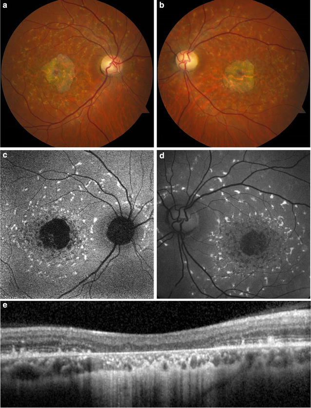
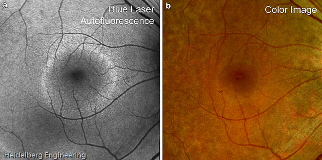
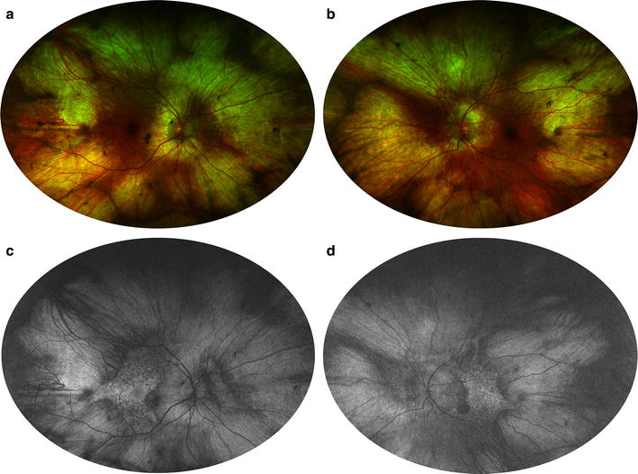
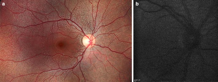
Fundus autofluorescence (FAF) is a non-invasive retinal imaging modality used in clinical practice to provide a density map of lipofuscin, the predominant ocular fluorophore, in the retinal pigment epithelium. Multiple commercially available imaging systems, including the fundus camera, the confocal scanning laser ophthalmoscope, and the ultra-widefield imaging device, are available to the clinician. Each offers unique advantages for evaluating various retinal diseases. The clinical applications of FAF continue to expand. It is now an essential tool for evaluating age related macular degeneration, macular dystrophies, retinitis pigmentosa, white dot syndromes, retinal drug toxicities, and various other retinal disorders. FAF may detect abnormalities beyond those detected on funduscopic exam, fluorescein angiography, or optical coherence tomography, and can be used to elucidate disease pathogenesis, form genotype-phenotype correlations, diagnose and monitor disease, and evaluate novel therapies. Given its ease of use, non-invasive nature, and value in characterizing retinal disease, FAF enjoys increasing clinical relevance. This review summarizes common ocular fluorophores, imaging modalities, and FAF findings for a wide spectrum of retinal disorders.
Overview
The claims and statistics of this paper haven’t been checked yet.
Run the check — free, no account needed. It reads the open-access text and checks the sources it cites.
Integrity signals
Registry
Cites
Statistics
Figures
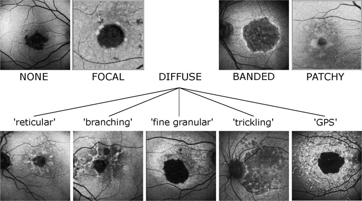
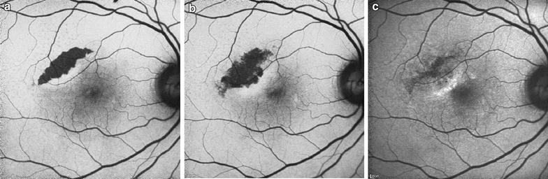
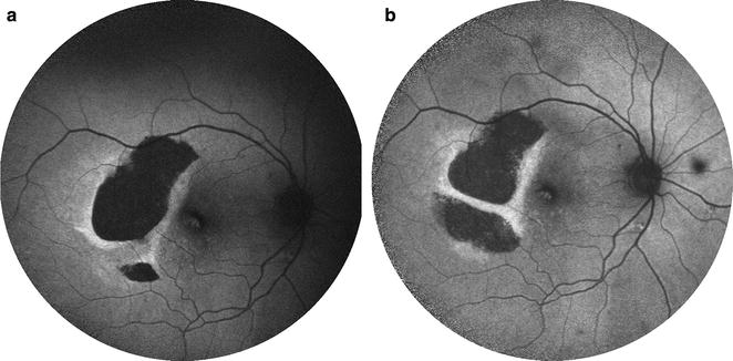
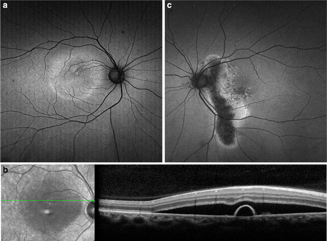
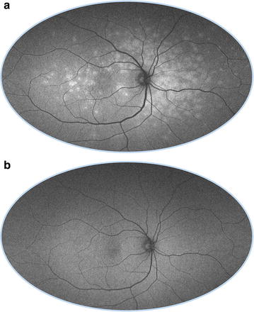
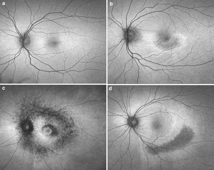
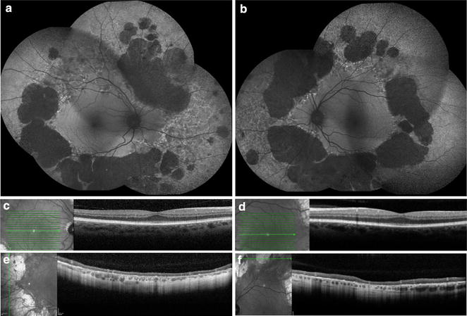
Verification
Claims the paper makes
The claims and statistics of this paper haven’t been checked yet.
Run the check — free, no account needed. It reads the open-access text and checks the sources it cites.
Claims other papers make about this one
Each is checked against this paper’s own text: does it say what the citing paper says it does?
The source backs part of the claim, not all of it — or backs it for a different population, condition or comparison.
In the citing paper
“As A2E can not be further metabolized and as RPE cells are postmitotic, A2E and related bisretinoids accumulate in the RPE in the form of lipofuscin deposits.”
From The Role of Vitamin A in Retinal Diseases (2022) · cites this paper as [75]
Experimental — accuracy not yet measuredThe source backs part of the claim, not all of it — or backs it for a different population, condition or comparison.
In the citing paper
“Although abundant lipofuscin accumulation is involved in the aetiology of certain retinal diseases (e.g., AMD, Best disease and ABCA4-retinopathy), it accumulates at a lower level also with age in the RPE of healthy eyes [9,72,73,74,75].”
From The Role of Vitamin A in Retinal Diseases (2022) · cites this paper as [75]
Experimental — accuracy not yet measuredThe source backs part of the claim, not all of it — or backs it for a different population, condition or comparison.
In the citing paper
“Lipofuscin is an autofluorescent cellular waste product that compromises the function and viability of RPE cells and ultimately results in photoreceptor degeneration and a loss of vision [54,69,73,74,75].”
From The Role of Vitamin A in Retinal Diseases (2022) · cites this paper as [75]
Experimental — accuracy not yet measured
Self-check
Percentages, sums and reported differences re-computed from the paper’s own numbers.
The self-check needs the paper’s full text.
Citations
Cites
Clinical Classification of Age-related Macular Degeneration
Ferris, F. L., Wilkinson, C. P., Bird, A. C., Chakravarthy, U., Chew, E. Y., Csaky, K. G., & Sadda, S. R. (2013) · Ophthalmology · doi:10.1016/j.ophtha.2012.10.036 · Cited by 1,868
Stereoscopic Atlas of Macular Diseases: Diagnosis and Treatment
(1990) · Archives of Ophthalmology · doi:10.1001/archopht.1990.01070060042026 · Cited by 1,014
Delori, F. C., Dorey, C. K., Staurenghi, G., Arend, O., Goger, D. G., & Weiter, J. J. (1995) · PubMed · Cited by 938
Revised Recommendations on Screening for Chloroquine and Hydroxychloroquine Retinopathy
Marmor, M. F., Kellner, U., Lai, T. Y. Y., Lyons, J. S., & Mieler, W. F. (2011) · Ophthalmology · doi:10.1016/j.ophtha.2010.11.017 · Cited by 703
Holz, F. G., Bindewald-Wittich, A., Fleckenstein, M., Dreyhaupt, J., Scholl, H. P. N., & Schmitz-Valckenberg, S. (2007) · American Journal of Ophthalmology · doi:10.1016/j.ajo.2006.11.041 · Cited by 619
Sparrow, J. R., Nakanishi, K., & Parish, C. A. (2000) · PubMed · Cited by 566
Retinal pigment epithelial lipofuscin and melanin and choroidal melanin in human eyes.
Weiter, J. J., Delori, F. C., Wing, G. L., & Fitch, K. A. (1986) · PubMed · Cited by 565
Aging human RPE: morphometric analysis of macular, equatorial, and peripheral cells.
Feeney-Burns, L., Hilderbrand, E. S., & Eldridge, S. (1984) · PubMed · Cited by 561
RPE65 is the isomerohydrolase in the retinoid visual cycle
Moiseyev, G., Chen, Y., Takahashi, Y., Wu, B. X., & Ma, J.-X. (2005) · Proceedings of the National Academy of Sciences · doi:10.1073/pnas.0503460102 · Cited by 536
Prevalence of Age-Related Macular Degeneration in the US Population
Klein, R. (2011) · Archives of Ophthalmology · doi:10.1001/archophthalmol.2010.318 · Cited by 523
Research works printed but not linked: 2
- Park SP, Siringo FS, Pensec N, et al. Comparison of fundus autofluorescence between fundus camera and confocal scanning laser ophthalmoscope-based systems. Ophthalmic Surgery Lasers Imaging Retina. 2013;44(6):536–43. · DOI
- Espina M, Barteselli G, Ma F, et al. Noncontact ultra-wide field lens system by Heidelberg spectralis. Invest Ophthalmol Vis Sci. 2014;55(13):1615.
Cited by
Sorted by citations
Items 201–236 of 236
Harel, I. D., Garg, I., & Delori, F. C. (2021) · Springer eBooks · doi:10.1007/978-3-319-90495-5_143-1
Dithmar, S., & Celik, N. (2020) · Springer eBooks · doi:10.1007/978-3-030-28384-1_13
Harel, I. D., Garg, I., & Delori, F. C. (2021) · Springer eBooks · doi:10.1007/978-3-319-90495-5_143-2
Mitchell, M. E., Cook, L. C., Shiers, S. I., Tavares‐Ferreira, D., Akopian, A. N., Dussor, G. O., & Price, T. J. (2022) · bioRxiv (Cold Spring Harbor Laboratory) · doi:10.1101/2022.09.15.508178
Astaneh, M. A., Hedayati, M., & Shariati, M. M. (2022) · Research Square · doi:10.21203/rs.3.rs-2281336/v1
Inflammasomes in the pathobiology of degenerative retinopathies
Silke, E., Cahill, M. T., Campbell, M., & Doyle, S. (2022) · Elsevier eBooks · doi:10.1016/b978-0-323-91802-2.00021-9 · Cited by 1
The significance of hyper-reflective spots in OCT imaging in retinal diseases
Darwish, A. (2022) · Advances in Ophthalmology & Visual System · doi:10.15406/aovs.2022.12.00417
Optic Disc Drusen Resulting in Disqualifying Field Loss
Binkley, R. C. (2023) · Optometric Clinical Practice · doi:10.37685/uiwlibraries.2575-7717.5.1.1002
Shariati, M. M. (2022) · Journal of Clinical Images and Medical Case Reports · doi:10.52768/2766-7820/2191
Dithmar, S., & Celik, N. (2024) · Springer eBooks · doi:10.1007/978-3-031-35055-9_13
Okonkwo, O. N., Agweye, C. T., Akanbi, T., & Utam, U. A. (2024) · IntechOpen eBooks · doi:10.5772/intechopen.1004859
Roadmap to a large area multispectral fluorescence imaging system
Lima, N. A., & Sawyer, T. W. (2024) · Optical Engineering · doi:10.1117/1.oe.63.5.055105
Park, S. H., Kang, T. S., Kim, M. J., & Kim, B. J. (2024) · Research Square · doi:10.21203/rs.3.rs-4280073/v1
Krema, H. (2025) · Springer eBooks · doi:10.1007/978-3-031-82953-6_6
Retinal Imaging and Function Testing for Clinicians
Quillen, D. A. (2025) · Springer eBooks · doi:10.1007/978-3-031-66092-4_2
Imaging in Inherited Retinal Dystrophies
Russell, M. W., Traboulsi, E. I., & Ehlers, J. P. (2025) · doi:10.1093/med/9780197659403.003.0027
Age-Related Macular Degeneration Screening—What Is Next?
Ranetti, A., & Stanca, H. T. (2023) · Romanian Journal of Preventive Medicine · doi:10.3390/rjpm2010047
12-YEAR FOLLOW-UP OF A BENIGN YELLOW DOT MACULOPATHY CASE
Nunziata, A., Fabbro, S. D., Antropoli, A., Bianco, L., Arrigo, A., Bandello, F. M., Mansour, A. M., & Parodi, M. B. (2025) · Retinal Cases & Brief Reports · doi:10.1097/icb.0000000000001810
Varela, M. D., Aychoua, N., Rashid, M., Lopes, A., & Michaelides, M. (2025) · Ophthalmology Science · doi:10.1016/j.xops.2025.101026
Targeted Supramolecular Senolytics by Enzyme-Responsive Disassembly and Intracellular Polymerization
Kim, S., Lee, J., Cho, Y., Park, H., Oh, S. H., Kim, J., Kim, D., Sim, Y., Seu, M., Kim, C., et al. (2026) · ACS Applied Materials & Interfaces · doi:10.1021/acsami.5c18350
Menna, F., Pinelli, C., Luca, L. D., Meduri, A., Baldascino, A., Lupo, S., & Vingolo, E. M. (2026) · Biomedicines · doi:10.3390/biomedicines14040782
Molleti, S., Monsalve, P., Sather, R., Moon, J. Y., Ihinger, J., & Montezuma, S. R. (2025) · Molecular vision · doi:10.63500/mv_v31_454
Comparison of Clarus, Optos, and Heidelberg Systems for Geographic Atrophy Area Measurements
Bogost, J., Saunders, T. F., Heathcote, J., Pak, J. W., Lopez, D., Froines, C. P., Blodi, B., Channa, R., Schildroth, K. R., Kurt, K., et al. (2026) · Ophthalmology Science · doi:10.1016/j.xops.2026.101207
Siraz, S., Kamanda, H., Nabil, A. S., Gholami, S., Rao, N. T., Ong, S. S., & Alam, M. N. (2026) · medRxiv · doi:10.64898/2026.06.17.26355882
Retinal Fingerprints in Alzheimer’s Disease
Barbosa, A. S., Fonseca, E. A., Cunha, R. S., de Miranda, H. L. S., Campos, J. L. E., Romano-Silva, M. A., Moreira, L. M., & Jório, A. (2026) · IntechOpen eBooks · doi:10.5772/intechopen.1015654
van Olden, M. C. S., Pas, J. A. A. H., Yzer, S., Hoyng, C. B., & Theelen, T. (2026) · Ophthalmology Science · doi:10.1016/j.xops.2026.101331
Trinh, H., Ibrahim, M. M., Charng, J., Tajbakhsh, Z., Chen, F. K., Mian, A. S., & Alam, K. (2026) · Journal of Ophthalmology · doi:10.1155/joph/8810684
Medghalchi, A., Kanafi, A. R., Alizadeh, Y., Baghersalimi, A., Doorandish, M., Rad, A. H., Kianmehr, S., & Darbandi, B. (2026) · Health Science Reports · doi:10.1002/hsr2.73053
Viggiano, P., Pignataro, M. G., Termite, A. C., Boscia, G., Savastano, A., Evangelista, F., Alessio, G., Boscia, F., Pető, T., & Chakravarthy, U. (2026) · Progress in Retinal and Eye Research · doi:10.1016/j.preteyeres.2026.101512
Monici, G., Cascio, F. L., Parisi, G., Marolo, P., Reibaldi, M., & Borrelli, E. (2026) · Neuroretina · doi:10.1007/s44448-026-00005-x
Fragiotta, S., Pippo, M. D., Ciancimino, C., Rispoli, F., Scuderi, G., & Abdolrahimzadeh, S. (2026) · Research Square · doi:10.21203/rs.3.rs-10870553/v1
Assessing oxidative stress in retinal tissue using multispectral autofluorescence imaging
Knab, A., Marupally, A. G., Anwer, A. G., Habibalahi, A., Pattamatta, U., Carnt, N. A., Casson, R. J., Goldys, E. M., & White, A. J. R. (2026) · Free Radical Biology and Medicine · doi:10.1016/j.freeradbiomed.2026.09.017
Huang, S. J., & Bitencourt, F. N. (2026) · Springer eBooks · doi:10.1007/978-3-032-23052-2_7
Hwang, Y., Jamil, M. U., Takahashi, H., Marmalidou, A., Won, J., Curcio, C. A., Fujimoto, J. G., Waheed, N. K., & Zhao, P. Y. (2026) · Ophthalmology Science · doi:10.1016/j.xops.2026.101445
Metadata
- Authors
- Yung, M., Klufas, M. A., & Sarraf, D.
- Affiliations
- University of California, Los Angeles (US)
- Corresponding author
- Sarraf, David
- Venue
- International Journal of Retina and Vitreous
- Published
- 2016-04-08
- Volume
- 2
- Issue
- 1
- Pages
- 12-12
- ISSN
- 2056-9920 (ISSN-L)
- PMC
- PMC5088473
- OpenAlex
- W2329521208
- Cited by
- 236
- References
- 198
- Citations by year
- 2026: 15 · 2025: 23 · 2024: 22 · 2023: 20 · 2022: 29 · 2021: 40 · 2020: 31 · 2019: 24 · 2018: 22 · 2017: 8 · 2016: 1
- FWCI
- 3.58
- Type
- journal article
- Language
- en
- Open access
- gold
- doi.org
- Licence
- cc-by
- Locations
- International Journal of Retina and Vitreous (publishedVersion, cc-by)
- PubMed Central (publishedVersion, cc-by)
- PubMed (publishedVersion)
- Funders
- Regeneron Pharmaceuticals
- Genentech
- Topics
- Health Sciences › Medicine › Ophthalmology › Retinal Diseases and Treatments
- Health Sciences › Medicine › Radiology, Nuclear Medicine and Imaging › Retinal Imaging and Analysis
- Health Sciences › Medicine › Ophthalmology › Retinal Development and Disorders
- Keywords
- fundus autofluorescence, lipofuscin, retinal pigment epithelium, age-related macular degeneration, macular dystrophy, retinitis pigmentosa, white dot syndromes, ultra-widefield imaging, confocal scanning laser ophthalmoscopy, genotype-phenotype correlation
About these checks
Registry, retraction, Cites and statistics results are computed, not judged, with one exception: a printed reference that no index lists is read once by a model before it counts as a citation error. Claims are extracted by a model and kept only when their quote appears verbatim in the text; each cited source is then checked, with a model’s help, for whether it says what the claim relies on. Those verdicts can be wrong — read the claim’s quote and the cited source before relying on one.
Statistics and arithmetic are recomputed, not judged.
Confidence bands are shown only for fields calibrated on our annotated test set. None is calibrated yet, so no band is shown.
What counts as a claim ›How we measure confidence ›How we check this ›
Are you an author, or do you see a problem with this page? Request a review
