Cited by 76

The DOI matches its Crossref record; 3 of the 3 references it cites were found in a registry or an index.

Consumption of Acidic Water Alters the Gut Microbiome and Decreases the Risk of Diabetes in NOD Mice

Wolf, Kyle J., Daft, Joseph G., Tanner, Scott M., Hartmann, Riley, Khafipour, Ehsan, Lorenz, Robin G.·2014·Journal of Histochemistry & Cytochemistry

Abstract

Infant formula and breastfeeding are environmental factors that influence the incidence of Type 1 Diabetes (T1D) as well as the acidity of newborn diets. To determine if altering the intestinal microbiome is one mechanism through which an acidic liquid plays a role in T1D, we placed non-obese diabetic (NOD)/ShiLtJt mice on neutral (N) or acidified H 2 O and monitored the impact on microbial composition and diabetes incidence. NOD-N mice showed an increased development of diabetes, while exhibiting a decrease in Firmicutes and an increase in Bacteroidetes, Actinobacteria, and Proteobacteria from as early as 2 weeks of age. NOD-N mice had a decrease in the levels of Foxp3 expression in CD4 + Foxp3 + cells, as well as decreased CD4 + IL17 + cells, and a lower ratio of IL17/IFNγ CD4+ T-cells. Our data clearly indicates that a change in the acidity of liquids consumed dramatically alters the intestinal microbiome, the presence of protective Th17 and Treg cells, and the incidence of diabetes. This data suggests that early dietary manipulation of intestinal microbiota may be a novel mechanism to delay T1D onset in genetically pre-disposed individuals.

Overview

The claims and statistics of this paper haven’t been checked yet.

Run the check — free, no account needed. It reads the open-access text and checks the sources it cites.

A few free checks a day · papers already verified open instantly · results are public

Integrity signals

Registry

Crossref · 10.1369/0022155413519650

Cites

3 of 3 references verified0 citation errors · 0 not checkable

Statistics

Figures

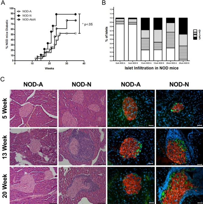
Figure 1. NOD mice on neutral drinking water have an increased incidence of diabetes and alterations in the diversity of their fecal microbiota. (A) Incidence of diabetes in NOD female mice on neutral water (NOD-N, open circles, n=9), acidified water (NOD-A, filled circles, n=23) or water switched from acidified to neutral at weaning (NOD-AtoN, half-filled circles, n=13). Data represent three individual experiments. Significance was determined by the Mantel-Cox Test. (B) Analysis of lymphocytic infiltrate and islet destruction in NOD mice on neutral or acidified water. 0 = no infiltration, 1 = peri-insulitis, 2 = <50% of islet infiltrated, 3 = >50% of islet infiltrated. At 20 weeks, the NOD-N mice had significantly more infiltrated islets (score of 2 or 3; p=0.03). An average of 80 islets per group was counted. (C) Representative H&E (left two panels; scale bar=100 µm) and immunofluorescence (right two panels; red = insulin, green = glucagon, blue = nucleus; scale bar =10 µm) images of pancreatic islets from NOD-A or NOD-N mice at the indicated ages.
Figure 1. NOD mice on neutral drinking water have an increased incidence of diabetes and alterations in the diversity of their fecal microbiota. (A) Incidence of diabetes in NOD female mice on neutral water (NOD-N, open circles, n=9), acidified water (NOD-A, filled circles, n=23) or water switched from acidified to neutral at weaning (NOD-AtoN, half-filled circles, n=13). Data represent three individual experiments. Significance was determined by the Mantel-Cox Test. (B) Analysis of lymphocytic infiltrate and islet destruction in NOD mice on neutral or acidified water. 0 = no infiltration, 1 = peri-insulitis, 2 = <50% of islet infiltrated, 3 = >50% of islet infiltrated. At 20 weeks, the NOD-N mice had significantly more infiltrated islets (score of 2 or 3; p=0.03). An average of 80 islets per group was counted. (C) Representative H&E (left two panels; scale bar=100 µm) and immunofluorescence (right two panels; red = insulin, green = glucagon, blue = nucleus; scale bar =10 µm) images of pancreatic islets from NOD-A or NOD-N mice at the indicated ages.CC BY-NC · PubMed Central
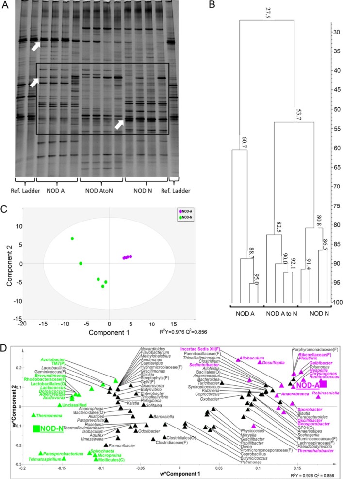
Figure 2. Impact of neutral or acidified drinking water on the fecal microbiota composition of NOD-N and NOD-A mice. (A) A representative denaturing gradient gel electrophoresis (DGGE) analysis of banding patterns from 10-week-old female mice on neutral water (NOD-N, right), acidified water (NOD-A, left) or water switched from acidified to neutral at weaning (NOD-AtoN, middle). Top arrow points to a band identified by sequencing as Lactobacillus johnsonii. Middle arrow points to a band identified as Clostridia Cluster XIV species. Bottom arrow points to a band identified as Bacteroides sp. (B) Banding similarity analysis of the representative samples from 10-week-old NOD-A, NOD-AtoN and NOD-N mice indicates that their microbiota share a 27.5% similarity. NOD-AtoN and NOD-N mice share a higher degree of similarity (53.7%) comparedwith NOD-A mice. Banding analysis was conducted using the Pearson correlation analysis. Figure S1 demonstrates that NOD-A mice have a lower pH throughout their GI tract. Pyrosequencing data were subjected to partial least squares discriminant analysis (PLS-DA). (C) Score scatter plot representing individual animals from each treatment, grouped based on the composition of fecal microbiota. The R2 and Q2 of the model were 0.97 and 0.85, respectively. (D) Bacterial taxa plotted using weighted PLS component 1 and 2. Genera in the plot closer to either treatment are more strongly associated to it. Genera found to significantly contribute to the model prediction are shown in green (NOD-N) and magenta (NOD-A). When a sequence could not be classified to the genus level, the closest level of classification was given, followed by F (family), O (order), C (class), or P (phylum). Additional data showing differences between NOD-A and NOD-N mice at the genus level can be found in Figure S3 and Tables S2, S3 and S4.
Figure 2. Impact of neutral or acidified drinking water on the fecal microbiota composition of NOD-N and NOD-A mice. (A) A representative denaturing gradient gel electrophoresis (DGGE) analysis of banding patterns from 10-week-old female mice on neutral water (NOD-N, right), acidified water (NOD-A, left) or water switched from acidified to neutral at weaning (NOD-AtoN, middle). Top arrow points to a band identified by sequencing as Lactobacillus johnsonii. Middle arrow points to a band identified as Clostridia Cluster XIV species. Bottom arrow points to a band identified as Bacteroides sp. (B) Banding similarity analysis of the representative samples from 10-week-old NOD-A, NOD-AtoN and NOD-N mice indicates that their microbiota share a 27.5% similarity. NOD-AtoN and NOD-N mice share a higher degree of similarity (53.7%) comparedwith NOD-A mice. Banding analysis was conducted using the Pearson correlation analysis. Figure S1 demonstrates that NOD-A mice have a lower pH throughout their GI tract. Pyrosequencing data were subjected to partial least squares discriminant analysis (PLS-DA). (C) Score scatter plot representing individual animals from each treatment, grouped based on the composition of fecal microbiota. The R2 and Q2 of the model were 0.97 and 0.85, respectively. (D) Bacterial taxa plotted using weighted PLS component 1 and 2. Genera in the plot closer to either treatment are more strongly associated to it. Genera found to significantly contribute to the model prediction are shown in green (NOD-N) and magenta (NOD-A). When a sequence could not be classified to the genus level, the closest level of classification was given, followed by F (family), O (order), C (class), or P (phylum). Additional data showing differences between NOD-A and NOD-N mice at the genus level can be found in Figure S3 and Tables S2, S3 and S4.CC BY-NC · PubMed Central
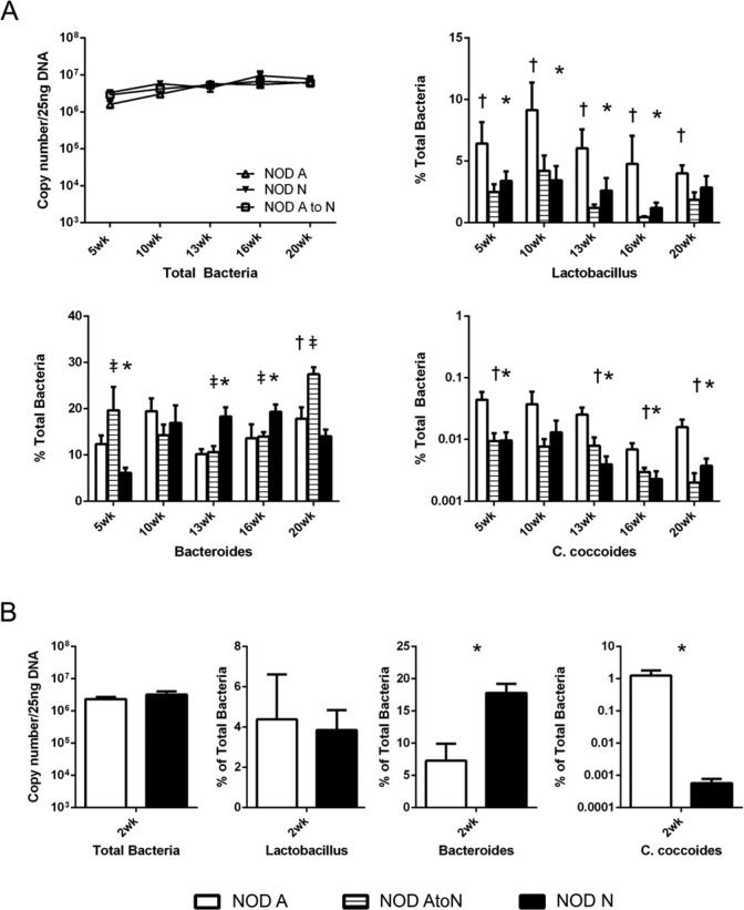
Figure 3. qRT-PCR analysis of bacteria copy numbers in feces of NOD mice shows a significant dysbiosis in mice on neutral drinking water. (A) qRT-PCR analysis of the total copy numbers of NOD mice on acidified (NOD-A), acidified to neutral (NOD-AtoN), or neutral water (NOD-N) drinking water (upper left) at pre-insulitis (5 weeks), post-insulitis, but before overt diabetes (10–13 weeks), and advanced progression of disease (16–20 weeks). Bacterial populations of Lactobacillus sp. (upper right), Bacteroides sp. (lower left), and C. coccoides (lower right) in the feces of female NOD-A (white, n=8–10), NOD-AtoN (striped, n=5), or NOD-N, black, n=8–10) mice. (B) Analysis of fecal microbial populations between 2-week-old NOD-A and NOD-N mice comparing numbers of total bacteria, Lactobacillus sp., Bacteroides sp., and C. coccoides (NOD-A, n=4, NOD-N, n=5). Significance was determined using Welches’ t-test at p<0.05. Significance is indicated by *- NOD-A vs NOD-N, †- NOD-A vs NOD-AtoN, and ‡- NOD-N vs NOD-AtoN.
Figure 3. qRT-PCR analysis of bacteria copy numbers in feces of NOD mice shows a significant dysbiosis in mice on neutral drinking water. (A) qRT-PCR analysis of the total copy numbers of NOD mice on acidified (NOD-A), acidified to neutral (NOD-AtoN), or neutral water (NOD-N) drinking water (upper left) at pre-insulitis (5 weeks), post-insulitis, but before overt diabetes (10–13 weeks), and advanced progression of disease (16–20 weeks). Bacterial populations of Lactobacillus sp. (upper right), Bacteroides sp. (lower left), and C. coccoides (lower right) in the feces of female NOD-A (white, n=8–10), NOD-AtoN (striped, n=5), or NOD-N, black, n=8–10) mice. (B) Analysis of fecal microbial populations between 2-week-old NOD-A and NOD-N mice comparing numbers of total bacteria, Lactobacillus sp., Bacteroides sp., and C. coccoides (NOD-A, n=4, NOD-N, n=5). Significance was determined using Welches’ t-test at p<0.05. Significance is indicated by *- NOD-A vs NOD-N, †- NOD-A vs NOD-AtoN, and ‡- NOD-N vs NOD-AtoN.CC BY-NC · PubMed Central
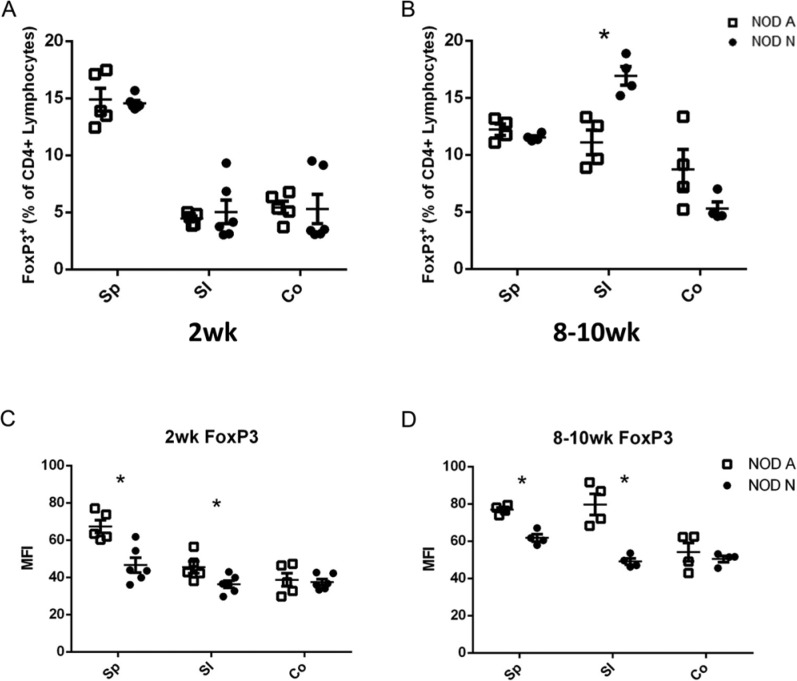
Figure 4. NOD mice on neutral drinking water (NOD-N) have similar percentages of CD4+Foxp3+ Tregs, but decreased expression levels of Foxp3 as mice on acidic water (NOD-A). (A and B) The percentage of CD4+Foxp3+ Tregs in splenocytes (Sp) and small intestinal (SI) and colonic (Co) lamina propria (LP) lymphocytes from NOD-A and NOD-N mice at 2 weeks (NOD-A, n=5, NOD-N, n=6, left panel) and 8–10 weeks (NOD-A, n=4, NOD-N, n=4, right panel). (C and D) Mean fluorescence intensity (MFI) of Foxp3 expression in 2-week- (left panel) and 8–10-week-old (right panel) NOD-A and NOD-N mice. Significance was determined using Welches’ t-test at p<0.5. Absolute numbers of cells in each population can be seen in Figure S2.
Figure 4. NOD mice on neutral drinking water (NOD-N) have similar percentages of CD4+Foxp3+ Tregs, but decreased expression levels of Foxp3 as mice on acidic water (NOD-A). (A and B) The percentage of CD4+Foxp3+ Tregs in splenocytes (Sp) and small intestinal (SI) and colonic (Co) lamina propria (LP) lymphocytes from NOD-A and NOD-N mice at 2 weeks (NOD-A, n=5, NOD-N, n=6, left panel) and 8–10 weeks (NOD-A, n=4, NOD-N, n=4, right panel). (C and D) Mean fluorescence intensity (MFI) of Foxp3 expression in 2-week- (left panel) and 8–10-week-old (right panel) NOD-A and NOD-N mice. Significance was determined using Welches’ t-test at p<0.5. Absolute numbers of cells in each population can be seen in Figure S2.CC BY-NC · PubMed Central
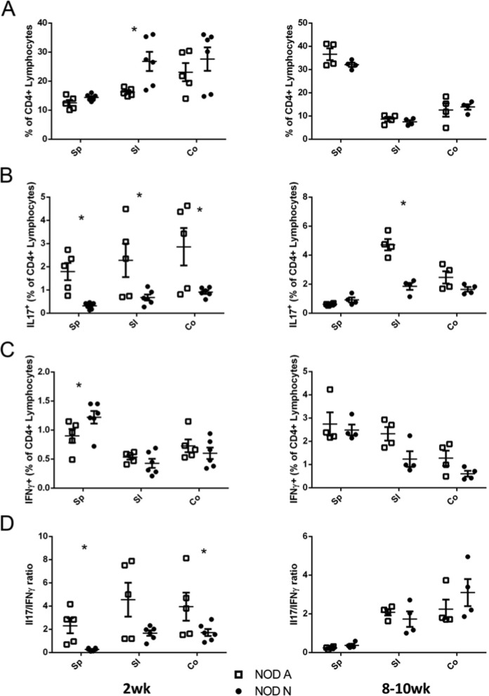
Figure 5. NOD mice on neutral drinking water (NOD-N) have decreased production of IL17 by CD4+ T cells as compared with mice on acidic water (NOD-A). (A) The percentage of CD4+ splenocytes and lymphocytes from the small intestine (SI) and colonic (Co) lamina propria (LP) of 2-week-old (left panel) and 8–10-week-old (right panel) NOD-A (open squares) and NOD-N (closed circles) mice. (B-C) Percentages of CD4+ lymphocytes that were also (B) IL17 or (C) IFNγ+. (D) Comparison of the ratio of CD4+IL17+ and CD4+IFNγ+ cells in 2-week-old (left panel) and 8–10-week-old (right panel) NOD-A and NOD-N mice. At 2 weeks: NOD-A, n=5; NOD-N, n=6. At 8–10 weeks: NOD-A, n=4; NOD-N, n=4). Significance was determined using Welches’ t-test at p<0.5. Absolute numbers of cells in each population can be seen in Figure S2.
Figure 5. NOD mice on neutral drinking water (NOD-N) have decreased production of IL17 by CD4+ T cells as compared with mice on acidic water (NOD-A). (A) The percentage of CD4+ splenocytes and lymphocytes from the small intestine (SI) and colonic (Co) lamina propria (LP) of 2-week-old (left panel) and 8–10-week-old (right panel) NOD-A (open squares) and NOD-N (closed circles) mice. (B-C) Percentages of CD4+ lymphocytes that were also (B) IL17 or (C) IFNγ+. (D) Comparison of the ratio of CD4+IL17+ and CD4+IFNγ+ cells in 2-week-old (left panel) and 8–10-week-old (right panel) NOD-A and NOD-N mice. At 2 weeks: NOD-A, n=5; NOD-N, n=6. At 8–10 weeks: NOD-A, n=4; NOD-N, n=4). Significance was determined using Welches’ t-test at p<0.5. Absolute numbers of cells in each population can be seen in Figure S2.CC BY-NC · PubMed Central

Verification

Claims the paper makes

The claims and statistics of this paper haven’t been checked yet.

Run the check — free, no account needed. It reads the open-access text and checks the sources it cites.

A few free checks a day · papers already verified open instantly · results are public

Claims other papers make about this one

Each is checked against this paper’s own text: does it say what the citing paper says it does?

Self-check

Percentages, sums and reported differences re-computed from the paper’s own numbers.

The self-check needs the paper’s full text.

Citations

Cites

Research works printed but not linked: 48

  1. 10.1007/s00125-012-2672-4 · DOI
  2. 10.1128/IAI.01432-07 · DOI
  3. 10.1016/j.coi.2011.10.002 · DOI
  4. 10.4049/jimmunol.0900576 · DOI
  5. 10.1017/S0007114508094658 · DOI · Paper page
  6. 10.3390/nu4081095 · DOI
  7. 10.4049/jimmunol.1200955 · DOI
  8. 10.1038/nmeth.f.303 · DOI · Paper page
  9. 10.1111/j.1365-2249.2005.02942.x · DOI
  10. 10.1084/jem.139.5.1189 · DOI
  11. 10.4049/jimmunol.182.1.148 · DOI
  12. 10.1111/j.1467-789X.2010.00845.x · DOI
  13. Dimmitt RA, 2010, J Pediatr Gastroenterol Nutr, 51, 262, 10.1097/MPG.0b013e3181e1a114 · DOI
  14. 10.2337/diabetes.53.2.366 · DOI
  15. 10.1093/bioinformatics/btr381 · DOI · Paper page
  16. 10.2337/db08-1113 · DOI
  17. 10.2337/db11-0090 · DOI
  18. 10.1038/ismej.2010.92 · DOI · Paper page
  19. 10.3389/fcimb.2012.00094 · DOI
  20. Hall JE, 1980, Lab Anim Sci, 30, 643
  21. 10.1016/j.clim.2012.12.001 · DOI
  22. Hermann LM, 1982, Lab Anim Sci, 32, 603
  23. 10.1016/j.cell.2009.09.033 · DOI · Paper page
  24. 10.1073/pnas.1108924108 · DOI
  25. 10.4049/jimmunol.1001864 · DOI
  26. 10.1038/ni.2416 · DOI · Paper page
  27. Leiter E, 1997, Current Protocols in Immunology
  28. 10.4049/jimmunol.1001860 · DOI
  29. 10.1111/j.1600-065X.2011.01084.x · DOI
  30. 10.2337/diab.37.12.1625 · DOI · Paper page
  31. 10.1046/j.1462-5822.2001.00090.x · DOI · Paper page
  32. 10.1016/j.ecl.2010.05.008 · DOI
  33. 10.4049/jimmunol.0902822 · DOI
  34. 10.1097/00005176-199210000-00004 · DOI
  35. 10.1016/j.it.2012.02.008 · DOI
  36. 10.1111/j.1753-4887.2012.00513.x · DOI
  37. 10.1038/ismej.2009.5 · DOI
  38. 10.1128/AEM.01541-09 · DOI
  39. 10.1369/0022155411416242 · DOI
  40. 10.1097/00005176-200403000-00012 · DOI
  41. 10.1002/dmrr.945 · DOI
  42. 10.1038/mi.2012.73 · DOI
  43. 10.3945/jn.112.166231 · DOI
  44. 10.1073/pnas.0509006102 · DOI
  45. 10.1084/jem.20120504 · DOI · Paper page
  46. 10.2337/db08-0331 · DOI
  47. 10.1371/journal.pone.0010507 · DOI
  48. 10.4049/jimmunol.1000462 · DOI · Paper page

Cited by

Sorted by citations

See all 76

Metadata

Authors
Wolf, K. J., Daft, J. G., Tanner, S. M., Hartmann, R., Khafipour, E., & Lorenz, R. G.
Venue
Journal of Histochemistry & Cytochemistry
Published
2014-01-22
Volume
62
Issue
4
Pages
237-250
ISSN
  • 0022-1554
  • 1551-5044
Cited by
76
References
3
Language
en

About these checks

Registry, retraction, Cites and statistics results are computed, not judged, with one exception: a printed reference that no index lists is read once by a model before it counts as a citation error. Claims are extracted by a model and kept only when their quote appears verbatim in the text; each cited source is then checked, with a model’s help, for whether it says what the claim relies on. Those verdicts can be wrong — read the claim’s quote and the cited source before relying on one.

Statistics and arithmetic are recomputed, not judged.

Confidence bands are shown only for fields calibrated on our annotated test set. None is calibrated yet, so no band is shown.

What counts as a claim ›How we measure confidence ›How we check this ›

Are you an author, or do you see a problem with this page? Request a review