

The DOI matches its Crossref record; 59 of the 59 references it cites were found in a registry or an index.
The Mechanochemistry of Endocytosis
Open accessAbstract
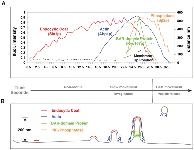
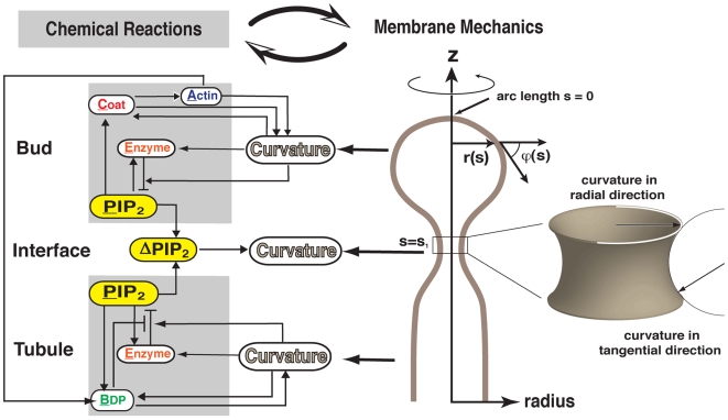
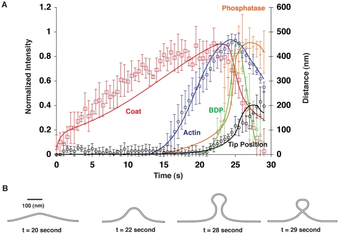
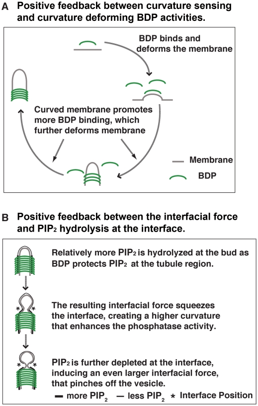
Endocytic vesicle formation is a complex process that couples sequential protein recruitment and lipid modifications with dramatic shape transformations of the plasma membrane. Although individual molecular players have been studied intensively, how they all fit into a coherent picture of endocytosis remains unclear. That is, how the proper temporal and spatial coordination of endocytic events is achieved and what drives vesicle scission are not known. Drawing upon detailed knowledge from experiments in yeast, we develop the first integrated mechanochemical model that quantitatively recapitulates the temporal and spatial progression of endocytic events leading to vesicle scission. The central idea is that membrane curvature is coupled to the accompanying biochemical reactions. This coupling ensures that the process is robust and culminates in an interfacial force that pinches off the vesicle. Calculated phase diagrams reproduce endocytic mutant phenotypes observed in experiments and predict unique testable endocytic phenotypes in yeast and mammalian cells. The combination of experiments and theory in this work suggest a unified mechanism for endocytic vesicle formation across eukaryotes.
Overview
The claims and statistics of this paper haven’t been checked yet.
Run the check — free, no account needed. It reads the open-access text and checks the sources it cites.
Integrity signals
Registry
Cites
Statistics
Figures
Verification
Claims the paper makes
The claims and statistics of this paper haven’t been checked yet.
Run the check — free, no account needed. It reads the open-access text and checks the sources it cites.
Claims other papers make about this one
Each is checked against this paper’s own text: does it say what the citing paper says it does?
The source backs part of the claim, not all of it — or backs it for a different population, condition or comparison.
In the citing paper
“Thereafter, endocytic proteins such as Synaptojanin 1, Endophilin A, Syndapin 1, and Dynamin 1 can further curve the membrane and generate vesicles47–50.”
From Membrane compression by synaptic vesicle exocytosis triggers ultrafast endocytosis (2023) · cites this paper as [47]
Experimental — accuracy not yet measured
Self-check
Percentages, sums and reported differences re-computed from the paper’s own numbers.
The self-check needs the paper’s full text.
Citations
Cites
Intermolecular and Surface Forces
(2011) · Elsevier eBooks · doi:10.1016/b978-0-12-391927-4.10024-6 · Cited by 17,242
Intermolecular and Surface Forces
(2011) · Elsevier eBooks · doi:10.1016/c2011-0-05119-0 · Cited by 9,159
Regulated portals of entry into the cell
Conner, S. D., & Schmid, S. L. (2003) · Nature · doi:10.1038/nature01451 · Cited by 3,958
Phosphoinositides in cell regulation and membrane dynamics
Di Paolo, G., & De Camilli, P. (2006) · Nature · doi:10.1038/nature05185 · Cited by 2,790
Membrane curvature and mechanisms of dynamic cell membrane remodelling
McMahon, H. T., & Gallop, J. L. (2005) · Nature · doi:10.1038/nature04396 · Cited by 2,205
BAR Domains as Sensors of Membrane Curvature: The Amphiphysin BAR Structure
Peter, B. J., Kent, H. M., Mills, I. G., Vallis, Y., Butler, P. J. G., Evans, P. R., & McMahon, H. T. (2004) · Science · doi:10.1126/science.1092586 · Cited by 1,636
The dynamin superfamily: universal membrane tubulation and fission molecules?
Praefcke, G. J. K., & McMahon, H. T. (2004) · Nature Reviews Molecular Cell Biology · doi:10.1038/nrm1313 · Cited by 1,370
Structure and dynamics of membranes
Lipowsky, R., & Sackmann, E. (1995) · Cited by 956
PIP2and Proteins: Interactions, Organization, and Information Flow
McLaughlin, S., Wang, J., Gambhir, A., & Murray, D. (2002) · Annual Review of Biophysics and Biomolecular Structure · doi:10.1146/annurev.biophys.31.082901.134259 · Cited by 837
A Modular Design for the Clathrin- and Actin-Mediated Endocytosis Machinery
Kaksonen, M., Toret, C. P., & Drubin, D. G. (2005) · Cell · doi:10.1016/j.cell.2005.09.024 · Cited by 726
Self-citation
Cited by
Sorted by citations
Dynamin, a membrane-remodelling GTPase
Ferguson, S. M., & Camilli, P. V. D. (2012) · Nature Reviews Molecular Cell Biology · doi:10.1038/nrm3266 · Cited by 1,027
A High Precision Survey of the Molecular Dynamics of Mammalian Clathrin-Mediated Endocytosis
Taylor, M. J., Perrais, D., & Merrifield, C. J. (2011) · PLoS Biology · doi:10.1371/journal.pbio.1000604 · Cited by 754
Regulation of the Actin Cytoskeleton-Plasma Membrane Interplay by Phosphoinositides
Saarikangas, J., Zhao, H., & Lappalainen, P. (2010) · Physiological Reviews · doi:10.1152/physrev.00036.2009 · Cited by 515
Saheki, Y., & De Camilli, P. (2012) · Cold Spring Harbor Perspectives in Biology · doi:10.1101/cshperspect.a005645 · Cited by 457
Mechanisms of Membrane Curvature Sensing
Antonny, B. (2011) · Annual Review of Biochemistry · doi:10.1146/annurev-biochem-052809-155121 · Cited by 442
Metadata
- Authors
- Liu, J., Sun, Y., Drubin, D. G., & Oster, G. F.
- Affiliations
- University of California, Berkeley (US)
- Corresponding author
- Drubin, David G.
- Oster, George F.
- Venue
- PLoS Biology
- Publisher
- Public Library of Science (PLoS)
- Published
- 2009-09-29
- Volume
- 7
- Issue
- 9
- Pages
- e1000204
- ISSN
- 1544-9173 (ISSN-L)
- 1545-7885
- PMC
- PMC2742711
- OpenAlex
- W2019040974
- Cited by
- 222
- References
- 59
- Citations by year
- 2026: 1 · 2025: 7 · 2024: 8 · 2023: 6 · 2022: 9 · 2021: 11 · 2020: 6 · 2019: 9 · 2018: 15 · 2017: 15 · 2016: 10 · 2015: 16 · 2014: 21 · 2013: 17 · 2012: 24
- FWCI
- 10.53
- Type
- journal article
- Language
- en
- Open access
- gold
- doi.org
- Locations
- PLoS Biology (publishedVersion, cc-by)
- PubMed Central (publishedVersion, cc-by)
- PubMed (publishedVersion)
- DOAJ (DOAJ: Directory of Open Access Journals) (submittedVersion)
- Figshare (submittedVersion, cc-by)
- Funders
- National Science Foundation (0414039)
- National Institute of General Medical Sciences (1 R01 GM 50399, R01GM042759, R01GM050399, 1 R01 GM 42759)
- Topics
- Life Sciences › Biochemistry, Genetics and Molecular Biology › Cell Biology › Cellular transport and secretion
- Life Sciences › Biochemistry, Genetics and Molecular Biology › Molecular Biology › Lipid Membrane Structure and Behavior
- Life Sciences › Biochemistry, Genetics and Molecular Biology › Molecular Biology › Fungal and yeast genetics research
- Keywords
- endocytosis, mechanochemistry, vesicle scission, membrane curvature, protein recruitment, lipid modification, interfacial forces, mechanochemical model, phase diagram, vesicle formation
- MeSH terms
- Actins, Animals, Biomechanical Phenomena, Cell Membrane, Endocytosis, Saccharomycetales, Hydrolysis, Lipids, Mammals, Models, Biological, Phosphoric Monoester Hydrolases, Time Factors, Protein Structure, Tertiary, Phosphatidylinositol 4,5-Diphosphate, Transport Vesicles, Feedback, Physiological, Saccharomyces cerevisiae Proteins
About these checks
Registry, retraction, Cites and statistics results are computed, not judged, with one exception: a printed reference that no index lists is read once by a model before it counts as a citation error. Claims are extracted by a model and kept only when their quote appears verbatim in the text; each cited source is then checked, with a model’s help, for whether it says what the claim relies on. Those verdicts can be wrong — read the claim’s quote and the cited source before relying on one.
Statistics and arithmetic are recomputed, not judged.
Confidence bands are shown only for fields calibrated on our annotated test set. None is calibrated yet, so no band is shown.
What counts as a claim ›How we measure confidence ›How we check this ›
Are you an author, or do you see a problem with this page? Request a review
