

The DOI matches its Crossref record; 65 of the 67 references it cites were found in a registry or an index.
Persistent Amyloidosis following Suppression of Aβ Production in a Transgenic Model of Alzheimer Disease
Open accessAbstract
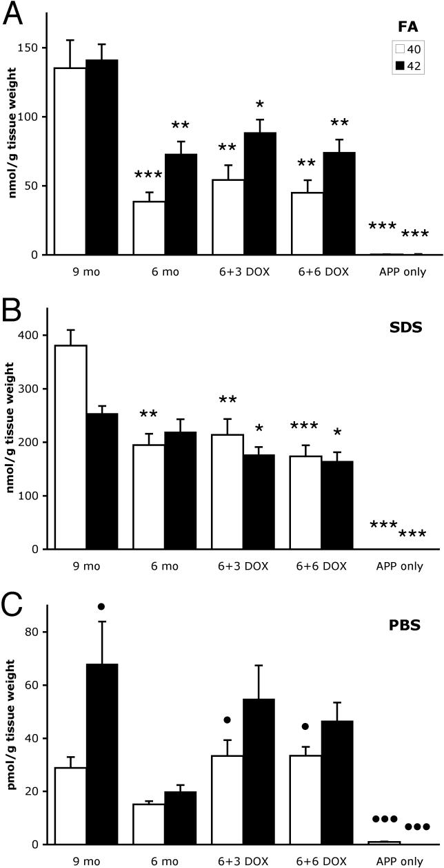
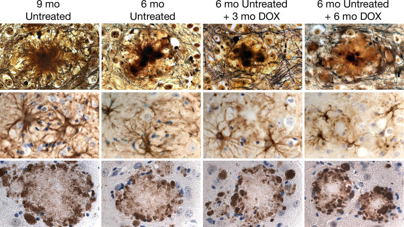
BACKGROUND: The proteases (secretases) that cleave amyloid-beta (Abeta) peptide from the amyloid precursor protein (APP) have been the focus of considerable investigation in the development of treatments for Alzheimer disease. The prediction has been that reducing Abeta production in the brain, even after the onset of clinical symptoms and the development of associated pathology, will facilitate the repair of damaged tissue and removal of amyloid lesions. However, no long-term studies using animal models of amyloid pathology have yet been performed to test this hypothesis. METHODS AND FINDINGS: We have generated a transgenic mouse model that genetically mimics the arrest of Abeta production expected from treatment with secretase inhibitors. These mice overexpress mutant APP from a vector that can be regulated by doxycycline. Under normal conditions, high-level expression of APP quickly induces fulminant amyloid pathology. We show that doxycycline administration inhibits transgenic APP expression by greater than 95% and reduces Abeta production to levels found in nontransgenic mice. Suppression of transgenic Abeta synthesis in this model abruptly halts the progression of amyloid pathology. However, formation and disaggregation of amyloid deposits appear to be in disequilibrium as the plaques require far longer to disperse than to assemble. Mice in which APP synthesis was suppressed for as long as 6 mo after the formation of Abeta deposits retain a considerable amyloid load, with little sign of active clearance. CONCLUSION: This study demonstrates that amyloid lesions in transgenic mice are highly stable structures in vivo that are slow to disaggregate. Our findings suggest that arresting Abeta production in patients with Alzheimer disease should halt the progression of pathology, but that early treatment may be imperative, as it appears that amyloid deposits, once formed, will require additional intervention to clear.
Overview
The claims and statistics of this paper haven’t been checked yet.
Run the check — free, no account needed. It reads the open-access text and checks the sources it cites.
Integrity signals
Registry
Cites
Statistics
Figures
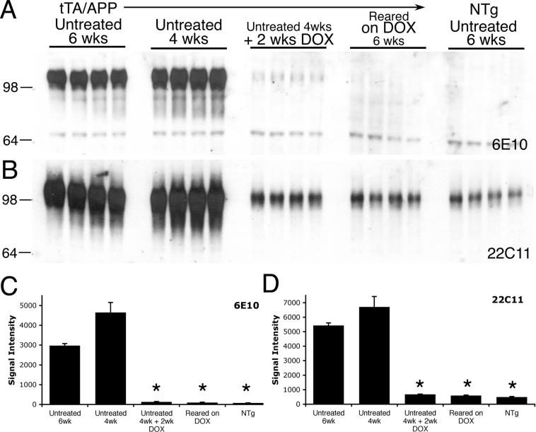
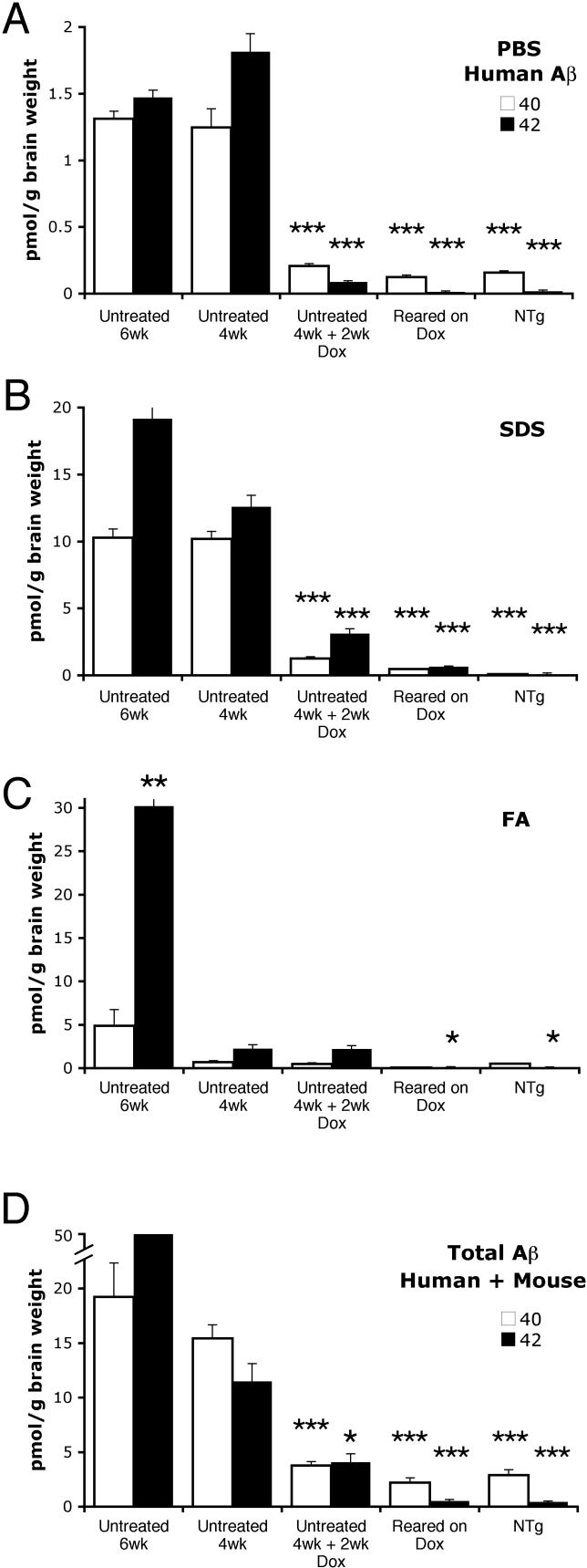
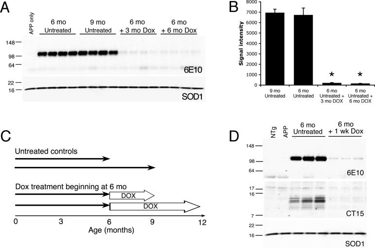
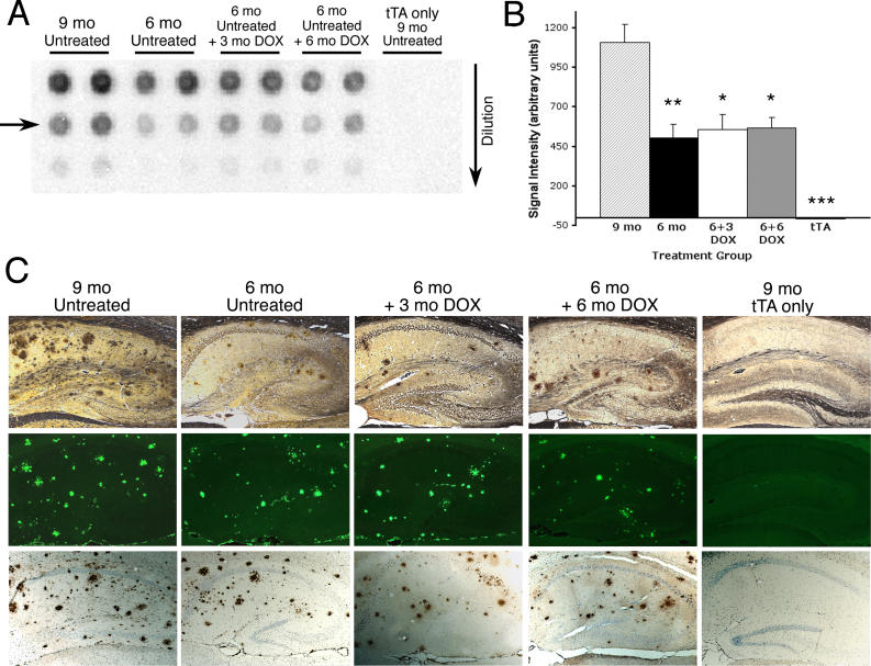
Verification
Claims the paper makes
The claims and statistics of this paper haven’t been checked yet.
Run the check — free, no account needed. It reads the open-access text and checks the sources it cites.
Claims other papers make about this one
Each is checked against this paper’s own text: does it say what the citing paper says it does?
In the citing paper
“Despite this response, however, Aβ clearance and plaque modulation/dynamics is unaffected10–12”
From Sustained microglial depletion with CSF1R inhibitor impairs parenchymal plaque development in an Alzheimer’s disease model (2019) · cites this paper as [12]
Self-check
Percentages, sums and reported differences re-computed from the paper’s own numbers.
The self-check needs the paper’s full text.
Citations
Cites
Amyloid oligomers exacerbate tau pathology in a mouse model of tauopathy
Selenica, M.-L. B., Brownlow, M., Jimenez, J. P., Lee, D. C., Pena, G., Dickey, C. A., Gordon, M. N., & Morgan, D. (2013) · Neurodegenerative Diseases · doi:10.1159/000337230 · Cited by 10,359
Church, G. M., & Gilbert, W. (1984) · Proceedings of the National Academy of Sciences · doi:10.1073/pnas.81.7.1991 · Cited by 8,213
Alzheimer's disease: the amyloid cascade hypothesis
Hardy, J. A., & Higgins, G. A. (1992) · Science · doi:10.1126/science.1566067 · Cited by 7,484
Tight control of gene expression in mammalian cells by tetracycline-responsive promoters.
Gossen, M., & Bujard, H. (1992) · Proceedings of the National Academy of Sciences · doi:10.1073/pnas.89.12.5547 · Cited by 4,943
Correlative Memory Deficits, Aβ Elevation, and Amyloid Plaques in Transgenic Mice
Hsiao, K., Chapman, P., Nilsen, S., Eckman, C., Harigaya, Y., Younkin, S., Yang, F., & Cole, G. (1996) · Science · doi:10.1126/science.274.5284.99 · Cited by 4,210
Self-citationTau Suppression in a Neurodegenerative Mouse Model Improves Memory Function
SantaCruz, K., Lewis, J., Spires, T., Paulson, J., Kotilinek, L., Ingelsson, M., Guimaraes, A., DeTure, M., Ramsden, M., McGowan, E., et al. (2005) · Science · doi:10.1126/science.1113694 · Cited by 2,014
Bard, F., Cannon, C., Barbour, R., Burke, R.-L., Games, D., Grajeda, H., Guido, T., Hu, K., Huang, J., Johnson-Wood, K., et al. (2000) · Nature Medicine · doi:10.1038/78682 · Cited by 2,012
Jankowsky, J. L., Fadale, D. J., Anderson, J., Xu, G. M., Gonzales, V., Jenkins, N. A., Copeland, N. G., Lee, M. K., Younkin, L. H., Wagner, S. L., et al. (2004) · Human Molecular Genetics · doi:10.1093/hmg/ddh019 · Cited by 1,575
Self-citationControl of memory formation through regulated expression of a CaMKII transgene
Mayford, M., Bach, M. E., Huang, Y.-Y., Wang, L., Hawkins, R. D., & Kandel, E. R. (1996) · Science · doi:10.1126/science.274.5293.1678 · Cited by 1,550
Borchelt, D. R., Thinakaran, G., Eckman, C. B., Lee, M. K., Davenport, F., Ratovitsky, T., Prada, C.-M., Kim, G., Seekins, S., Yager, D., et al. (1996) · Neuron · doi:10.1016/s0896-6273(00)80230-5 · Cited by 1,478
Self-citation
Research works printed but not linked: 2
- Cumming, 2004, Curr Opin Drug Discov Devel, 7, 536
- Campbell, 1987, Soc Neurosci Abstr, 13, 189.9
Cited by
Sorted by citations
Karran, E., Mercken, M., & Strooper, B. D. (2011) · Nature Reviews Drug Discovery · doi:10.1038/nrd3505 · Cited by 2,188
ApoE-Directed Therapeutics Rapidly Clear β-Amyloid and Reverse Deficits in AD Mouse Models
Cramer, P. E., Cirrito, J. R., Wesson, D. W., Lee, C. Y. D., Karlo, J. C., Zinn, A. E., Casali, B. T., Restivo, J. L., Goebel, W. D., James, M. J., et al. (2012) · Science · doi:10.1126/science.1217697 · Cited by 1,050
Spangenberg, E., Severson, P. L., Hohsfield, L. A., Crapser, J., Zhang, J., Burton, E. A., Zhang, Y., Spevak, W., Lin, J., Phan, N. Y., et al. (2019) · Nature Communications · doi:10.1038/s41467-019-11674-z · Cited by 939
Protein aggregation diseases: pathogenicity and therapeutic perspectives
Aguzzi, A., & O’Connor, T. (2010) · Nature Reviews Drug Discovery · doi:10.1038/nrd3050 · Cited by 766
Lian, H., Yang, L., Cole, A., Sun, L., Chiang, A. C.-A., Fowler, S. W., Shim, D. J., Rodriguez-Rivera, J., Taglialatela, G., Jankowsky, J. L., et al. (2015) · Neuron · doi:10.1016/j.neuron.2014.11.018 · Cited by 697
Self-citation
Metadata
- Authors
- Jankowsky, J. L., Slunt, H. H., Gonzales, V., Savonenko, A. V., Wen, J. C., Jenkins, N. A., Copeland, N. G., Younkin, L. H., Lester, H. A., Younkin, S. G., & Borchelt, D. R.
- Affiliations
- California Institute of Technology (US)
- Johns Hopkins University (US)
- National Cancer Institute (US)
- Mayo Clinic (US)
- Corresponding author
- Jankowsky, Joanna L
- Borchelt, David R
- Venue
- PLoS Medicine
- Publisher
- Public Library of Science (PLoS)
- Published
- 2005-11-15
- Volume
- 2
- Issue
- 12
- Pages
- e355
- ISSN
- 1549-1277 (ISSN-L)
- 1549-1676
- PMC
- PMC1283364
- OpenAlex
- W2152864425
- Cited by
- 237
- References
- 67
- Citations by year
- 2026: 3 · 2025: 6 · 2024: 23 · 2023: 10 · 2022: 14 · 2021: 7 · 2020: 5 · 2019: 9 · 2018: 9 · 2017: 14 · 2016: 19 · 2015: 9 · 2014: 13 · 2013: 8 · 2012: 16
- FWCI
- 6.19
- Type
- journal article
- Language
- en
- Open access
- gold
- doi.org
- Locations
- PLoS Medicine (publishedVersion, cc-by)
- Europe PMC (PubMed Central) (publishedVersion, cc-by)
- PubMed Central (publishedVersion, cc-by)
- PubMed (publishedVersion)
- CaltechAUTHORS (California Institute of Technology) (acceptedVersion)
- CaltechAUTHORS (California Institute of Technology) (submittedVersion, cc-by)
- DOAJ (DOAJ: Directory of Open Access Journals) (submittedVersion, cc-by-sa)
- Figshare (submittedVersion, cc-by)
- Funders
- Alzheimer's Association
- Johns Hopkins University
- National Alliance for Research on Schizophrenia and Depression
- Rose Hills Foundation
- National Institute on Aging (K01 AG26144-01, P50 AGO5146-20, K01AG026144, P01AG015453, P50AG005146, R01AG006656)
- National Cancer Institute
- National Institute of Neurological Disorders and Stroke (R01NS047225)
- Topics
- Life Sciences › Neuroscience › Cellular and Molecular Neuroscience › Alzheimer's disease research and treatments
- Health Sciences › Medicine › Psychiatry and Mental health › Dementia and Cognitive Impairment Research
- Life Sciences › Neuroscience › Neurology › Neuroinflammation and Neurodegeneration Mechanisms
- Keywords
- Alzheimer's disease, amyloid-beta, amyloid plaques, amyloidosis, amyloid precursor protein, secretase inhibitors, transgenic mouse model, amyloid clearance
- MeSH terms
- Alzheimer Disease, Animals, Humans, Mice, Inbred C57BL, Mice, Transgenic, Amyloid beta-Peptides, Amyloid beta-Protein Precursor, Gene Expression Profiling, Mice, Plaque, Amyloid
About these checks
Registry, retraction, Cites and statistics results are computed, not judged, with one exception: a printed reference that no index lists is read once by a model before it counts as a citation error. Claims are extracted by a model and kept only when their quote appears verbatim in the text; each cited source is then checked, with a model’s help, for whether it says what the claim relies on. Those verdicts can be wrong — read the claim’s quote and the cited source before relying on one.
Statistics and arithmetic are recomputed, not judged.
Confidence bands are shown only for fields calibrated on our annotated test set. None is calibrated yet, so no band is shown.
What counts as a claim ›How we measure confidence ›How we check this ›
Are you an author, or do you see a problem with this page? Request a review
