

The DOI matches its Crossref record; 65 of the 67 references it cites were found in a registry or an index.
Persistent Amyloidosis following Suppression of Aβ Production in a Transgenic Model of Alzheimer Disease
Open accessAbstract
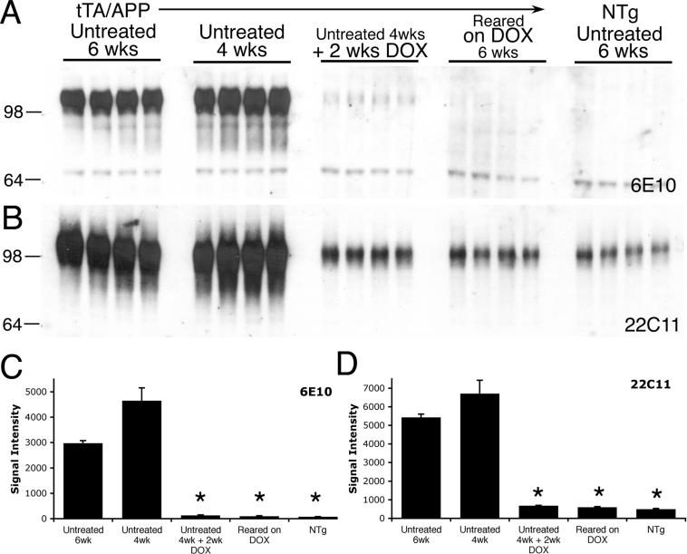
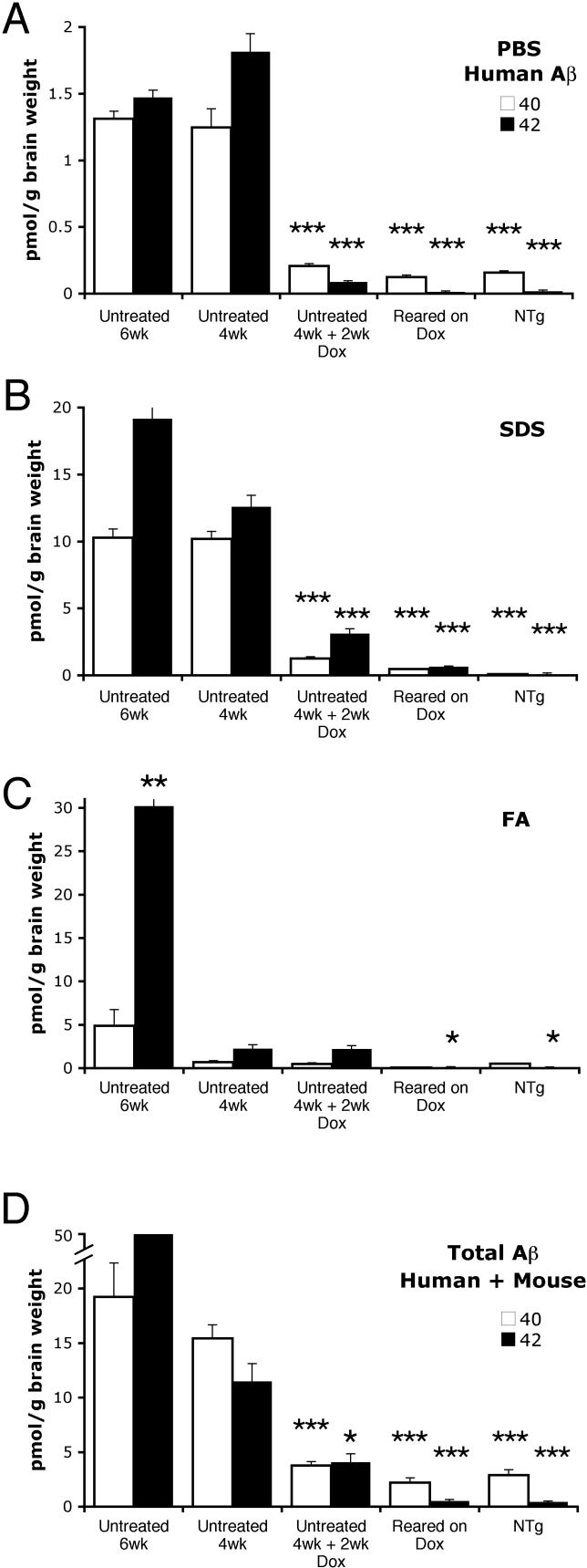
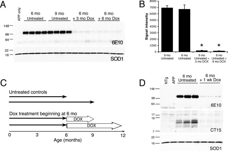
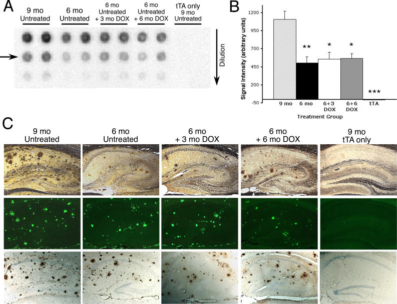
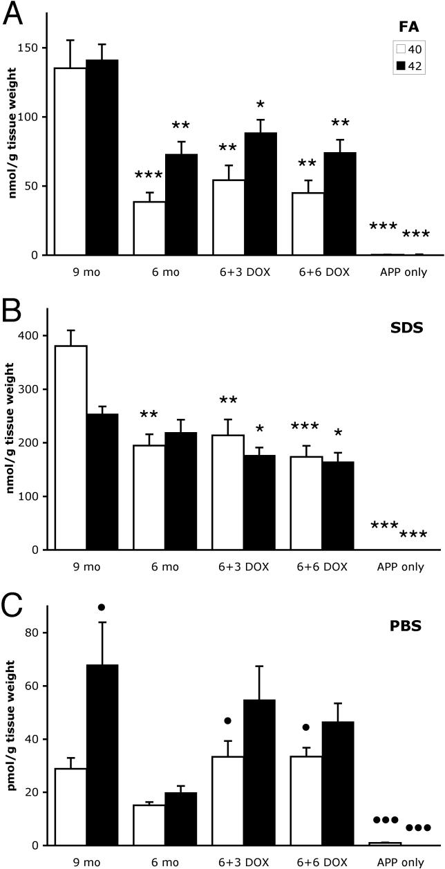
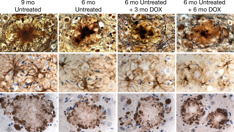
BACKGROUND: The proteases (secretases) that cleave amyloid-beta (Abeta) peptide from the amyloid precursor protein (APP) have been the focus of considerable investigation in the development of treatments for Alzheimer disease. The prediction has been that reducing Abeta production in the brain, even after the onset of clinical symptoms and the development of associated pathology, will facilitate the repair of damaged tissue and removal of amyloid lesions. However, no long-term studies using animal models of amyloid pathology have yet been performed to test this hypothesis. METHODS AND FINDINGS: We have generated a transgenic mouse model that genetically mimics the arrest of Abeta production expected from treatment with secretase inhibitors. These mice overexpress mutant APP from a vector that can be regulated by doxycycline. Under normal conditions, high-level expression of APP quickly induces fulminant amyloid pathology. We show that doxycycline administration inhibits transgenic APP expression by greater than 95% and reduces Abeta production to levels found in nontransgenic mice. Suppression of transgenic Abeta synthesis in this model abruptly halts the progression of amyloid pathology. However, formation and disaggregation of amyloid deposits appear to be in disequilibrium as the plaques require far longer to disperse than to assemble. Mice in which APP synthesis was suppressed for as long as 6 mo after the formation of Abeta deposits retain a considerable amyloid load, with little sign of active clearance. CONCLUSION: This study demonstrates that amyloid lesions in transgenic mice are highly stable structures in vivo that are slow to disaggregate. Our findings suggest that arresting Abeta production in patients with Alzheimer disease should halt the progression of pathology, but that early treatment may be imperative, as it appears that amyloid deposits, once formed, will require additional intervention to clear.
Overview
The claims and statistics of this paper haven’t been checked yet.
Run the check — free, no account needed. It reads the open-access text and checks the sources it cites.
Integrity signals
Registry
Cites
Statistics
Figures
Verification
Claims the paper makes
The claims and statistics of this paper haven’t been checked yet.
Run the check — free, no account needed. It reads the open-access text and checks the sources it cites.
Claims other papers make about this one
Each is checked against this paper’s own text: does it say what the citing paper says it does?
In the citing paper
“Despite this response, however, Aβ clearance and plaque modulation/dynamics is unaffected10–12”
From Sustained microglial depletion with CSF1R inhibitor impairs parenchymal plaque development in an Alzheimer’s disease model (2019) · cites this paper as [12]
Self-check
Percentages, sums and reported differences re-computed from the paper’s own numbers.
The self-check needs the paper’s full text.
Citations
Cites
Amyloid oligomers exacerbate tau pathology in a mouse model of tauopathy
Selenica, M.-L. B., Brownlow, M., Jimenez, J. P., Lee, D. C., Pena, G., Dickey, C. A., Gordon, M. N., & Morgan, D. (2013) · Neurodegenerative Diseases · doi:10.1159/000337230 · Cited by 10,359
Church, G. M., & Gilbert, W. (1984) · Proceedings of the National Academy of Sciences · doi:10.1073/pnas.81.7.1991 · Cited by 8,213
Alzheimer's disease: the amyloid cascade hypothesis
Hardy, J. A., & Higgins, G. A. (1992) · Science · doi:10.1126/science.1566067 · Cited by 7,484
Tight control of gene expression in mammalian cells by tetracycline-responsive promoters.
Gossen, M., & Bujard, H. (1992) · Proceedings of the National Academy of Sciences · doi:10.1073/pnas.89.12.5547 · Cited by 4,943
Correlative Memory Deficits, Aβ Elevation, and Amyloid Plaques in Transgenic Mice
Hsiao, K., Chapman, P., Nilsen, S., Eckman, C., Harigaya, Y., Younkin, S., Yang, F., & Cole, G. (1996) · Science · doi:10.1126/science.274.5284.99 · Cited by 4,210
Self-citationTau Suppression in a Neurodegenerative Mouse Model Improves Memory Function
SantaCruz, K., Lewis, J., Spires, T., Paulson, J., Kotilinek, L., Ingelsson, M., Guimaraes, A., DeTure, M., Ramsden, M., McGowan, E., et al. (2005) · Science · doi:10.1126/science.1113694 · Cited by 2,014
Bard, F., Cannon, C., Barbour, R., Burke, R.-L., Games, D., Grajeda, H., Guido, T., Hu, K., Huang, J., Johnson-Wood, K., et al. (2000) · Nature Medicine · doi:10.1038/78682 · Cited by 2,012
Jankowsky, J. L., Fadale, D. J., Anderson, J., Xu, G. M., Gonzales, V., Jenkins, N. A., Copeland, N. G., Lee, M. K., Younkin, L. H., Wagner, S. L., et al. (2004) · Human Molecular Genetics · doi:10.1093/hmg/ddh019 · Cited by 1,575
Self-citationControl of memory formation through regulated expression of a CaMKII transgene
Mayford, M., Bach, M. E., Huang, Y.-Y., Wang, L., Hawkins, R. D., & Kandel, E. R. (1996) · Science · doi:10.1126/science.274.5293.1678 · Cited by 1,550
Borchelt, D. R., Thinakaran, G., Eckman, C. B., Lee, M. K., Davenport, F., Ratovitsky, T., Prada, C.-M., Kim, G., Seekins, S., Yager, D., et al. (1996) · Neuron · doi:10.1016/s0896-6273(00)80230-5 · Cited by 1,478
Self-citation
Research works printed but not linked: 2
- Cumming, 2004, Curr Opin Drug Discov Devel, 7, 536
- Campbell, 1987, Soc Neurosci Abstr, 13, 189.9
Cited by
Sorted by citations
Karran, E., Mercken, M., & Strooper, B. D. (2011) · Nature Reviews Drug Discovery · doi:10.1038/nrd3505 · Cited by 2,188
ApoE-Directed Therapeutics Rapidly Clear β-Amyloid and Reverse Deficits in AD Mouse Models
Cramer, P. E., Cirrito, J. R., Wesson, D. W., Lee, C. Y. D., Karlo, J. C., Zinn, A. E., Casali, B. T., Restivo, J. L., Goebel, W. D., James, M. J., et al. (2012) · Science · doi:10.1126/science.1217697 · Cited by 1,050
Spangenberg, E., Severson, P. L., Hohsfield, L. A., Crapser, J., Zhang, J., Burton, E. A., Zhang, Y., Spevak, W., Lin, J., Phan, N. Y., et al. (2019) · Nature Communications · doi:10.1038/s41467-019-11674-z · Cited by 939
Protein aggregation diseases: pathogenicity and therapeutic perspectives
Aguzzi, A., & O’Connor, T. (2010) · Nature Reviews Drug Discovery · doi:10.1038/nrd3050 · Cited by 766
Lian, H., Yang, L., Cole, A., Sun, L., Chiang, A. C.-A., Fowler, S. W., Shim, D. J., Rodriguez-Rivera, J., Taglialatela, G., Jankowsky, J. L., et al. (2015) · Neuron · doi:10.1016/j.neuron.2014.11.018 · Cited by 697
Self-citationSpangenberg, E. E., Lee, R. J., Najafi, A. R., Rice, R. A., Elmore, M. R. P., Blurton-Jones, M., West, B. L., & Green, K. N. (2016) · Brain · doi:10.1093/brain/aww016 · Cited by 695
Lian, H., Litvinchuk, A., Chiang, A. C.-A., Aithmitti, N., Jankowsky, J. L., & Zheng, H. (2016) · The Journal of Neuroscience · doi:10.1523/jneurosci.2117-15.2016 · Cited by 597
Self-citationType I interferon response drives neuroinflammation and synapse loss in Alzheimer disease
Roy, E. R., Wang, B., Wan, Y.-W., Chiu, G., Cole, A., Yin, Z., Propson, N. E., Xu, Y., Jankowsky, J. L., Liu, Z., et al. (2020) · Journal of Clinical Investigation · doi:10.1172/jci133737 · Cited by 563
Self-citationAmyloid-Beta: A Crucial Factor in Alzheimer's Disease
Sadigh‐Eteghad, S., Sabermarouf, B., Majdi, A., Talebi, M., Farhoudi, M., & Mahmoudi, J. (2014) · Medical Principles and Practice · doi:10.1159/000369101 · Cited by 563
Dynamics of the Microglial/Amyloid Interaction Indicate a Role in Plaque Maintenance
Bolmont, T., Haiss, F., Eicke, D., Radde, R., Mathis, C. A., Klunk, W. E., Kohsaka, S., Jucker, M., & Calhoun, M. E. (2008) · The Journal of Neuroscience · doi:10.1523/jneurosci.4814-07.2008 · Cited by 526
Lin, X. Z., Parisiadou, L., Gu, X., Wang, L., Shim, H., Sun, L., Xie, C., Long, C.-X., Yang, W.-J., Ding, J., et al. (2009) · Neuron · doi:10.1016/j.neuron.2009.11.006 · Cited by 532
Practical considerations for choosing a mouse model of Alzheimer’s disease
Jankowsky, J. L., & Zheng, H. (2017) · Molecular Neurodegeneration · doi:10.1186/s13024-017-0231-7 · Cited by 449
Self-citationAlzheimer disease models and human neuropathology: similarities and differences
Duyckaerts, C., Potier, M.-C., & Delatour, B. (2008) · Acta Neuropathologica · doi:10.1007/s00401-007-0312-8 · Cited by 375
Dysregulation of the ALS-associated gene TDP-43 leads to neuronal death and degeneration in mice
Igaz, L. M., Kwong, L. K., Lee, E. B., Chen‐Plotkin, A. S., Swanson, E., Unger, T. L., Malunda, J. A., Xu, Y., Winton, M. J., Trojanowski, J. Q., et al. (2011) · Journal of Clinical Investigation · doi:10.1172/jci44867 · Cited by 448
Inflammation in Alzheimer’s disease: Lessons learned from microglia-depletion models
Spangenberg, E. E., & Green, K. N. (2017) · Brain, Behavior, and Immunity · doi:10.1016/j.bbi.2016.07.003 · Cited by 352
Njie, E. G., Boelen, E., Stassen, F. R., Steinbusch, H. W. M., Borchelt, D. R., & Streit, W. J. (2012) · Neurobiology of Aging · doi:10.1016/j.neurobiolaging.2010.05.008 · Cited by 335
Self-citationHarris, J. A., Devidze, N., Verret, L., Ho, K., Halabisky, B., Thwin, M. T., Kim, D., Hamto, P., Lo, I., Yu, G.-Q., et al. (2010) · Neuron · doi:10.1016/j.neuron.2010.10.020 · Cited by 319
Buckingham, S. D., Jones, A. K., Brown, L. A., & Sattelle, D. B. (2009) · Pharmacological Reviews · doi:10.1124/pr.108.000562 · Cited by 312
Parisiadou, L., Xie, C., Cho, H. J., Lin, X. Z., Gu, X., Long, C., Lobbestael, E., Baekelandt, V., Taymans, J., Sun, L., et al. (2009) · Journal of Neuroscience · doi:10.1523/jneurosci.3799-09.2009 · Cited by 316
Parkin-mediated mitophagy in mutant hAPP neurons and Alzheimer's disease patient brains
Ye, X., Sun, X., Starovoytov, V., & Cai, Q. (2015) · Human Molecular Genetics · doi:10.1093/hmg/ddv056 · Cited by 274
Characterizing the Appearance and Growth of Amyloid Plaques in APP/PS1 Mice
Yan, P., Bero, A. W., Cirrito, J. R., Xiao, Q., Hu, X., Wang, Y., Gonzales, E., Holtzman, D. M., & Lee, J.-M. (2009) · The Journal of Neuroscience · doi:10.1523/jneurosci.2637-09.2009 · Cited by 264
Molecular mechanisms for Alzheimer's disease: implications for neuroimaging and therapeutics
Masters, C. L., Cappai, R., Barnham, K. J., & Villemagne, V. L. (2006) · Journal of Neurochemistry · doi:10.1111/j.1471-4159.2006.03989.x · Cited by 242
Liu, Y., Yoo, M.-J., Savonenko, A., Stirling, W., Price, D. L., Borchelt, D. R., Mamounas, L., Lyons, W. E., Blue, M. E., & Lee, M. K. (2008) · The Journal of Neuroscience · doi:10.1523/jneurosci.4218-08.2008 · Cited by 206
Self-citationAβ ion channels. Prospects for treating Alzheimer's disease with Aβ channel blockers
Arispe, N. J., Díaz, J. C., & Šimáková, O. (2007) · Biochimica et Biophysica Acta (BBA) - Biomembranes · doi:10.1016/j.bbamem.2007.03.014 · Cited by 196
Amyloid-β Peptide Remnants in AN-1792-Immunized Alzheimer's Disease Patients
Patton, R., Kalback, W. M., Esh, C. L., Kokjohn, T. A., Vickle, G. D. V., Luehrs, D. C., Kuo, Y., Lopez, J. C., Brune, D. C., Ferrer, I., et al. (2006) · American Journal Of Pathology · doi:10.2353/ajpath.2006.060269 · Cited by 195
Goure, W. F., Krafft, G. A., Jerecic, J., & Hefti, F. (2014) · Alzheimer's Research & Therapy · doi:10.1186/alzrt272 · Cited by 190
Mori, T., Koyama, N., Guillot‐Sestier, M., Tan, J., & Town, T. (2013) · PLoS ONE · doi:10.1371/journal.pone.0055774 · Cited by 191
Wang, L., Xie, C., Greggio, E., Parisiadou, L., Shim, H., Sun, L., Chandran, J. S., Lin, X. Z., Lai, C., Yang, W.-J., et al. (2008) · Journal of Neuroscience · doi:10.1523/jneurosci.0185-08.2008 · Cited by 189
Mori, T., Rezai-Zadeh, K. P., Koyama, N., Arendash, G. W., Yamaguchi, H., Kakuda, N., Horikoshi‐Sakuraba, Y., Tan, J., & Town, T. (2012) · Journal of Biological Chemistry · doi:10.1074/jbc.m111.294025 · Cited by 189
Kitazawa, M., Medeiros, R., & M. LaFerla, F. (2012) · Current Pharmaceutical Design · doi:10.2174/138161212799315786 · Cited by 178
Born, H. A., Kim, J.-Y., Savjani, R. R., Das, P., Dabaghian, Y. A., Guo, Q., Yoo, J. W., Schuler, D. R., Cirrito, J. R., Zheng, H., et al. (2014) · The Journal of Neuroscience · doi:10.1523/jneurosci.5171-13.2014 · Cited by 178
Self-citationTammineni, P., Ye, X., Feng, T., Aikal, D., & Cai, Q. (2017) · eLife · doi:10.7554/elife.21776 · Cited by 175
The Basal Forebrain Cholinergic Projection System in Mice
Záborszky, L., van den Pol, A. N., & Gyengési, E. (2011) · Elsevier eBooks · doi:10.1016/b978-0-12-369497-3.10028-7 · Cited by 173
Microglial degeneration in the aging brain - bad news for neurons?
Streit, W. (2008) · Frontiers in Bioscience · doi:10.2741/2937 · Cited by 150
Yoo, M., Gu, X., Xu, X.-M., Kim, J. Y., Carlson, B. A., Patterson, A. D., Cai, H., Gladyshev, V. N., & Hatfield, D. L. (2009) · Antioxidants and Redox Signaling · doi:10.1089/ars.2009.2891 · Cited by 148
Plaques, Tangles, and Memory Loss in Mouse Models of Neurodegeneration
Eriksen, J. L., & Janus, C. G. (2007) · Behavior Genetics · doi:10.1007/s10519-006-9118-z · Cited by 146
Quaternary Structure Defines a Large Class of Amyloid-β Oligomers Neutralized by Sequestration
Liu, P., Reed, M. N., Kotilinek, L., Grant, M. K. O., Forster, C. L., Qiang, W., Shapiro, S. L., Reichl, J. H., Chiang, A. C. A., Jankowsky, J. L., et al. (2015) · Cell Reports · doi:10.1016/j.celrep.2015.05.021 · Cited by 148
Self-citationMasters, C. L., & Beyreuther, K. (2006) · Brain · doi:10.1093/brain/awl251 · Cited by 145
Rodent Aβ Modulates the Solubility and Distribution of Amyloid Deposits in Transgenic Mice
Jankowsky, J. L., Younkin, L. H., Gonzales, V., Fadale, D. J., Slunt, H. H., Lester, H. A., Younkin, S. G., & Borchelt, D. R. (2007) · Journal of Biological Chemistry · doi:10.1074/jbc.m611050200 · Cited by 122
Self-citationThe case for soluble Aβ oligomers as a drug target in Alzheimer's disease
Hefti, F. F., Goure, W. F., Jerecìć, J., Iverson, K. S., Walicke, P. A., & Krafft, G. A. (2013) · Trends in Pharmacological Sciences · doi:10.1016/j.tips.2013.03.002 · Cited by 122
Wang, A., Das, P., Switzer, R. C., Golde, T. E., & Jankowsky, J. L. (2011) · The Journal of Neuroscience · doi:10.1523/jneurosci.5077-10.2011 · Cited by 118
Self-citationDisease modifying therapy for AD?1
Golde, T. E. (2006) · Journal of Neurochemistry · doi:10.1111/j.1471-4159.2006.04211.x · Cited by 117
Kiyota, T., Yamamoto, M., Xiong, H., Lambert, M. P., Klein, W. L., Gendelman, H. E., Ransohoff, R. M., & Ikezu, T. (2009) · PLoS ONE · doi:10.1371/journal.pone.0006197 · Cited by 114
The neuritic plaque facilitates pathological conversion of tau in an Alzheimer’s disease mouse model
Li, T., Braunstein, K. E., Zhang, J., Lau, A., Sibener, L., Deeble, C., & Wong, P. C. (2016) · Nature Communications · doi:10.1038/ncomms12082 · Cited by 114
-Syn Suppression Reverses Synaptic and Memory Defects in a Mouse Model of Dementia with Lewy Bodies
Lim, Y., Kehm, V. M., Lee, E. B., Soper, J. H., Li, C., Trojanowski, J. Q., & Lee, V. M. Y. (2011) · Journal of Neuroscience · doi:10.1523/jneurosci.0618-11.2011 · Cited by 119
Cellular Regulation of Amyloid Formation in Aging and Disease
Stroo, E., Koopman, M., Nollen, E. A. A., & Mata‐Cabana, A. (2017) · Frontiers in Neuroscience · doi:10.3389/fnins.2017.00064 · Cited by 119
Modeling human neurodegenerative diseases in transgenic systems
Sosa, M. A. G., Gasperi, R. D., & Elder, G. A. (2011) · Human Genetics · doi:10.1007/s00439-011-1119-1 · Cited by 114
Crews, L. A., Rockenstein, E. M., & Masliah, E. (2009) · Brain Structure and Function · doi:10.1007/s00429-009-0232-6 · Cited by 110
Han, H. J., Allen, C. C., Buchovecky, C. M., Yetman, M. J., Born, H. A., Marin, M. A., Rodgers, S. P., Song, B. J., Lu, H.-C., Justice, M. J., et al. (2012) · Journal of Neuroscience · doi:10.1523/jneurosci.0893-12.2012 · Cited by 105
Self-citationBonet-Costa, V., Herranz‐Pérez, V., Blanco-Gandía, M. C., Mas‐Bargues, C., Inglés, M., García-Tárraga, P., Rodríguez‐Arias, M., Miñarro, J., Borrás, C., García‐Verdugo, J. M., et al. (2016) · Journal of Alzheimer s Disease · doi:10.3233/jad-151020 · Cited by 104
Microglial Activation in Alzheimers Disease
Schlachetzki, J. C. M., & Hüll, M. (2009) · Current Alzheimer Research · doi:10.2174/156720509790147179 · Cited by 105
Golde, T. E., & Miller, V. M. (2009) · Alzheimer's Research & Therapy · doi:10.1186/alzrt5 · Cited by 98
The Pharmacological Properties and Therapeutic Use of Apomorphine
Ribarič, S. (2012) · Molecules · doi:10.3390/molecules17055289 · Cited by 101
Animal models of amyloid‐β‐related pathologies in Alzheimer’s disease
Philipson, O., Lord, A., Gumucio, A., O'Callaghan, P. W., Lannfelt, L., & Nilsson, L. N. G. (2010) · FEBS Journal · doi:10.1111/j.1742-4658.2010.07564.x · Cited by 101
Mechanisms of amyloid plaque pathogenesis
Fiala, J. C. (2007) · Acta Neuropathologica · doi:10.1007/s00401-007-0284-8 · Cited by 99
Abramowski, D., Wiederhold, K.-H., Furrer, U., Jaton, A.-L., Neuenschwander, A., Runser, M.-J., Danner, S., Reichwald, J., Ammaturo, D., Staab, D., et al. (2008) · The Journal of Pharmacology and Experimental Therapeutics · doi:10.1124/jpet.108.140327 · Cited by 89
Emerging Link between Alzheimer’s Disease and Homeostatic Synaptic Plasticity
Jang, S.-S., & Chung, H. J. (2016) · Neural Plasticity · doi:10.1155/2016/7969272 · Cited by 93
The neurotrophic compound J147 reverses cognitive impairment in aged Alzheimer's disease mice
Prior, M., Dargusch, R., Ehren, J. L., Chiruta, C., & Schubert, D. A. (2013) · Alzheimer s Research & Therapy · doi:10.1186/alzrt179 · Cited by 89
BACE1 inhibition more effectively suppresses initiation than progression of β-amyloid pathology
Peters, F., Salihoglu, H., Rodrigues, E. F., Herzog, É., Blume, T., Filser, S., Dorostkar, M. M., Shimshek, D. R., Brose, N., Neumann, U., et al. (2018) · Acta Neuropathologica · doi:10.1007/s00401-017-1804-9 · Cited by 86
Imaging of cerebrovascular pathology in animal models of Alzheimer's disease
Klohs, J., Rudin, M., Shimshek, D. R., & Beckmann, N. (2014) · Frontiers in Aging Neuroscience · doi:10.3389/fnagi.2014.00032 · Cited by 82
Kiyota, T., Machhi, J., Lu, Y., Dyavarshetty, B., Nemati, M., Yokoyama, I., Mosley, R. L., & Gendelman, H. E. (2018) · Journal of Neuroimmunology · doi:10.1016/j.jneuroim.2018.03.009 · Cited by 80
Fowler, S., Chiang, A. C. A., Savjani, R. R., Larson, M., Sherman, M. A., Schuler, D. R., Cirrito, J. R., Lesné, S., & Jankowsky, J. L. (2014) · Journal of Neuroscience · doi:10.1523/jneurosci.0572-14.2014 · Cited by 80
Self-citationMckean, N. E., Handley, R. R., & Snell, R. G. (2021) · International Journal of Molecular Sciences · doi:10.3390/ijms222313168 · Cited by 81
The Brain–Gut Axis, an Important Player in Alzheimer and Parkinson Disease: A Narrative Review
Caradonna, E., Nemni, R., Bifone, A., Gandolfo, P., Costantino, L., Giordano, L., Mormone, E., Macula, A., Cuomo, M., Difruscolo, R., et al. (2024) · Journal of Clinical Medicine · doi:10.3390/jcm13144130 · Cited by 80
Johnson, E. C. B., Ho, K., Yu, G.-Q., Das, M., Sanchez, P. E., Djukic, B., Lopez, I., Yu, X., Gill, M., Zhang, W., et al. (2020) · Molecular Neurodegeneration · doi:10.1186/s13024-020-00393-5 · Cited by 73
Garcı́a-Alloza, M., Subramanian, M., Thyssen, D. H., Borrelli, L. A., Fauq, A. H., Das, P., Golde, T. E., Hyman, B. T., & Bacskai, B. J. (2009) · Molecular Neurodegeneration · doi:10.1186/1750-1326-4-19 · Cited by 71
Rodgers, S. P., Born, H. A., Das, P., & Jankowsky, J. L. (2012) · Molecular Neurodegeneration · doi:10.1186/1750-1326-7-28 · Cited by 69
Self-citationPaulson, J. B., Ramsden, M., Forster, C. L., Sherman, M. A., McGowan, E. M., & Ashe, K. H. (2008) · American Journal Of Pathology · doi:10.2353/ajpath.2008.080175 · Cited by 69
Amyloid-β plaques disrupt axon initial segments
Marin, M. A., Ziburkus, J., Jankowsky, J. L., & Rasband, M. N. (2016) · Experimental Neurology · doi:10.1016/j.expneurol.2016.04.018 · Cited by 72
Self-citationKim, D. W., Tu, K. J., Wei, A., Lau, A., Gonzalez-Gil, A., Cao, T., Braunstein, K. E., Ling, J. P., Troncoso, J. C., Wong, P. C., et al. (2022) · Molecular Neurodegeneration · doi:10.1186/s13024-022-00589-x · Cited by 71
Murine Aβ over-production produces diffuse and compact Alzheimer-type amyloid deposits
Xu, G., Ran, Y., Fromholt, S. E., Fu, C., Yachnis, A. T., Golde, T. E., & Borchelt, D. R. (2015) · Acta Neuropathologica Communications · doi:10.1186/s40478-015-0252-9 · Cited by 65
Self-citationYe, X., & Cai, Q. (2014) · Cell Reports · doi:10.1016/j.celrep.2013.12.008 · Cited by 63
APP transgenic mice for modelling behavioural and psychological symptoms of dementia (BPSD)
Lalonde, R., Fukuchi, K., & Strazielle, C. (2012) · Neuroscience & Biobehavioral Reviews · doi:10.1016/j.neubiorev.2012.02.011 · Cited by 66
Ye, X., Feng, T., Tammineni, P., Chang, Q., Jeong, Y. Y., Margolis, D. J., Cai, H., Kusnecov, A. W., & Cai, Q. (2017) · Journal of Neuroscience · doi:10.1523/jneurosci.2851-16.2017 · Cited by 62
Validation of a rodent model of episodic memory
Zhou, W., & Crystal, J. D. (2011) · Animal Cognition · doi:10.1007/s10071-010-0367-0 · Cited by 60
Cornejo, F., Vruwink, M., Metz, C., Muñoz, P., Salgado, N., Poblete, J., Andrés, M. E., Eugenín, J., & von Bernhardi, R. (2018) · Brain, Behavior, and Immunity · doi:10.1016/j.bbi.2017.12.007 · Cited by 59
Huttunen, H. J., Havas, D., Peach, C., Barren, C., Duller, S., Xia, W., Frosch, M. P., Hutter-Paier, B., Windisch, M., & Kovacs, D. M. (2010) · Journal of Neuropathology & Experimental Neurology · doi:10.1097/nen.0b013e3181e77ed9 · Cited by 54
Targeting Alzheimer’s disease with multimodal polypeptide-based nanoconjugates
Duro‐Castaño, A., Borrás, C., Herranz‐Pérez, V., Blanco-Gandía, M. C., Conejos‐Sánchez, I., Armiñán, A., Mas‐Bargues, C., Inglés, M., Miñarro, J., Rodríguez‐Arias, M., et al. (2021) · Science Advances · doi:10.1126/sciadv.abf9180 · Cited by 58
Salazar, S. V., Gallardo, C., Kaufman, A. C., Herber, C. S., Haas, L. T., Robinson, S., Manson, J. C., Lee, M. K., & Strittmatter, S. M. (2017) · The Journal of Neuroscience · doi:10.1523/jneurosci.0722-17.2017 · Cited by 55
NSAIDs for the Chemoprevention of Alzheimer’s Disease
Szekely, C. A., Town, T., & Zandi, P. P. (2007) · Sub-cellular biochemistry/Subcellular biochemistry · doi:10.1007/1-4020-5688-5_11 · Cited by 53
Mori, T., Koyama, N., Segawa, T., Maeda, M., Maruyama, N., Kinoshita, N., Hou, H., Tan, J., & Town, T. (2014) · Journal of Biological Chemistry · doi:10.1074/jbc.m114.568212 · Cited by 51
The entorhinal cortex and neurotrophin signaling in Alzheimer’s disease and other disorders
Scharfman, H. E., & Chao, M. V. (2013) · Cognitive Neuroscience · doi:10.1080/17588928.2013.826184 · Cited by 52
Ohno, M. (2016) · Brain Research Bulletin · doi:10.1016/j.brainresbull.2016.04.007 · Cited by 51
Reversible Pathologic and Cognitive Phenotypes in an Inducible Model of Alzheimer-Amyloidosis
Melnikova, T., Fromholt, S., Kim, H.-S., Lee, D., Xu, G. M., Price, A. R., Moore, B. D., Golde, T. E., Felsenstein, K. M., Savonenko, A. V., et al. (2013) · Journal of Neuroscience · doi:10.1523/jneurosci.4251-12.2013 · Cited by 51
Self-citationWalker, A. K., Tripathy, K., Restrepo, C. R., Ge, G., Xu, Y., Kwong, L. K., Trojanowski, J. Q., & Lee, V. M. ‐. (2015) · Human Molecular Genetics · doi:10.1093/hmg/ddv424 · Cited by 50
Badea, A., Johnson, G. A., & Jankowsky, J. L. (2009) · NeuroImage · doi:10.1016/j.neuroimage.2009.12.070 · Cited by 47
Self-citationGenetic Mouse Models of Neurodegenerative Diseases
Trancikova, A. K., Ramonet, D., & Moore, D. J. (2011) · Progress in molecular biology and translational science · doi:10.1016/b978-0-12-384878-9.00012-1 · Cited by 47
Prionoid Proteins in the Pathogenesis of Neurodegenerative Diseases
Wells, C., Brennan, S. E., Keon, M., & Saksena, N. (2019) · Frontiers in Molecular Neuroscience · doi:10.3389/fnmol.2019.00271 · Cited by 46
Disturbances in the control of capillary flow in an aged APPswe/PS1ΔE9 model of Alzheimer's disease
Gutiérrez‐Jiménez, E., Angleys, H., Rasmussen, P. M., West, M. J., Catalini, L., Iversen, N., Jensen, M. S., Frische, S., & Østergaard, L. (2017) · Neurobiology of Aging · doi:10.1016/j.neurobiolaging.2017.10.006 · Cited by 44
A Novel Liposomal Nanoparticle for the Imaging of Amyloid Plaque by Magnetic Resonance Imaging
Tanifum, E. A., Ghaghada, K. B., Vollert, C. T., Head, E., Eriksen, J. L., & Annapragada, A. (2016) · Journal of Alzheimer s Disease · doi:10.3233/jad-151124 · Cited by 43
Zhao, R. J., Fowler, S., Chiang, A. C. A., Ji, D., & Jankowsky, J. L. (2014) · Hippocampus · doi:10.1002/hipo.22283 · Cited by 42
Self-citationA novel Alzheimer’s disease drug candidate targeting inflammation and fatty acid metabolism
Daugherty, D., Goldberg, J., Fischer, W. H., Dargusch, R., Maher, P. A., & Schubert, D. A. (2017) · Alzheimer s Research & Therapy · doi:10.1186/s13195-017-0277-3 · Cited by 42
HSV, Axonal Transport and Alzheimer‘s Disease:in Vitroandin VivoEvidence for Causal Relationships
Bearer, E. L. (2012) · Future Virology · doi:10.2217/fvl.12.81 · Cited by 41
Loss of presenilin function is associated with a selective gain of APP function
Deyts, C., Clutter, M., Herrera, S., Jovanovic, N., GODDI, A. A., & Parent, A. (2016) · eLife · doi:10.7554/elife.15645 · Cited by 41
Badea, A., Kane, L., Anderson, R. J., Qi, Y., Foster, M., Cofer, G. P., Medvitz, N., Buckley, A. F., Badea, A. K., Wetsel, W. C., et al. (2016) · NeuroImage · doi:10.1016/j.neuroimage.2016.08.014 · Cited by 39
Ageing Increases Vulnerability to Aβ42 Toxicity in Drosophila
Rogers, I., Kerr, F., Martinez, P., Hardy, J., Lovestone, S., & Partridge, L. (2012) · PLoS ONE · doi:10.1371/journal.pone.0040569 · Cited by 41
Nizari, S., Wells, J. A., Carare, R. O., Romero, I. A., & Hawkes, C. A. (2021) · Acta Neuropathologica Communications · doi:10.1186/s40478-020-01108-z · Cited by 40
Relevance of transgenic mouse models for Alzheimer's disease
Tai, L. M., Weng, J. M., LaDu, M. J., & Brady, S. T. (2020) · Progress in molecular biology and translational science · doi:10.1016/bs.pmbts.2020.07.007 · Cited by 39
Lim, Y., Kehm, V. M., Li, C., Trojanowski, J. Q., & Lee, V. M. ‐. (2009) · Experimental Neurology · doi:10.1016/j.expneurol.2009.10.005 · Cited by 36
Cheng, N., Cai, H., & Belluscio, L. (2011) · Journal of Neuroscience · doi:10.1523/jneurosci.1714-11.2011 · Cited by 36
Metadata
- Authors
- Jankowsky, J. L., Slunt, H. H., Gonzales, V., Savonenko, A. V., Wen, J. C., Jenkins, N. A., Copeland, N. G., Younkin, L. H., Lester, H. A., Younkin, S. G., & Borchelt, D. R.
- Affiliations
- California Institute of Technology (US)
- Johns Hopkins University (US)
- National Cancer Institute (US)
- Mayo Clinic (US)
- Corresponding author
- Jankowsky, Joanna L
- Borchelt, David R
- Venue
- PLoS Medicine
- Publisher
- Public Library of Science (PLoS)
- Published
- 2005-11-15
- Volume
- 2
- Issue
- 12
- Pages
- e355
- ISSN
- 1549-1277 (ISSN-L)
- 1549-1676
- PMC
- PMC1283364
- OpenAlex
- W2152864425
- Cited by
- 237
- References
- 67
- Citations by year
- 2026: 3 · 2025: 6 · 2024: 23 · 2023: 10 · 2022: 14 · 2021: 7 · 2020: 5 · 2019: 9 · 2018: 9 · 2017: 14 · 2016: 19 · 2015: 9 · 2014: 13 · 2013: 8 · 2012: 16
- FWCI
- 6.19
- Type
- journal article
- Language
- en
- Open access
- gold
- doi.org
- Locations
- PLoS Medicine (publishedVersion, cc-by)
- Europe PMC (PubMed Central) (publishedVersion, cc-by)
- PubMed Central (publishedVersion, cc-by)
- PubMed (publishedVersion)
- CaltechAUTHORS (California Institute of Technology) (acceptedVersion)
- CaltechAUTHORS (California Institute of Technology) (submittedVersion, cc-by)
- DOAJ (DOAJ: Directory of Open Access Journals) (submittedVersion, cc-by-sa)
- Figshare (submittedVersion, cc-by)
- Funders
- Alzheimer's Association
- Johns Hopkins University
- National Alliance for Research on Schizophrenia and Depression
- Rose Hills Foundation
- National Institute on Aging (K01 AG26144-01, P50 AGO5146-20, K01AG026144, P01AG015453, P50AG005146, R01AG006656)
- National Cancer Institute
- National Institute of Neurological Disorders and Stroke (R01NS047225)
- Topics
- Life Sciences › Neuroscience › Cellular and Molecular Neuroscience › Alzheimer's disease research and treatments
- Health Sciences › Medicine › Psychiatry and Mental health › Dementia and Cognitive Impairment Research
- Life Sciences › Neuroscience › Neurology › Neuroinflammation and Neurodegeneration Mechanisms
- Keywords
- Alzheimer's disease, amyloid-beta, amyloid plaques, amyloidosis, amyloid precursor protein, secretase inhibitors, transgenic mouse model, amyloid clearance
- MeSH terms
- Alzheimer Disease, Animals, Humans, Mice, Inbred C57BL, Mice, Transgenic, Amyloid beta-Peptides, Amyloid beta-Protein Precursor, Gene Expression Profiling, Mice, Plaque, Amyloid
About these checks
Registry, retraction, Cites and statistics results are computed, not judged, with one exception: a printed reference that no index lists is read once by a model before it counts as a citation error. Claims are extracted by a model and kept only when their quote appears verbatim in the text; each cited source is then checked, with a model’s help, for whether it says what the claim relies on. Those verdicts can be wrong — read the claim’s quote and the cited source before relying on one.
Statistics and arithmetic are recomputed, not judged.
Confidence bands are shown only for fields calibrated on our annotated test set. None is calibrated yet, so no band is shown.
What counts as a claim ›How we measure confidence ›How we check this ›
Are you an author, or do you see a problem with this page? Request a review
