

The DOI matches its Crossref record; 79 of the 79 references it cites were found in a registry or an index.
Heme Oxygenase-1 Accelerates Cutaneous Wound Healing in Mice
Open accessAbstract
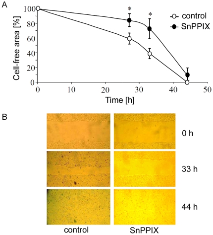
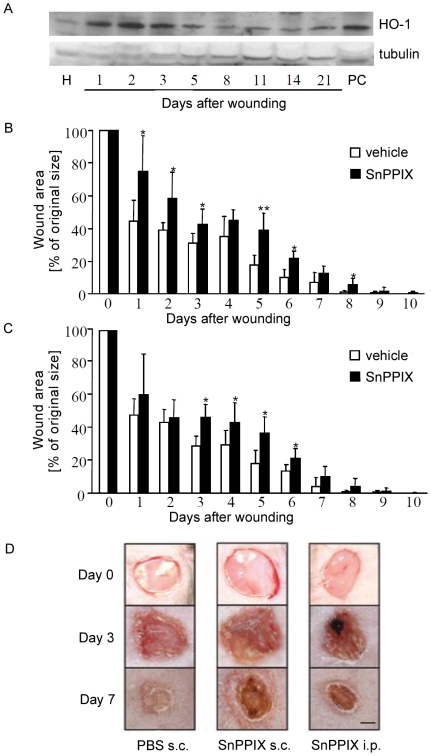
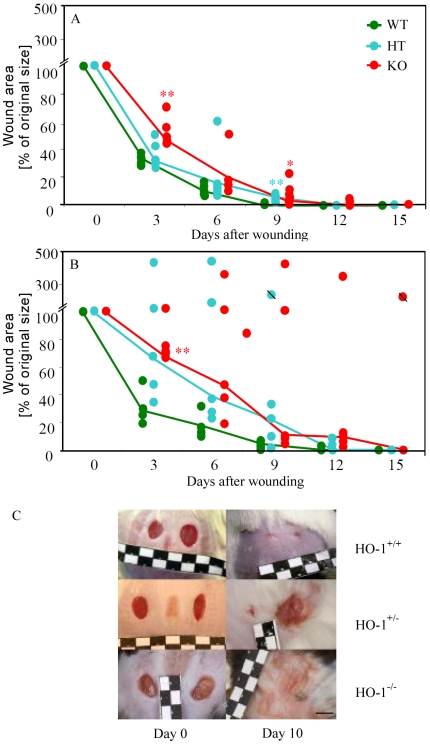
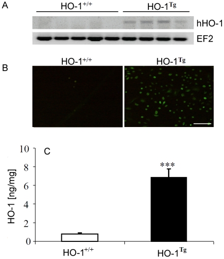
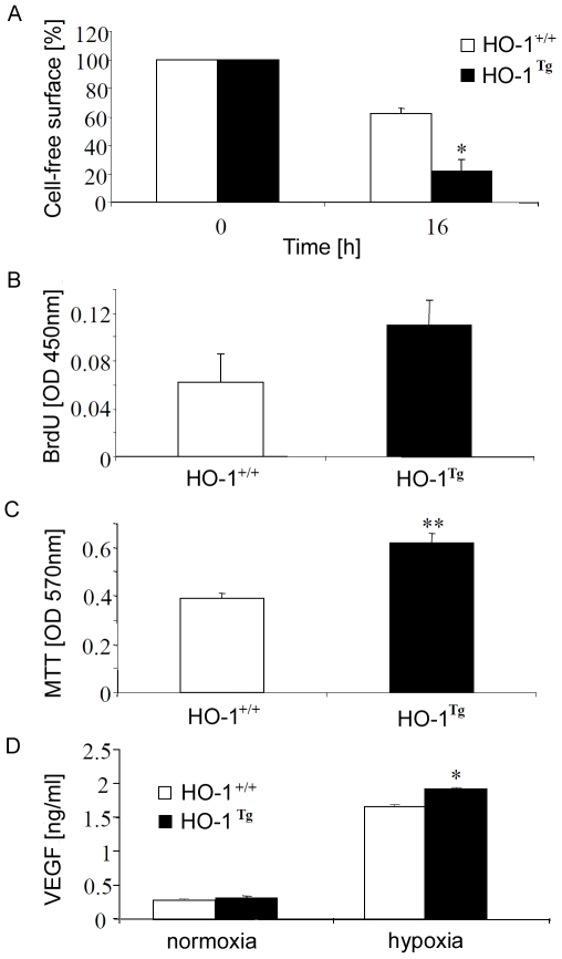
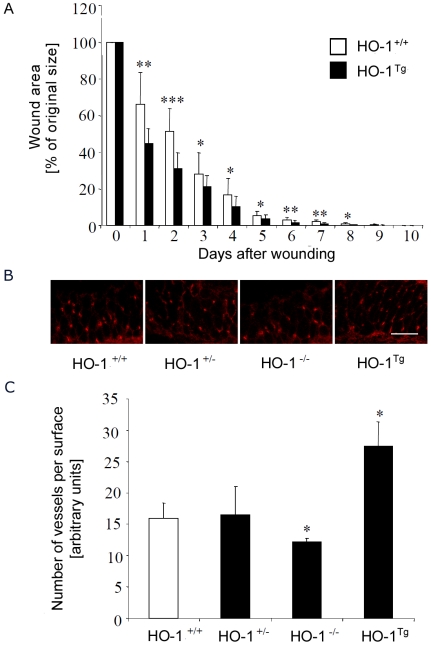
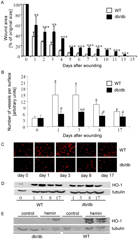
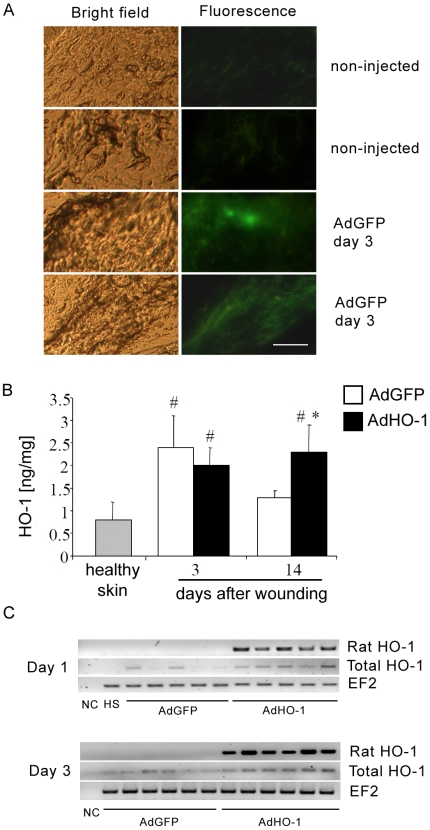
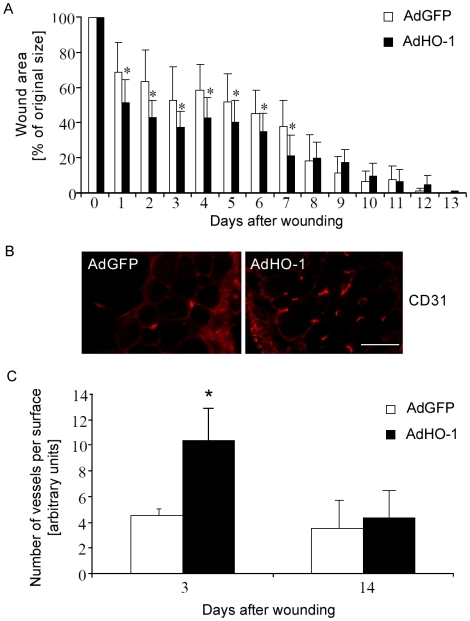
Heme oxygenase-1 (HO-1), a cytoprotective, pro-angiogenic and anti-inflammatory enzyme, is strongly induced in injured tissues. Our aim was to clarify its role in cutaneous wound healing. In wild type mice, maximal expression of HO-1 in the skin was observed on the 2(nd) and 3(rd) days after wounding. Inhibition of HO-1 by tin protoporphyrin-IX resulted in retardation of wound closure. Healing was also delayed in HO-1 deficient mice, where lack of HO-1 could lead to complete suppression of reepithelialization and to formation of extensive skin lesions, accompanied by impaired neovascularization. Experiments performed in transgenic mice bearing HO-1 under control of keratin 14 promoter showed that increased level of HO-1 in keratinocytes is enough to improve the neovascularization and hasten the closure of wounds. Importantly, induction of HO-1 in wounded skin was relatively weak and delayed in diabetic (db/db) mice, in which also angiogenesis and wound closure were impaired. In such animals local delivery of HO-1 transgene using adenoviral vectors accelerated the wound healing and increased the vascularization. In summary, induction of HO-1 is necessary for efficient wound closure and neovascularization. Impaired wound healing in diabetic mice may be associated with delayed HO-1 upregulation and can be improved by HO-1 gene transfer.
Overview
The claims and statistics of this paper haven’t been checked yet.
Run the check — free, no account needed. It reads the open-access text and checks the sources it cites.
Integrity signals
Registry
Cites
Statistics
Figures
Verification
Claims the paper makes
The claims and statistics of this paper haven’t been checked yet.
Run the check — free, no account needed. It reads the open-access text and checks the sources it cites.
Claims other papers make about this one
Each is checked against this paper’s own text: does it say what the citing paper says it does?
In the citing paper
“S. epidermidis association promoted the expression of genes associated specifically with angiogenesis, including angiogenin (Ang) and Hgf (47, 48), as well as genes that more broadly promote tissue repair, like Ceacam1, Grn, Hmox1, Igf1, and Pdgfa (49–53) (Fig. 3R).”
From MAIT cells are imprinted by the microbiota in early life and promote tissue repair (2019) · cites this paper as [53]
Self-check
Percentages, sums and reported differences re-computed from the paper’s own numbers.
The self-check needs the paper’s full text.
Citations
Cites
Gurtner, G. C., Werner, S., Barrandon, Y., & Longaker, M. T. (2008) · Nature · doi:10.1038/nature07039 · Cited by 6,542
Wound healing and its impairment in the diabetic foot
Falanga, V. (2005) · The Lancet · doi:10.1016/s0140-6736(05)67700-8 · Cited by 2,715
THE HEME OXYGENASE SYSTEM:A Regulator of Second Messenger Gases
Maines, M. D. (1997) · The Annual Review of Pharmacology and Toxicology · doi:10.1146/annurev.pharmtox.37.1.517 · Cited by 2,489
Heme oxygenase: Function, multiplicity, regulatory mechanisms, and clinical applications
Maines, M. D. (1988) · The FASEB Journal · doi:10.1096/fasebj.2.10.3290025 · Cited by 1,707
Reduced stress defense in heme oxygenase 1-deficient cells
Poss, K. D., & Tonegawa, S. (1997) · Proceedings of the National Academy of Sciences · doi:10.1073/pnas.94.20.10925 · Cited by 1,294
Heme oxygenase 1 is required for mammalian iron reutilization
Poss, K. D., & Tonegawa, S. (1997) · Proceedings of the National Academy of Sciences · doi:10.1073/pnas.94.20.10919 · Cited by 1,016
Physiology and healing dynamics of chronic cutaneous wounds
Stadelmann, W. K., Digenis, A. G., & Tobin, G. R. (1998) · The American Journal of Surgery · doi:10.1016/s0002-9610(98)00183-4 · Cited by 910
Galiano, R. D., Tepper, O. M., Pelo, C. R., Bhatt, K. A., Callaghan, M. J., Bastidas, N., Bunting, S., Steinmetz, H., & Gurtner, G. C. (2004) · American Journal Of Pathology · doi:10.1016/s0002-9440(10)63754-6 · Cited by 812
Heme Oxygenase-1 in Tumors: Is It a False Friend?
Józkowicz, A., Waś, H., & Dulak, J. (2007) · Antioxidants and Redox Signaling · doi:10.1089/ars.2007.1659 · Cited by 381
Self-citationBruce, C. R., Carey, A. L., Hawley, J. A., & Febbraio, M. A. (2003) · Diabetes · doi:10.2337/diabetes.52.9.2338 · Cited by 318
Cited by
Sorted by citations
Krzyszczyk, P., Schloss, R. S., Palmer, A. F., & Francois Berthiaume, B. (2018) · Frontiers in Physiology · doi:10.3389/fphys.2018.00419 · Cited by 1,606
Guan, Y., Niu, H. M., Liu, Z., Dang, Y., Shen, J., Zayed, M. A., Ma, L., & Guan, J. (2021) · Science Advances · doi:10.1126/sciadv.abj0153 · Cited by 654
MAIT cells are imprinted by the microbiota in early life and promote tissue repair
Constantinides, M. G., Link, V. M., Tamoutounour, S., Wong, A. C., Perez-Chaparro, P. J., Han, S.-J., Chen, Y. E., Li, K., Farhat, S., Weckel, A., et al. (2019) · Science · doi:10.1126/science.aax6624 · Cited by 567
Free radical biology and medicine
(2003) · Free Radical Biology and Medicine · doi:10.1016/s0891-5849(03)00484-2 · Cited by 551
Targeting heme oxygenase-1 and carbon monoxide for therapeutic modulation of inflammation
Ryter, S. W., & Choi, A. M. K. (2015) · Translational research · doi:10.1016/j.trsl.2015.06.011 · Cited by 363
Heme Oxygenase-1/Carbon Monoxide
Ryter, S. W., & Choi, A. M. K. (2009) · American Journal of Respiratory Cell and Molecular Biology · doi:10.1165/rcmb.2009-0170tr · Cited by 309
Heme Oxygenases in Cardiovascular Health and Disease
Ayer, A., Zarjou, A., Agarwal, A., & Stocker, R. (2016) · Physiological Reviews · doi:10.1152/physrev.00003.2016 · Cited by 248
HO-1 Induction in Cancer Progression: A Matter of Cell Adaptation
Nitti, M., Piras, S., Marinari, U. M., Moretta, L., Pronzato, M. A., & Furfaro, A. L. (2017) · Antioxidants · doi:10.3390/antiox6020029 · Cited by 222
Kim, Y., Pae, H., Park, J. E., Lee, Y. C., Woo, J. M., Kim, N., Choi, Y. K., Lee, B.-S., Kim, S. R., & Chung, H. (2010) · Antioxidants and Redox Signaling · doi:10.1089/ars.2010.3153 · Cited by 224
Heme Oxygenase-1 and Carbon Monoxide in the Heart
Otterbein, L. E., Foresti, R., & Motterlini, R. (2016) · Circulation Research · doi:10.1161/circresaha.116.306588 · Cited by 217
Curcumin-induced angiogenesis hastens wound healing in diabetic rats
Kant, V., Gopal, A., Kumar, D., Pathak, N. N., Ram, M., Jangir, B. L., Tandan, S. K., & Kumar, D. (2014) · Journal of Surgical Research · doi:10.1016/j.jss.2014.10.019 · Cited by 217
Cellular and molecular mechanisms of inflammation‐induced angiogenesis
Szade, A., Grochot‐Przeczek, A., Florczyk, U., Józkowicz, A., & Dulak, J. (2015) · IUBMB Life · doi:10.1002/iub.1358 · Cited by 203
Self-citationNew Insights into Intracellular Locations and Functions of Heme Oxygenase-1
Dunn, L., Midwinter, R. G., Ni, J., Hamid, H. A., Parish, C. R., & Stocker, R. (2013) · Antioxidants and Redox Signaling · doi:10.1089/ars.2013.5675 · Cited by 181
Vitamin C promotes wound healing through novel pleiotropic mechanisms
Mohammed, B. M., Fisher, B. J., Kraskauskas, D., Ward, S. I., Wayne, J. S., Brophy, D. F., Fowler, A. A., Yager, D. R., & Natarajan, R. (2015) · International Wound Journal · doi:10.1111/iwj.12484 · Cited by 177
Wound Administration of M2-Polarized Macrophages Does Not Improve Murine Cutaneous Healing Responses
Jetten, N., Roumans, N., Gijbels, M. J., Romano, A., Post, M. J., de Winther, M. P. J., van der Hulst, R. R. W. J., & Xanthoulea, S. (2014) · PLoS ONE · doi:10.1371/journal.pone.0102994 · Cited by 162
Skrzypek, K., Tertil, M., Gołda, S., Cieśla, M., Węglarczyk, K., Collet, G., Guichard, A., Kozakowska, M., Boczkowski, J., Waś, H., et al. (2013) · Antioxidants and Redox Signaling · doi:10.1089/ars.2013.5184 · Cited by 145
Self-citationHaem oxygenase-1: non-canonical roles in physiology and pathology
Grochot‐Przeczek, A., Dulak, J., & Józkowicz, A. (2011) · Clinical Science · doi:10.1042/cs20110147 · Cited by 145
Self-citationMaamoun, H., Benameur, T., Pintus, G., Munusamy, S., & Agouni, A. (2019) · Frontiers in Physiology · doi:10.3389/fphys.2019.00070 · Cited by 143
Lima, C. F., Pereira‐Wilson, C., & Rattan, S. I. S. (2010) · Molecular Nutrition & Food Research · doi:10.1002/mnfr.201000221 · Cited by 136
Macrophages are exploited from an innate wound healing response to facilitate cancer metastasis
Muliaditan, T., Caron, J., Okesola, M., Opzoomer, J. W., Kosti, P., Georgouli, M., Gordon, P., Lall, S. K., Kuzeva, D. M., Pedro, L., et al. (2018) · Nature Communications · doi:10.1038/s41467-018-05346-7 · Cited by 128
Lowe, D. T. (2017) · Complementary Therapies in Clinical Practice · doi:10.1016/j.ctcp.2017.09.008 · Cited by 122
Heme Oxygenase-1 Inhibits Myoblast Differentiation by Targeting Myomirs
Kozakowska, M., Cieśla, M., Stefanska, A., Skrzypek, K., Waś, H., Jaźwa, A., Grochot‐Przeczek, A., Kotlinowski, J., Szymula, A., Bartelik, A., et al. (2011) · Antioxidants and Redox Signaling · doi:10.1089/ars.2011.3964 · Cited by 119
Self-citationBeneficial Effects of Green Tea EGCG on Skin Wound Healing: A Comprehensive Review
Xu, F.-W., Lv, Y.-L., Zhong, Y., Xue, Y., Wang, Y., Zhang, L., Hu, X. M., & Tan, W. (2021) · Molecules · doi:10.3390/molecules26206123 · Cited by 127
Role of Heme Oxygenase-1 in Human Endothelial Cells
Taha, H., Skrzypek, K., Guevara, I., Nigisch, A., Mustafa, S., Grochot‐Przeczek, A., Ferdek, P. E., Waś, H., Kotlinowski, J., Kozakowska, M., et al. (2010) · Arteriosclerosis Thrombosis and Vascular Biology · doi:10.1161/atvbaha.110.207316 · Cited by 107
Self-citationRedox Functions of Heme Oxygenase-1 and Biliverdin Reductase in Diabetes
Rochette, L., Zeller, M., Cottin, Y., & Vergely, C. (2017) · Trends in Endocrinology and Metabolism · doi:10.1016/j.tem.2017.11.005 · Cited by 103
Therapeutic angiogenesis for revascularization in peripheral artery disease
Grochot‐Przeczek, A., Dulak, J., & Józkowicz, A. (2013) · Gene · doi:10.1016/j.gene.2013.03.097 · Cited by 90
Self-citationHeme Oxygenase, Inflammation, and Fibrosis: The Good, the Bad, and the Ugly?
Lundvig, D. M. S., Immenschuh, S., & Wagener, F. A. D. T. G. (2012) · Frontiers in Pharmacology · doi:10.3389/fphar.2012.00081 · Cited by 86
Topical application of substance P promotes wound healing in streptozotocin-induced diabetic rats
Kant, V., Kumar, D., Kumar, D., Prasad, R., Gopal, A., Pathak, N. N., Kumar, P., & Tandan, S. K. (2015) · Cytokine · doi:10.1016/j.cyto.2014.12.015 · Cited by 79
Cao, X., Chen, Y., Zhang, J., You, M., Wang, K., & Hu, F. (2017) · Phytomedicine · doi:10.1016/j.phymed.2017.06.001 · Cited by 79
The Multifunctional Role and Therapeutic Potential of HO-1 in the Vascular Endothelium
Calay, D., & Mason, J. C. (2013) · Antioxidants and Redox Signaling · doi:10.1089/ars.2013.5659 · Cited by 73
Proximal tubule-targeted heme oxygenase-1 in cisplatin-induced acute kidney injury
Bolisetty, S., Traylor, A., Joseph, R., Zarjou, A., & Agarwal, A. (2015) · American Journal of Physiology-Renal Physiology · doi:10.1152/ajprenal.00335.2015 · Cited by 70
Hormesis: Wound healing and keratinocytes
Calabrese, E. J., Dhawan, G., Kapoor, R., Agathokleous, E., & Calabrese, V. (2022) · Pharmacological Research · doi:10.1016/j.phrs.2022.106393 · Cited by 66
Hasegawa‐Moriyama, M., Kurimoto, T., Nakama, M., Godai, K., Kojima, M., Kuwaki, T., & Kanmura, Y. (2013) · Pain · doi:10.1016/j.pain.2013.04.039 · Cited by 63
Exendin-4, a glucagon-like peptide-1 analogue, accelerates diabetic wound healing
Roan, J., Cheng, H.-N., Young, C.-C., Lee, C.-J., Yeh, M., Luo, C.-Y., Tsai, Y.-S., & Lam, C.-F. (2016) · Journal of Surgical Research · doi:10.1016/j.jss.2016.09.024 · Cited by 60
Targeting angiogenesis in Duchenne muscular dystrophy
Podkalicka, P., Mucha, O., Dulak, J., & Łoboda, A. (2019) · Cellular and Molecular Life Sciences · doi:10.1007/s00018-019-03006-7 · Cited by 59
Self-citationHaem oxygenase-1 and cardiovascular disease: mechanisms and therapeutic potential
Chan, K. H., Ng, M., & Stocker, R. (2011) · Clinical Science · doi:10.1042/cs20100508 · Cited by 54
Grochot‐Przeczek, A., Kotlinowski, J., Kozakowska, M., Starowicz, K., Jagodzinska, J., Stachurska, A., Volger, O. L., Bukowska-Straková, K., Florczyk, U., Tertil, M., et al. (2013) · Antioxidants and Redox Signaling · doi:10.1089/ars.2013.5426 · Cited by 53
Self-citationNegi, G., Nakkina, V., Kamble, P., & Sharma, S. S. (2015) · Pharmacological Research · doi:10.1016/j.phrs.2015.09.014 · Cited by 52
Hemostatic and Wound Healing Properties ofChromolaena odorataLeaf Extract
Pandith, H., Zhang, X., Liggett, J. L., Min, K., Gritsanapan, W., & Baek, S. J. (2013) · ISRN Dermatology · doi:10.1155/2013/168269 · Cited by 50
Heme Oxygenase-1 Influences Satellite Cells and Progression of Duchenne Muscular Dystrophy in Mice
Pietraszek‐Gremplewicz, K., Kozakowska, M., Bronisz-Budzyńska, I., Cieśla, M., Mucha, O., Podkalicka, P., Madej, M., Głowniak-Kwitek, U., Szade, K., Stępniewski, J., et al. (2018) · Antioxidants and Redox Signaling · doi:10.1089/ars.2017.7435 · Cited by 50
Self-citationJaźwa, A., Stępniewski, J., Zamykal, M., Jagodzinska, J., Meloni, M., Emanueli, C., Józkowicz, A., & Dulak, J. (2012) · Cardiovascular Research · doi:10.1093/cvr/cvs284 · Cited by 49
Self-citationKnockdown of heme oxygenase-2 impairs corneal epithelial cell wound healing
Halilovic, A., Patil, K., Bellner, L., Marrazzo, G., Castellano, K., Cullaro, G., Dunn, M. W., & Schwartzman, M. L. (2010) · Journal of Cellular Physiology · doi:10.1002/jcp.22502 · Cited by 47
Waś, H., Sokołowska, M., Bartelik, A., Dominik, P., Skrzypek, K., Lackowska, B., Pratnicki, A., Grochot‐Przeczek, A., Taha, H., Kotlinowski, J., et al. (2011) · Free Radical Biology and Medicine · doi:10.1016/j.freeradbiomed.2011.07.025 · Cited by 47
Self-citationHormesis: wound healing and fibroblasts
Calabrese, E. J., Dhawan, G., Kapoor, R., Agathokleous, E., & Calabrese, V. (2022) · Pharmacological Research · doi:10.1016/j.phrs.2022.106449 · Cited by 46
Nazari, M., Taremi, S., Elahi, R., Mostanadi, P., & Esmaeilzadeh, A. (2024) · Stem Cell Reviews and Reports · doi:10.1007/s12015-024-10806-3 · Cited by 49
Wound healing activity of carbon monoxide liberated from CO-releasing molecule (CO-RM)
Ahanger, A. A., Prawez, S., Kumar, D., Prasad, R., Amarpal, Tandan, S. K., & Kumar, D. (2011) · Naunyn-Schmiedeberg s Archives of Pharmacology · doi:10.1007/s00210-011-0653-7 · Cited by 46
Park, N.-Y., & Lim, Y. (2011) · Nutrition & Metabolism · doi:10.1186/1743-7075-8-80 · Cited by 46
Costantini, E., Sinjari, B., D’Angelo, C., Murmura, G., Reale, M., & Caputi, S. (2019) · International Journal of Molecular Sciences · doi:10.3390/ijms20092108 · Cited by 42
Tomczyk, M., Kraszewska, I., Szade, K., Bukowska-Straková, K., Meloni, M., Józkowicz, A., Dulak, J., & Jaźwa, A. (2017) · Basic Research in Cardiology · doi:10.1007/s00395-017-0629-y · Cited by 41
Self-citationZrelli, H., Kusunoki, M., & Miyazaki, H. (2015) · Phytotherapy Research · doi:10.1002/ptr.5339 · Cited by 41
Godai, K., Hasegawa‐Moriyama, M., Kurimoto, T., Saito, T., Yamada, T., Sato, T., Kojima, M., & Kanmura, Y. (2014) · Molecular Pain · doi:10.1186/1744-8069-10-36 · Cited by 40
Antioxidant Activities of Natural Compounds from Caribbean Plants to Enhance Diabetic Wound Healing
Accipe, L., Abadie, A., Nevière, R., & Bercion, S. (2023) · Antioxidants · doi:10.3390/antiox12051079 · Cited by 40
Horiba, M., Kamiya, T., Hara, H., & Adachi, T. (2017) · Scientific Reports · doi:10.1038/srep42208 · Cited by 38
Redox regulation of gasotransmission in the vascular system: A focus on angiogenesis
Mistry, R. K., & Brewer, A. C. (2017) · Free Radical Biology and Medicine · doi:10.1016/j.freeradbiomed.2017.04.025 · Cited by 37
Heme oxygenase-1 in neovascularisation: A diabetic perspective
Grochot‐Przeczek, A., Dulak, J., & Józkowicz, A. (2010) · Thrombosis and Haemostasis · doi:10.1160/th09-12-0825 · Cited by 37
Self-citationAlmeida, A. S., Soares, N. L., Vieira, M. R., Gramsbergen, J. B., & Vieira, H. L. A. (2016) · PLoS ONE · doi:10.1371/journal.pone.0154781 · Cited by 35
Ali, Z. E., Ollivier, A., Manin, S., Rivard, M., Motterlini, R., & Foresti, R. (2020) · Redox Biology · doi:10.1016/j.redox.2020.101521 · Cited by 36
Critical Role of 5-Lipoxygenase and Heme Oxygenase-1 in Wound Healing
Brogliato, A. R., Moor, A. N., Kesl, S., Guilherme, R. F., Georgii, J. L., Peters-Golden, M. L., Canetti, C. D. A., Gould, L. J., & Benjamim, C. F. (2013) · Journal of Investigative Dermatology · doi:10.1038/jid.2013.493 · Cited by 35
Heme Oxygenase-1 as Therapeutic Target for Diabetic Foot Ulcers
Leal, E. C., & Carvalho, E. (2022) · International Journal of Molecular Sciences · doi:10.3390/ijms231912043 · Cited by 36
Liu, F.-C., Yu, H., Hwang, T., & Tsai, Y. (2012) · PLoS ONE · doi:10.1371/journal.pone.0053203 · Cited by 33
Argon Mitigates Impaired Wound Healing Process and Enhances Wound Healing In Vitro and In Vivo
Ning, J., Zhao, H., Chen, B., Mi, E., Yang, Z. L., Qing, W., Lam, K. W. J., Yi, B., Chen, Q., Gu, J., et al. (2019) · Theranostics · doi:10.7150/thno.29361 · Cited by 33
Heme oxygenase-1 in protozoan infections: A tale of resistance and disease tolerance
Silva, R. C. M. C., Travassos, L. H., Paiva, C. N., & Bozza, M. T. (2020) · PLoS Pathogens · doi:10.1371/journal.ppat.1008599 · Cited by 29
Hemin attenuated oxidative stress and inflammation to improve wound healing in diabetic rats
Kumar, D., Jena, G. R., Ram, M., Lingaraju, M. C., Singh, V., Prasad, R., Kumawat, S., Kant, V., Gupta, P., Kumar, D., et al. (2019) · Naunyn-Schmiedeberg s Archives of Pharmacology · doi:10.1007/s00210-019-01682-7 · Cited by 31
The Heme-Heme Oxygenase System in Wound Healing; Implications for Scar Formation
Wagener, F. A. D. T. G., Scharstuhl, A., Tyrrell, R. M., den Hoff, J. W. V., Józkowicz, A., Dulak, J., Rüssel, F. G. M., & Kuijpers‐Jagtman, A. M. (2010) · Current Drug Targets · doi:10.2174/1389450111009011571 · Cited by 31
Self-citationPro-healing potential of hemin: An inducer of heme oxygenase-1
Ahanger, A. A., Prawez, S., Leo, M. D., Kathirvel, K., Kumar, D., Tandan, S. K., & Malik, J. K. (2010) · European Journal of Pharmacology · doi:10.1016/j.ejphar.2010.06.048 · Cited by 30
Novel Insights into the Vasoprotective Role of Heme Oxygenase‐1
Marcantoni, E., Francesco, L. D., Dovizio, M., Bruno, A., & Patrignani, P. (2012) · International Journal of Hypertension · doi:10.1155/2012/127910 · Cited by 31
Bauer, A., Mylroie, H., Thornton, C. C., Calay, D., Birdsey, G. M., Kiprianos, A. P., Wilson, G. K., Soares, M. P., Yin, X., Mayr, M., et al. (2016) · Scientific Reports · doi:10.1038/srep29417 · Cited by 29
Krzyszczyk, P., Kang, H. J., Kumar, S., Meng, Y., O'Reggio, M. D., Patel, K. P., Pires, I. S., Yarmush, M. L., Schloss, R. S., Palmer, A. F., et al. (2020) · Wound Repair and Regeneration · doi:10.1111/wrr.12814 · Cited by 28
The metabolic landscape in chronic rotator cuff tear reveals tissue‐region‐specific signatures
Olie, C. S., van Zeijl, R. J. M., el Abdellaoui, S., Kolk, A., Overbeek, C. L., Nelissen, R. G. H. H., Heijs, B., & Raz, V. (2021) · Journal of Cachexia Sarcopenia and Muscle · doi:10.1002/jcsm.12873 · Cited by 27
Choi, D. S., Kim, S., Lim, Y., Gwon, H., Park, J. S., Nho, Y., & Kwon, J. (2012) · Tissue Engineering and Regenerative Medicine · doi:10.1007/s13770-012-0036-2 · Cited by 27
Cremers, N. A. J., SUTTORP, M. J., Gerritsen, M. M., Wong, R. J., Breda, C. V. R., van Dam, G. M., Brouwer, K. M., Kuijpers‐Jagtman, A. M., Carels, C. E. L., Lundvig, D. M. S., et al. (2015) · Frontiers in Medicine · doi:10.3389/fmed.2015.00086 · Cited by 27
Nowak, W. N., Borys, S., Kusińska, K., Bukowska-Straková, K., Witek, P., Koblik, T., Józkowicz, A., Malecki, M. T., & Dulak, J. (2013) · Journal of Diabetes Investigation · doi:10.1111/jdi.12131 · Cited by 24
Self-citationLoPresti, S. T., Popovic, B., Kulkarni, M., Skillen, C. D., & Brown, B. N. (2019) · Biomaterials · doi:10.1016/j.biomaterials.2019.119376 · Cited by 24
Tendera, M., Cybulski, W., Zuba‐Surma, E. K., Szade, K., Florczyk, U., Kozakowska, M., Szymula, A., Krzych, Ł. J., Pasławska, U., Pasławski, R., et al. (2012) · Thrombosis and Haemostasis · doi:10.1160/th12-05-0303 · Cited by 23
Self-citationDeSouza-Vieira, T. S., Iniguez, E., Serafim, T. D., de Castro, W., Karmakar, S., Disotuar, M. M., Cecílio, P., Lacsina, J. R., Meneses, C., Nagata, B. M., et al. (2020) · Cell Reports · doi:10.1016/j.celrep.2020.108317 · Cited by 22
Kozakowska, M., Kotlinowski, J., Grochot‐Przeczek, A., Cieśla, M., Pilecki, B., Derlacz, R., Dulak, J., & Józkowicz, A. (2015) · Stem Cell Research & Therapy · doi:10.1186/s13287-015-0063-8 · Cited by 22
Self-citationHolowiecki, A., O'Shields, B., & Jenny, M. J. (2016) · Toxicology and Applied Pharmacology · doi:10.1016/j.taap.2016.09.022 · Cited by 22
Mangiferin alleviates hyperglycemia-induced endothelial impairment via Nrf2 signaling pathway
Jayasuriya, R., & Ramkumar, K. M. (2022) · European Journal of Pharmacology · doi:10.1016/j.ejphar.2022.175359 · Cited by 19
Peripheral Nerve Block Facilitates Acute Inflammatory Responses Induced by Surgical Incision in Mice
Yamada, T., Hasegawa‐Moriyama, M., Kurimoto, T., Saito, T., Kuwaki, T., & Kanmura, Y. (2016) · Regional Anesthesia & Pain Medicine · doi:10.1097/aap.0000000000000458 · Cited by 19
Cytoprotective responses in HaCaT keratinocytes exposed to high doses of curcumin
Lundvig, D. M. S., Pennings, S. W. C., Brouwer, K. M., Mtaya, M., Mugonzibwa, E. A., Kuijpers‐Jagtman, A. M., Wagener, F. A. D. T. G., & den Hoff, J. W. V. (2015) · Experimental Cell Research · doi:10.1016/j.yexcr.2015.06.002 · Cited by 19
Heme catabolism and heme oxygenase-1-expressing myeloid cells in pathophysiology
Consonni, F. M., Incerti, M., Bertolotti, M., Ballerini, G., Garlatti, V., & Sica, A. (2024) · Frontiers in Immunology · doi:10.3389/fimmu.2024.1433113 · Cited by 22
Carbon Monoxide in Plasma and Agriculture: Just a Foe or a Potential Friend?
Carbone, E., & Douat, C. (2018) · Plasma Medicine · doi:10.1615/plasmamed.2018024519 · Cited by 17
Honey Therapy in Diabetic Foot Ulcers: A Promising Strategy for Effective Wound Healing
Bezerra, A. B. G., Fonseca, H., Rodrigues, F., Delerue-Matos, C. M., Gouvinhas, I., & Garcia, J. (2023) · Applied Sciences · doi:10.3390/app132312820 · Cited by 22
Khorsandi, K., Fekrazad, R., & Hamblin, M. R. (2021) · Journal of Biophotonics · doi:10.1002/jbio.202100005 · Cited by 17
Kotlinowski, J., Grochot‐Przeczek, A., Taha, H., Kozakowska, M., Pilecki, B., Skrzypek, K., Bartelik, A., Derlacz, R., Horrevoets, A. J. G., Pap, A., et al. (2014) · Cardiovascular Diabetology · doi:10.1186/s12933-014-0150-7 · Cited by 16
Self-citationLiu, F.-C., Hwang, T., Lau, Y., & Yu, H. (2011) · PLoS ONE · doi:10.1371/journal.pone.0025907 · Cited by 15
The Role of Heme Oxygenase-1 in Remote Ischemic and Anesthetic Organ Conditioning
Bauer, I., & Raupach, A. (2019) · Antioxidants · doi:10.3390/antiox8090403 · Cited by 15
Zheng, R., Heck, D. E., Black, A. T., Gow, A. J., Laskin, D. L., & Laskin, J. D. (2013) · Free Radical Biology and Medicine · doi:10.1016/j.freeradbiomed.2013.10.011 · Cited by 14
Terenzi, D. C., Bakbak, E., Teoh, H., Krishnaraj, A., Puar, P., Rotstein, O. D., Cosentino, F., Goldenberg, R., Verma, S., & Hess, D. C. (2023) · Cardiovascular Research · doi:10.1093/cvr/cvae016 · Cited by 14
Kaminski, T. W., Sivanantham, A., Mozhenkova, A., Smith, A., Ungarala, R., Dubey, R. K., Shrestha, B., Hanway, C., Katoch, O., Tejero, J., et al. (2024) · American Journal of Physiology-Cell Physiology · doi:10.1152/ajpcell.00386.2023 · Cited by 13
Delayed cutaneous wound closure in HO ‐2 deficient mice despite normal HO ‐1 expression
Lundvig, D. M. S., Scharstuhl, A., Cremers, N. A. J., Pennings, S. W. C., Paske, J. T., van Rheden, R. E. M., Breda, C. V. R., Regan, R. F., Rüssel, F. G. M., Carels, C. E. L., et al. (2014) · Journal of Cellular and Molecular Medicine · doi:10.1111/jcmm.12389 · Cited by 13
Emad, A. M., Mahrous, E. A., Rasheed, D. M., Gomaa, F. A. M., Hamdan, A. M., Selim, H. M. R. M., Yousef, E. M., Abo-Zalam, H. B., El-Gazar, A. A., & Ragab, G. M. (2024) · Pharmaceuticals · doi:10.3390/ph17060733 · Cited by 14
Jennewein, M., Bubel, M., Guthörl, S., Metzger, W., WEIGERT, M. G., Pohlemann, T., & Oberringer, M. (2016) · Cell and Tissue Research · doi:10.1007/s00441-016-2391-0 · Cited by 12
Li, Q., Liang, S., Lai, Q., Shen, L., Zhang, Y., & Guo, R. (2021) · Experimental and Therapeutic Medicine · doi:10.3892/etm.2021.10646 · Cited by 10
Unknown biological effects of l-glucose, ALA, and PUFA
Yamada, K., Sato, D., Nakamura, T., Amano, H., & Morimoto, Y. (2017) · The Journal of Physiological Sciences · doi:10.1007/s12576-017-0544-x · Cited by 9
Bednarz, A., Lipiński, P., Starzyński, R. R., Tomczyk, M., Kraszewska, I., Herman, S., Kowalski, K., Gruca, E., Jończy, A., Mazgaj, R., et al. (2020) · International Journal of Molecular Sciences · doi:10.3390/ijms21207754 · Cited by 7
Heme oxygenase-2 suppresses acute inflammation and improves the survival of skin allografts
Chen, R., Wang, Z., Chen, Q., Zhang, T.-Y., Zhu, Y., Xian, X., & Han, X. (2018) · International Immunopharmacology · doi:10.1016/j.intimp.2018.08.009 · Cited by 8
Effects of Remote Ischemic Preconditioning on Heme Oxygenase-1 Expression and Cutaneous Wound Repair
Cremers, N. A. J., Wever, K. E., Wong, R. J., van Rheden, R. E. M., Vermeij, E. A., Dam, G. M. V., Carels, C. E. L., Lundvig, D. M. S., & Wagener, F. A. D. T. G. (2017) · International Journal of Molecular Sciences · doi:10.3390/ijms18020438 · Cited by 7
HIF‐2α/ARNT complex regulates hair development via induction of p21 Waf1/Cip1 and p27 Kip1
Imamura, Y., Tomita, S., Imanishi, M., Kihira, Y., Ikeda, Y., Ishizawa, K., Tsuchiya, K., & Tamaki, T. (2014) · The FASEB Journal · doi:10.1096/fj.13-244079 · Cited by 6
Paradoxical effects of heme arginate on survival of myocutaneous flaps
Edmunds, M.-C., Czopek, A., Wigmore, S. J., & Kluth, D. (2013) · American Journal of Physiology-Regulatory, Integrative and Comparative Physiology · doi:10.1152/ajpregu.00240.2013 · Cited by 6
Metadata
- Authors
- Grochot-Przeczek, A., Lach, R., Mis, J., Skrzypek, K., Gozdecka, M., Sroczynska, P., Dubiel, M., Rutkowski, A., Kozakowska, M., Zagorska, A., Walczynski, J., Was, H., Kotlinowski, J., Drukala, J., Kurowski, K., Kieda, C., Herault, Y., Dulak, J., & Jozkowicz, A.
- Affiliations
- Jagiellonian University (PL)
- Adamed (Poland) (PL)
- Venue
- PLoS ONE
- Publisher
- Public Library of Science (PLoS)
- Published
- 2009-06-04
- Volume
- 4
- Issue
- 6
- Pages
- e5803
- ISSN
- 1932-6203 (ISSN-L)
- PMC
- PMC2686151
- OpenAlex
- W2135022152
- Cited by
- 131
- References
- 79
- Citations by year
- 2026: 2 · 2025: 1 · 2024: 5 · 2023: 5 · 2022: 7 · 2021: 9 · 2020: 5 · 2019: 9 · 2018: 9 · 2017: 12 · 2016: 8 · 2015: 10 · 2014: 8 · 2013: 14 · 2012: 7
- FWCI
- 3.49
- Type
- journal article
- Language
- en
- Open access
- gold
- doi.org
- Locations
- PLoS ONE (publishedVersion, cc-by)
- PubMed Central (publishedVersion, cc-by)
- PubMed (publishedVersion)
- Homo Politicus (Academy of Humanities and Economics in Lodz) (submittedVersion, cc-by)
- HAL (submittedVersion)
- DOAJ (DOAJ: Directory of Open Access Journals) (submittedVersion, cc-by-sa)
- Funders
- Wellcome Trust
- European Commission
- Fundacja na rzecz Nauki Polskiej
- Institut National Du Cancer
- Topics
- Life Sciences › Biochemistry, Genetics and Molecular Biology › Molecular Biology › Heme Oxygenase-1 and Carbon Monoxide
- Health Sciences › Medicine › Surgery › Wound Healing and Treatments
- Health Sciences › Medicine › Hepatology › Liver physiology and pathology
- Keywords
- heme oxygenase-1, skin wound healing, neovascularization, angiogenesis, re-epithelialization, tin protoporphyrin IX, keratinocytes, diabetic wound healing, db/db mice, adenoviral gene transfer, transgenic mice
- MeSH terms
- Adenoviridae, Animals, Anti-Inflammatory Agents, Diabetes Mellitus, Experimental, Humans, Inflammation, Keratins, Mice, Inbred C57BL, Promoter Regions, Genetic, Wound Healing, Gene Transfer Techniques, Transgenes, Angiogenesis Inhibitors, Mice, Heme Oxygenase-1
About these checks
Registry, retraction, Cites and statistics results are computed, not judged, with one exception: a printed reference that no index lists is read once by a model before it counts as a citation error. Claims are extracted by a model and kept only when their quote appears verbatim in the text; each cited source is then checked, with a model’s help, for whether it says what the claim relies on. Those verdicts can be wrong — read the claim’s quote and the cited source before relying on one.
Statistics and arithmetic are recomputed, not judged.
Confidence bands are shown only for fields calibrated on our annotated test set. None is calibrated yet, so no band is shown.
What counts as a claim ›How we measure confidence ›How we check this ›
Are you an author, or do you see a problem with this page? Request a review
