

The DOI matches its Crossref record; 90 of the 90 references it cites were found in a registry or an index.
BDNF Activates mTOR to Regulate GluR1 Expression Required for Memory Formation
Open accessAbstract
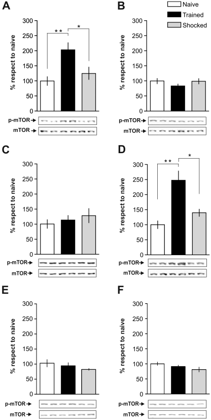
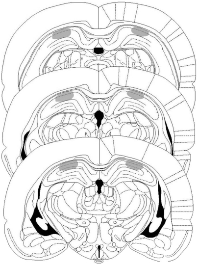
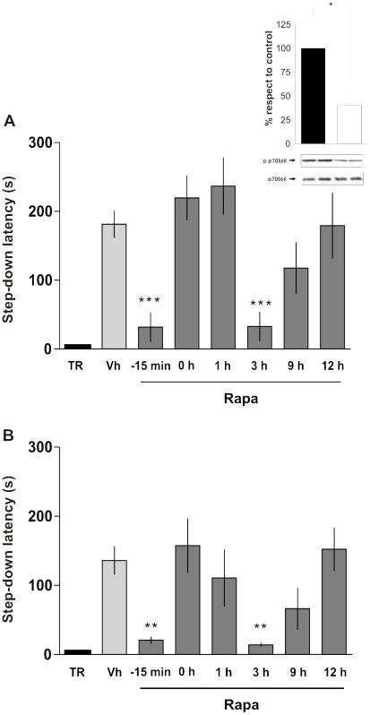
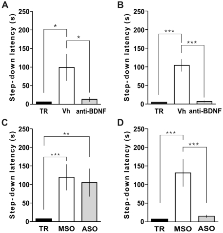
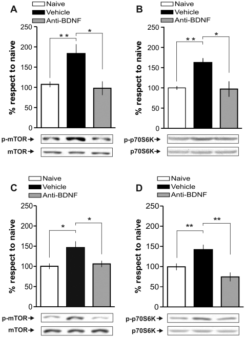
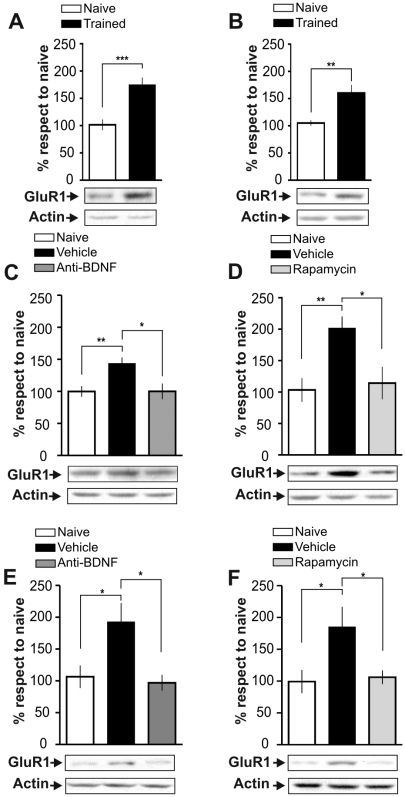
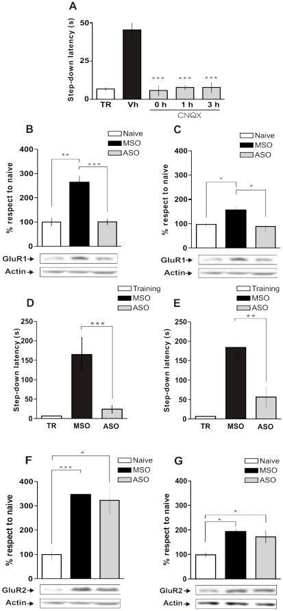
BACKGROUND: The mammalian target of Rapamycin (mTOR) kinase plays a key role in translational control of a subset of mRNAs through regulation of its initiation step. In neurons, mTOR is present at the synaptic region, where it modulates the activity-dependent expression of locally-translated proteins independently of mRNA synthesis. Indeed, mTOR is necessary for different forms of synaptic plasticity and long-term memory (LTM) formation. However, little is known about the time course of mTOR activation and the extracellular signals governing this process or the identity of the proteins whose translation is regulated by this kinase, during mnemonic processing. METHODOLOGY/PRINCIPAL FINDINGS: Here we show that consolidation of inhibitory avoidance (IA) LTM entails mTOR activation in the dorsal hippocampus at the moment of and 3 h after training and is associated with a rapid and rapamycin-sensitive increase in AMPA receptor GluR1 subunit expression, which was also blocked by intra-hippocampal delivery of GluR1 antisense oligonucleotides (ASO). In addition, we found that pre- or post-training administration of function-blocking anti-BDNF antibodies into dorsal CA1 hampered IA LTM retention, abolished the learning-induced biphasic activation of mTOR and its readout, p70S6K and blocked GluR1 expression, indicating that BDNF is an upstream factor controlling mTOR signaling during fear-memory consolidation. Interestingly, BDNF ASO hindered LTM retention only when given into dorsal CA1 1 h after but not 2 h before training, suggesting that BDNF controls the biphasic requirement of mTOR during LTM consolidation through different mechanisms: an early one involving BDNF already available at the moment of training, and a late one, happening around 3 h post-training that needs de novo synthesis of this neurotrophin. CONCLUSIONS/SIGNIFICANCE: IN CONCLUSION, OUR FINDINGS DEMONSTRATE THAT: 1) mTOR-mediated mRNA translation is required for memory consolidation during at least two restricted time windows; 2) this kinase acts downstream BDNF in the hippocampus and; 3) it controls the increase of synaptic GluR1 necessary for memory consolidation.
Overview
The claims and statistics of this paper haven’t been checked yet.
Run the check — free, no account needed. It reads the open-access text and checks the sources it cites.
Integrity signals
Registry
Cites
Statistics
Figures
Verification
Claims the paper makes
The claims and statistics of this paper haven’t been checked yet.
Run the check — free, no account needed. It reads the open-access text and checks the sources it cites.
Claims other papers make about this one
Each is checked against this paper’s own text: does it say what the citing paper says it does?
The cited source says what the claim attributes to it.
In the citing paper
“Previous work has shown that BDNF is a potent activator of mTOR signaling (Jourdi et al., 2009; Takei et al., 2004; Slipczuk et al., 2009), and mTOR is a key integrator for many signaling pathways (see Costa-Mattioli and Monteggia, 2013).”
From BDNF - a key transducer of antidepressant effects (2016) · cites this paper as Slipczuk et al., 2009
Self-check
Percentages, sums and reported differences re-computed from the paper’s own numbers.
The self-check needs the paper’s full text.
Citations
Cites
The rat brain in stereotaxic coordinates
Palkovits, M. (1983) · Neuropeptides · doi:10.1016/0143-4179(83)90049-5 · Cited by 28,959
The Rat Brain in Stereotaxic Coordinates
Paxinos, G., & Watson, C. (1983) · Elsevier eBooks · doi:10.1016/c2009-0-63235-9 · Cited by 28,585
Molecular Mechanisms of Depression: Perspectives on New Treatment Strategies
Lang, U. E., & Borgwardt, S. (2013) · Cellular Physiology and Biochemistry · doi:10.1159/000350094 · Cited by 8,526
AChE-stained horizontal sections of the rat brain in stereotaxic coordinates
Paxinos, G., WATSON, C. R. R., & Emson, P. C. (1980) · Journal of Neuroscience Methods · doi:10.1016/0165-0270(80)90021-7 · Cited by 4,514
Memory - A century of consolidation
McGaugh, J. L. (2000) · Science · doi:10.1126/science.287.5451.248 · Cited by 4,250
Upstream and downstream of mTOR
Hay, N., & Sonenberg, N. (2004) · Genes & Development · doi:10.1101/gad.1212704 · Cited by 4,181
da Luz, A. C. D. D. S., Dias, G. P., Bevilaqua, M. C. D. N., Cocks, G. D., Gardino, P. F., Thuret, S., & Nardi, A. E. (2013) · Neuropsychobiology · doi:10.1159/000353269 · Cited by 4,015
Regulation of Translation Initiation in Eukaryotes: Mechanisms and Biological Targets
Sonenberg, N., & Hinnebusch, A. G. (2009) · Cell · doi:10.1016/j.cell.2009.01.042 · Cited by 3,304
Kim, D., Sarbassov, D. D., Ali, S. M., King, J. E., Latek, R. R., Erdjument‐Bromage, H., Tempst, P., & Sabatini, D. M. (2002) · Cell · doi:10.1016/s0092-8674(02)00808-5 · Cited by 3,072
Targets for Cell Cycle Arrest by the Immunosuppressant Rapamycin in Yeast
Heitman, J., Movva, N. R., & Hall, M. N. (1991) · Science · doi:10.1126/science.1715094 · Cited by 2,055
Cited by
Sorted by citations
mTOR signaling: At the crossroads of plasticity, memory and disease
Hoeffer, C. A., & Klann, E. (2010) · Trends in Neurosciences · doi:10.1016/j.tins.2009.11.003 · Cited by 1,128
BDNF - a key transducer of antidepressant effects
Björkholm, C., & Monteggia, L. M. (2016) · Neuropharmacology · doi:10.1016/j.neuropharm.2015.10.034 · Cited by 958
“Previous work has shown that BDNF is a potent activator of mTOR signaling (Jourdi et al., 2009; Takei et al., 2004; Slipczuk et al., 2009), and mTOR is a key integrator for many signaling pathways (see Costa-Mattioli and Monteggia, 2013).”
BackgroundA simple role for BDNF in learning and memory?
Cunha, C. (2010) · Frontiers in Molecular Neuroscience · doi:10.3389/neuro.02.001.2010 · Cited by 830
BDNF mediates adaptive brain and body responses to energetic challenges
Marosi, K., & Mattson, M. P. (2014) · Trends in Endocrinology & Metabolism · doi:10.1016/j.tem.2013.10.006 · Cited by 568
Signaling pathways underlying the rapid antidepressant actions of ketamine
Duman, R. S., Li, N., Liu, R.-J., Duric, V., & Aghajanian, G. (2012) · Neuropharmacology · doi:10.1016/j.neuropharm.2011.08.044 · Cited by 537
Metadata
- Authors
- Slipczuk, L., Bekinschtein, P., Katche, C., Cammarota, M., Izquierdo, I., & Medina, J. H.
- Affiliations
- Universidad de Buenos Aires (AR)
- Pontifícia Universidade Católica do Rio Grande do Sul (BR)
- Corresponding author
- Medina, Jorge H.
- Venue
- PLoS ONE
- Publisher
- Public Library of Science (PLoS)
- Published
- 2009-06-23
- Volume
- 4
- Issue
- 6
- Pages
- e6007
- ISSN
- 1932-6203 (ISSN-L)
- PMC
- PMC2695538
- OpenAlex
- W2035139662
- Cited by
- 246
- References
- 90
- Citations by year
- 2026: 3 · 2025: 3 · 2024: 8 · 2023: 6 · 2022: 13 · 2021: 10 · 2020: 19 · 2019: 1 · 2018: 14 · 2017: 21 · 2016: 19 · 2015: 20 · 2014: 23 · 2013: 19 · 2012: 29
- FWCI
- 10.85
- Type
- journal article
- Language
- en
- Open access
- gold
- doi.org
- Locations
- PLoS ONE (publishedVersion, cc-by)
- PubMed Central (publishedVersion, cc-by)
- PubMed (publishedVersion)
- DOAJ (DOAJ: Directory of Open Access Journals) (submittedVersion, cc-by-sa)
- Greater South Information System
- Topics
- Life Sciences › Neuroscience › Cognitive Neuroscience › Memory and Neural Mechanisms
- Life Sciences › Neuroscience › Cellular and Molecular Neuroscience › Neuroscience and Neuropharmacology Research
- Life Sciences › Biochemistry, Genetics and Molecular Biology › Molecular Biology › PI3K/AKT/mTOR signaling in cancer
- Keywords
- brain-derived neurotrophic factor, mTOR signaling, GluR1, AMPA receptors, long-term memory consolidation, passive avoidance, fear memory, hippocampus, CA1, p70S6K, antisense oligonucleotides
- MeSH terms
- Animals, Hippocampus, Male, Memory, Models, Biological, Neuronal Plasticity, Protein Kinases, RNA, Messenger, Synapses, Time Factors, Rats, Wistar, Receptors, AMPA, Brain-Derived Neurotrophic Factor, Rats, TOR Serine-Threonine Kinases
About these checks
Registry, retraction, Cites and statistics results are computed, not judged, with one exception: a printed reference that no index lists is read once by a model before it counts as a citation error. Claims are extracted by a model and kept only when their quote appears verbatim in the text; each cited source is then checked, with a model’s help, for whether it says what the claim relies on. Those verdicts can be wrong — read the claim’s quote and the cited source before relying on one.
Statistics and arithmetic are recomputed, not judged.
Confidence bands are shown only for fields calibrated on our annotated test set. None is calibrated yet, so no band is shown.
What counts as a claim ›How we measure confidence ›How we check this ›
Are you an author, or do you see a problem with this page? Request a review
