

Human Engineered Heart Tissue as a Versatile Tool in Basic Research and Preclinical Toxicology
Open accessThe DOI matches its Crossref record; 31 of the 31 references it cites were found in a registry or an index.
Abstract
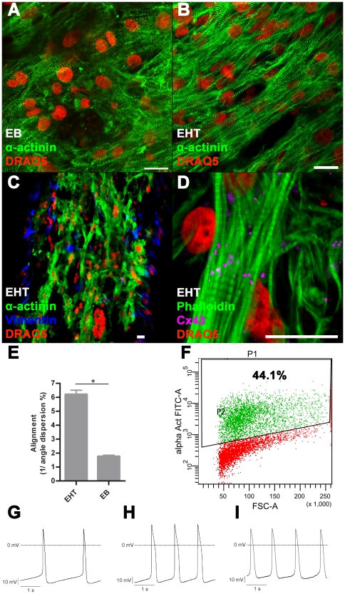
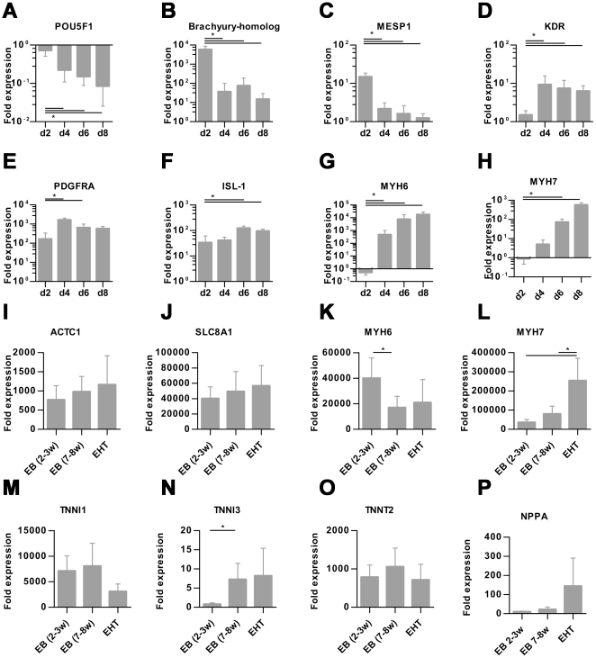
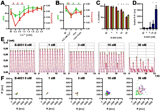
Human embryonic stem cell (hESC) progenies hold great promise as surrogates for human primary cells, particularly if the latter are not available as in the case of cardiomyocytes. However, high content experimental platforms are lacking that allow the function of hESC-derived cardiomyocytes to be studied under relatively physiological and standardized conditions. Here we describe a simple and robust protocol for the generation of fibrin-based human engineered heart tissue (hEHT) in a 24-well format using an unselected population of differentiated human embryonic stem cells containing 30-40% α-actinin-positive cardiac myocytes. Human EHTs started to show coherent contractions 5-10 days after casting, reached regular (mean 0.5 Hz) and strong (mean 100 µN) contractions for up to 8 weeks. They displayed a dense network of longitudinally oriented, interconnected and cross-striated cardiomyocytes. Spontaneous hEHT contractions were analyzed by automated video-optical recording and showed chronotropic responses to calcium and the β-adrenergic agonist isoprenaline. The proarrhythmic compounds E-4031, quinidine, procainamide, cisapride, and sertindole exerted robust, concentration-dependent and reversible decreases in relaxation velocity and irregular beating at concentrations that recapitulate findings in hERG channel assays. In conclusion this study establishes hEHT as a simple in vitro model for heart research.
Overview
The claims and statistics of this paper haven’t been checked yet.
Free: it reads the open-access text and checks the first 5 claims that cite a source we can check. Create a free account to check every claim.
Integrity signals
Registry
Cites
Statistics
Figures

Verification
Claims the paper makes
The claims and statistics of this paper haven’t been checked yet.
Free: it reads the open-access text and checks the first 5 claims that cite a source we can check. Create a free account to check every claim.
Claims other papers make about this one
Each is checked against this paper’s own text: does it say what the citing paper says it does?
The source backs part of the claim, not all of it — or backs it for a different population, condition or comparison.
In the citing paper
“but not pressure- and volume-based metrics directly representing pump performance of the heart”
From Bioengineering an electro-mechanically functional miniature ventricular heart chamber from human pluripotent stem cells (2018) · cites this paper as [16]
Experimental — accuracy not yet measuredThe cited source says what the claim attributes to it.
In the citing paper
“engineered cardiac thin films, micro-tissues and strips allow the assessment of twitch force [9,12–19]”
From Bioengineering an electro-mechanically functional miniature ventricular heart chamber from human pluripotent stem cells (2018) · cites this paper as [16]
Experimental — accuracy not yet measured
Self-check
Percentages, sums and reported differences re-computed from the paper’s own numbers.
The self-check needs the paper’s full text.
Citations
Cites
Proceedings of the National Academy of Sciences of the United States of America
Dowling, J. E., & Wald, G. (2009) · Nutrition Reviews · doi:10.1111/j.1753-4887.1981.tb06752.x · Cited by 17,893
Laflamme, M. A., Chen, K. Y., Naumova, A. V., Muskheli, V., Fugate, J. A., Dupras, S. K., Reinecke, H., Xu, C., Hassanipour, M., Police, S., et al. (2007) · Nature Biotechnology · doi:10.1038/nbt1327 · Cited by 2,206
Feeder-free growth of undifferentiated human embryonic stem cells
Xu, C., Inokuma, M. S., Denham, J., Golds, K., Kundu, P., Gold, J. D., & Carpenter, M. K. (2001) · Nature Biotechnology · doi:10.1038/nbt1001-971 · Cited by 1,870
Kehat, I., Kenyagin‐Karsenti, D., Snir, M., Segev, H., Amit, M., Gepstein, A., Livne, E., Binah, O., Itskovitz‐Eldor, J., & Gepstein, L. (2001) · Journal of Clinical Investigation · doi:10.1172/jci12131 · Cited by 1,814
Cai, C.-L., Liang, X., Shi, Y., Chu, P.-H., Pfaff, S. L., Chen, J., & Evans, S. (2003) · Developmental Cell · doi:10.1016/s1534-5807(03)00363-0 · Cited by 1,610
Kehat, I., Kenyagin‐Karsenti, D., Snir, M., et al. (2001) · Journal of Clinical Investigation · doi:10.1172/jci200112131 · Cited by 1,536
REDFERN, W., CARLSSON, L., DAVIS, A., LYNCH, W., MACKENZIE, I., PALETHORPE, S., SIEGL, P., STRANG, I., SULLIVAN, A., & WALLIS, R. (2003) · Cardiovascular Research · doi:10.1016/s0008-6363(02)00846-5 · Cited by 1,532
Human cardiovascular progenitor cells develop from a KDR+ embryonic-stem-cell-derived population
Yang, L., Soonpaa, M. H., Adler, E. D., Roepke, T. K., Kattman, S. J., Kennedy, M., Henckaerts, E., Bonham, K., Abbott, G. W., Linden, R. M., et al. (2008) · Nature · doi:10.1038/nature06894 · Cited by 1,389 · cited 3×
“Cardiomyocyte differentiation was performed with a modified EB-based, three-stage, growth factor cocktail protocol [2].”
MethodInfluentialEngineered heart tissue grafts improve systolic and diastolic function in infarcted rat hearts
Zimmermann, W.-H., Melnychenko, I., Wasmeier, G., Didié, M., Naito, H., Nixdorff, U., Hess, A., Budinsky, L., Brune, K., Michaelis, B., et al. (2006) · Nature Medicine · doi:10.1038/nm1394 · Cited by 993
Self-citationTissue Engineering of a Differentiated Cardiac Muscle Construct
Zimmermann, W.-H., Schneiderbanger, K., Schubert, P., Didié, M., Münzel, F., Heubach, J. F., Kostin, S., Neuhuber, W. L., & Eschenhagen, T. (2002) · Circulation Research · doi:10.1161/hh0202.103644 · Cited by 917
Self-citation
Research works printed but not linked: 2
- [3]Mummery C Ward-van Oostwaard D Doevendans P Spijker R Brink S van den 2003 Differentiation of human embryonic stem cells to cardiomyocytes: role of coculture with visceral endoderm-like cells. Circulation 107 2733 40 12742992
- [12]FDA 2005 Guidance for Industry S7B Nonclinical Evaluation of the Potential for Delayed Ventricular Repolarization (QT Interval Prolongation) by Human Pharmaceuticals. 16237859
Cited by
Advances in organ-on-a-chip engineering
Zhang, B., Korolj, A., Lai, B. F. L., & Radisic, M. (2018) · Nature Reviews Materials · doi:10.1038/s41578-018-0034-7 · Cited by 1,017
Biowire: a platform for maturation of human pluripotent stem cell–derived cardiomyocytes
Nunes, S. S., Miklas, J. W., Liu, J., Aschar-Sobbi, R., Xiao, Y., Zhang, B., Jiang, J., Massé, S., Gagliardi, M., Hsieh, A., et al. (2013) · Nature Methods · doi:10.1038/nmeth.2524 · Cited by 952
Cardiomyocyte maturation: advances in knowledge and implications for regenerative medicine
Karbassi, E., Fenix, A., Marchiano, S., Muraoka, N., Nakamura, K., Yang, X., & Murry, C. E. (2020) · Nature Reviews Cardiology · doi:10.1038/s41569-019-0331-x · Cited by 755
Tiburcy, M., Hudson, J. E., Balfanz, P., Schlick, S., Meyer, T., Chang Liao, M.-L., Levent, E., Raad, F., Zeidler, S., Wingender, E., et al. (2017) · Circulation · doi:10.1161/circulationaha.116.024145 · Cited by 645
Self-citationA Platform for Generation of Chamber-Specific Cardiac Tissues and Disease Modeling
Zhao, Y., Rafatian, N., Feric, N. T., Cox, B. J., Aschar-Sobbi, R., Wang, E. Y., Aggarwal, P., Zhang, B., Conant, G., Ronaldson-Bouchard, K., et al. (2019) · Cell · doi:10.1016/j.cell.2018.11.042 · Cited by 610
Metadata
- Authors
- Schaaf, S., Shibamiya, A., Mewe, M., Eder, A., Stöhr, A., Hirt, M. N., Rau, T., Zimmermann, W.-H., Conradi, L., Eschenhagen, T., & Hansen, A.
- Affiliations
- University Medical Center Hamburg-Eppendorf (DE)
- Corresponding author
- Hansen, Arne
- Venue
- PLoS ONE
- Publisher
- Public Library of Science (PLoS)
- Published
- 2011-10-20
- Volume
- 6
- Issue
- 10
- Pages
- e26397
- ISSN
- 1932-6203 (ISSN-L)
- PMC
- PMC3197640
- OpenAlex
- W1969832018
- Cited by
- 365
- References
- 30
- Citations by year
- 2026: 11 · 2025: 7 · 2024: 15 · 2023: 19 · 2022: 17 · 2021: 20 · 2020: 24 · 2019: 26 · 2018: 32 · 2017: 39 · 2016: 34 · 2015: 35 · 2014: 37 · 2013: 27 · 2012: 20
- FWCI
- 10.92
- Type
- journal article
- Language
- en
- Open access
- gold
- doi.org
- Locations
- PLoS ONE (publishedVersion, cc-by)
- Europe PMC (PubMed Central) (publishedVersion, cc-by)
- PubMed Central (publishedVersion, cc-by)
- PubMed (publishedVersion)
- DOAJ (DOAJ: Directory of Open Access Journals) (submittedVersion, cc-by-sa)
- Figshare (submittedVersion, cc-by)
- GRO (submittedVersion)
- Funders
- European Commission
- Deutsche Forschungsgemeinschaft (ZI 708/7-1, HA 3423/3-1)
- Deutsche Herzstiftung (F/13/10)
- Topics
- Life Sciences › Biochemistry, Genetics and Molecular Biology › Molecular Biology › Pluripotent Stem Cells Research
- Health Sciences › Medicine › Surgery › Tissue Engineering and Regenerative Medicine
- Health Sciences › Medicine › Cardiology and Cardiovascular Medicine › Cardiac electrophysiology and arrhythmias
- Keywords
- human embryonic stem cells, cardiomyocytes, cardiomyocyte contraction, spontaneous beating, proarrhythmic drugs, hERG, calcium-induced contraction, beta-adrenergic stimulation, isoproterenol, E-4031, quinidine, procainamide, cisapride, sertindole, preclinical toxicology, high-content screening
- MeSH terms
- Arrhythmias, Cardiac, Cell Differentiation, Drug Evaluation, Preclinical, Fluorescent Antibody Technique, Heart, Humans, Muscle Contraction, Myocardium, RNA, Messenger, Reproducibility of Results, Toxicity Tests, Tissue Engineering, Myocytes, Cardiac, Embryonic Stem Cells, Electrophysiological Phenomena, HEK293 Cells
About these checks
Registry, retraction, Cites and statistics results are computed, not judged, with one exception: a printed reference that no index lists is read once by a model before it counts as a citation error. Claims are extracted by a model and kept only when their quote appears verbatim in the text; each cited source is then checked, with a model’s help, for whether it says what the claim relies on. Those verdicts can be wrong — read the claim’s quote and the cited source before relying on one.
Statistics and arithmetic are recomputed, not judged.
Confidence bands are shown only for fields calibrated on our annotated test set. None is calibrated yet, so no band is shown.
What counts as a claim ›How we measure confidence ›How we check this ›
Are you an author, or do you see a problem with this page? Request a review
How to cite
Cite CoChat for the checks on this page, and the paper for its own findings.
- This page
- CoChat. (2026). Citation check: Human Engineered Heart Tissue as a Versatile Tool in Basic Research and Preclinical Toxicology. https://app.cochat.ai/papers/10.1371/journal.pone.0026397
- The paper
- Schaaf, S., Shibamiya, A., Mewe, M., Eder, A., Stöhr, A., Hirt, M. N., Rau, T., Zimmermann, W.-H., Conradi, L., Eschenhagen, T., & Hansen, A. (2011). Human Engineered Heart Tissue as a Versatile Tool in Basic Research and Preclinical Toxicology. PLoS ONE. https://doi.org/10.1371/journal.pone.0026397
Data sources
Bibliographic data: Semantic Scholar, OpenAlex, Crossref.
Contains information from the Semantic Scholar Academic Graph Datasets, made available under the ODC Attribution License (ODC-BY 1.0).
Semantic Scholar’s data is described in: Kinney, R., et al. (2023). The Semantic Scholar Open Data Platform. arXiv:2301.10140.
