

Human Engineered Heart Tissue as a Versatile Tool in Basic Research and Preclinical Toxicology
Open accessThe DOI matches its Crossref record; 31 of the 31 references it cites were found in a registry or an index.
Abstract
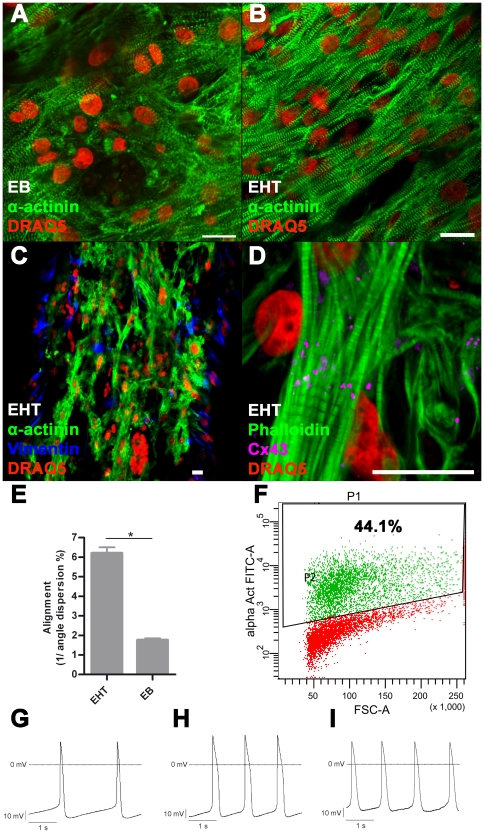
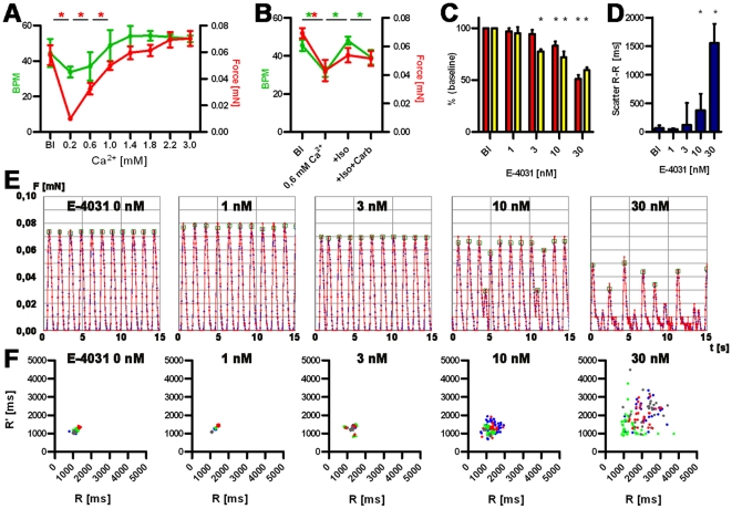
Human embryonic stem cell (hESC) progenies hold great promise as surrogates for human primary cells, particularly if the latter are not available as in the case of cardiomyocytes. However, high content experimental platforms are lacking that allow the function of hESC-derived cardiomyocytes to be studied under relatively physiological and standardized conditions. Here we describe a simple and robust protocol for the generation of fibrin-based human engineered heart tissue (hEHT) in a 24-well format using an unselected population of differentiated human embryonic stem cells containing 30-40% α-actinin-positive cardiac myocytes. Human EHTs started to show coherent contractions 5-10 days after casting, reached regular (mean 0.5 Hz) and strong (mean 100 µN) contractions for up to 8 weeks. They displayed a dense network of longitudinally oriented, interconnected and cross-striated cardiomyocytes. Spontaneous hEHT contractions were analyzed by automated video-optical recording and showed chronotropic responses to calcium and the β-adrenergic agonist isoprenaline. The proarrhythmic compounds E-4031, quinidine, procainamide, cisapride, and sertindole exerted robust, concentration-dependent and reversible decreases in relaxation velocity and irregular beating at concentrations that recapitulate findings in hERG channel assays. In conclusion this study establishes hEHT as a simple in vitro model for heart research.
Overview
The claims and statistics of this paper haven’t been checked yet.
Free: it reads the open-access text and checks the first 5 claims that cite a source we can check. Create a free account to check every claim.
Integrity signals
Registry
Cites
Statistics
Figures

Verification
Claims the paper makes
The claims and statistics of this paper haven’t been checked yet.
Free: it reads the open-access text and checks the first 5 claims that cite a source we can check. Create a free account to check every claim.
Claims other papers make about this one
Each is checked against this paper’s own text: does it say what the citing paper says it does?
The source backs part of the claim, not all of it — or backs it for a different population, condition or comparison.
In the citing paper
“but not pressure- and volume-based metrics directly representing pump performance of the heart”
From Bioengineering an electro-mechanically functional miniature ventricular heart chamber from human pluripotent stem cells (2018) · cites this paper as [16]
Experimental — accuracy not yet measuredThe cited source says what the claim attributes to it.
In the citing paper
“engineered cardiac thin films, micro-tissues and strips allow the assessment of twitch force [9,12–19]”
From Bioengineering an electro-mechanically functional miniature ventricular heart chamber from human pluripotent stem cells (2018) · cites this paper as [16]
Experimental — accuracy not yet measured
Self-check
Percentages, sums and reported differences re-computed from the paper’s own numbers.
The self-check needs the paper’s full text.
Citations
Cites
Proceedings of the National Academy of Sciences of the United States of America
Dowling, J. E., & Wald, G. (2009) · Nutrition Reviews · doi:10.1111/j.1753-4887.1981.tb06752.x · Cited by 17,893
Laflamme, M. A., Chen, K. Y., Naumova, A. V., Muskheli, V., Fugate, J. A., Dupras, S. K., Reinecke, H., Xu, C., Hassanipour, M., Police, S., et al. (2007) · Nature Biotechnology · doi:10.1038/nbt1327 · Cited by 2,206 · cited 2×
“One of the current limitations of hESC-derived cardiac myocytes is their immature and unorganized phenotype in the EB format or 2D cultures [3,5].”
BackgroundFeeder-free growth of undifferentiated human embryonic stem cells
Xu, C., Inokuma, M. S., Denham, J., Golds, K., Kundu, P., Gold, J. D., & Carpenter, M. K. (2001) · Nature Biotechnology · doi:10.1038/nbt1001-971 · Cited by 1,870 · cited 1×
“with CF1-MEF conditioned medium according to published protocols [28].”
MethodKehat, I., Kenyagin‐Karsenti, D., Snir, M., Segev, H., Amit, M., Gepstein, A., Livne, E., Binah, O., Itskovitz‐Eldor, J., & Gepstein, L. (2001) · Journal of Clinical Investigation · doi:10.1172/jci12131 · Cited by 1,814 · cited 1×
“differentiation in serum containing media yielding 8% of beating embryoid bodies (EBs) [1].”
BackgroundCai, C.-L., Liang, X., Shi, Y., Chu, P.-H., Pfaff, S. L., Chen, J., & Evans, S. (2003) · Developmental Cell · doi:10.1016/s1534-5807(03)00363-0 · Cited by 1,610 · cited 1×
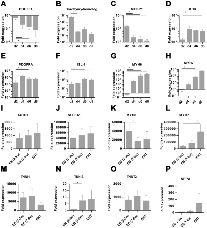
“Islet-1 (ISL1) a progenitor marker of the development of the secondary heart field [21] was significantly upregulated on day 6 and 8.”
BackgroundKehat, I., Kenyagin‐Karsenti, D., Snir, M., et al. (2001) · Journal of Clinical Investigation · doi:10.1172/jci200112131 · Cited by 1,536
REDFERN, W., CARLSSON, L., DAVIS, A., LYNCH, W., MACKENZIE, I., PALETHORPE, S., SIEGL, P., STRANG, I., SULLIVAN, A., & WALLIS, R. (2003) · Cardiovascular Research · doi:10.1016/s0008-6363(02)00846-5 · Cited by 1,532 · cited 2×
“Concentrations were chosen according to published IC50 in hERG assays [23,24].”
MethodHuman cardiovascular progenitor cells develop from a KDR+ embryonic-stem-cell-derived population
Yang, L., Soonpaa, M. H., Adler, E. D., Roepke, T. K., Kattman, S. J., Kennedy, M., Henckaerts, E., Bonham, K., Abbott, G. W., Linden, R. M., et al. (2008) · Nature · doi:10.1038/nature06894 · Cited by 1,389 · cited 3×
“Cardiomyocyte differentiation was performed with a modified EB-based, three-stage, growth factor cocktail protocol [2].”
MethodInfluentialEngineered heart tissue grafts improve systolic and diastolic function in infarcted rat hearts
Zimmermann, W.-H., Melnychenko, I., Wasmeier, G., Didié, M., Naito, H., Nixdorff, U., Hess, A., Budinsky, L., Brune, K., Michaelis, B., et al. (2006) · Nature Medicine · doi:10.1038/nm1394 · Cited by 993 · cited 1×
“The principal usefulness of these techniques for in vitro testing [17,18] and cardiac repair [19] has been demonstrated.”
BackgroundSelf-citationTissue Engineering of a Differentiated Cardiac Muscle Construct
Zimmermann, W.-H., Schneiderbanger, K., Schubert, P., Didié, M., Münzel, F., Heubach, J. F., Kostin, S., Neuhuber, W. L., & Eschenhagen, T. (2002) · Circulation Research · doi:10.1161/hh0202.103644 · Cited by 917 · cited 1×
“EHTs from neonatal rat hearts develop a high degree of cellular differentiation, longitudinal orientation, intercellular coupling and force generation [15,16].”
BackgroundSelf-citation
Research works printed but not linked: 2
- [3]Mummery C Ward-van Oostwaard D Doevendans P Spijker R Brink S van den 2003 Differentiation of human embryonic stem cells to cardiomyocytes: role of coculture with visceral endoderm-like cells. Circulation 107 2733 40 12742992
- [12]FDA 2005 Guidance for Industry S7B Nonclinical Evaluation of the Potential for Delayed Ventricular Repolarization (QT Interval Prolongation) by Human Pharmaceuticals. 16237859
Cited by
Advances in organ-on-a-chip engineering
Zhang, B., Korolj, A., Lai, B. F. L., & Radisic, M. (2018) · Nature Reviews Materials · doi:10.1038/s41578-018-0034-7 · Cited by 1,017
Biowire: a platform for maturation of human pluripotent stem cell–derived cardiomyocytes
Nunes, S. S., Miklas, J. W., Liu, J., Aschar-Sobbi, R., Xiao, Y., Zhang, B., Jiang, J., Massé, S., Gagliardi, M., Hsieh, A., et al. (2013) · Nature Methods · doi:10.1038/nmeth.2524 · Cited by 952
Cardiomyocyte maturation: advances in knowledge and implications for regenerative medicine
Karbassi, E., Fenix, A., Marchiano, S., Muraoka, N., Nakamura, K., Yang, X., & Murry, C. E. (2020) · Nature Reviews Cardiology · doi:10.1038/s41569-019-0331-x · Cited by 755
Tiburcy, M., Hudson, J. E., Balfanz, P., Schlick, S., Meyer, T., Chang Liao, M.-L., Levent, E., Raad, F., Zeidler, S., Wingender, E., et al. (2017) · Circulation · doi:10.1161/circulationaha.116.024145 · Cited by 645
Self-citationA Platform for Generation of Chamber-Specific Cardiac Tissues and Disease Modeling
Zhao, Y., Rafatian, N., Feric, N. T., Cox, B. J., Aschar-Sobbi, R., Wang, E. Y., Aggarwal, P., Zhang, B., Conant, G., Ronaldson-Bouchard, K., et al. (2019) · Cell · doi:10.1016/j.cell.2018.11.042 · Cited by 610
“Although ideal platforms should yield distinct atrial and ventricular tissues, most approaches using human pluripotent stem cells (hPSC) have focused on generating ventricular myocardium (Ahn et al., 2018; Eschenhagen et al., 2012; Lemoine et al., 2017; Lind et al., 2017a; MacQueen et al., 2018; Nunes et al., 2013; Sc…”
Zhang, D., Shadrin, I. Y., Lam, J., Xian, H.-Q., Snodgrass, H. R., & Bursac, N. (2013) · Biomaterials · doi:10.1016/j.biomaterials.2013.04.026 · Cited by 565
Screening out irrelevant cell-based models of disease
Horvath, P., Aulner, N., Bickle, M., Davies, A. M., Nery, E. D., Ebner, D., Montoya, M. C., Östling, P., Pietiäinen, V., Price, L. S., et al. (2016) · Nature Reviews Drug Discovery · doi:10.1038/nrd.2016.175 · Cited by 525
Mills, R. J., Titmarsh, D. M., Koenig, X., Parker, B. L., Ryall, J. G., Quaife-Ryan, G. A., Voges, H. K., Hodson, M. P., Ferguson, C., Drowley, L., et al. (2017) · Proceedings of the National Academy of Sciences · doi:10.1073/pnas.1707316114 · Cited by 478
Ruan, J.-L., Tulloch, N. L., Razumova, M. V., Saiget, M., Muskheli, V., Pabon, L., Reinecke, H., Regnier, M., & Murry, C. E. (2016) · Circulation · doi:10.1161/circulationaha.114.014998 · Cited by 467
Marsano, A., Conficconi, C., Lemme, M., Occhetta, P., Gaudiello, E., Votta, E., Cerino, G., Redaelli, A., & Rasponi, M. (2016) · Lab on a Chip · doi:10.1039/c5lc01356a · Cited by 447
Organ-on-a-chip devices advance to market
Zhang, B., & Radisic, M. (2017) · Lab on a Chip · doi:10.1039/c6lc01554a · Cited by 437
Induced pluripotent stem cells: the new patient?
Bellin, M., Marchetto, M. C., Gage, F. H., & Mummery, C. L. (2012) · Nature Reviews Molecular Cell Biology · doi:10.1038/nrm3448 · Cited by 431
Cardiac Tissue Engineering: State of the Art
Hirt, M. N., Hansen, A., & Eschenhagen, T. (2014) · Circulation Research · doi:10.1161/circresaha.114.300522 · Cited by 425
Self-citationShadrin, I. Y., Allen, B. W., Qian, Y., Jackman, C. P., Carlson, A. L., Juhas, M. E., & Bursac, N. (2017) · Nature Communications · doi:10.1038/s41467-017-01946-x · Cited by 420
Human Engineered Heart Tissue: Analysis of Contractile Force
Mannhardt, I., Breckwoldt, K., Letuffe-Brenière, D., Schaaf, S., Schulz, H., Neuber, C., Benzin, A., Werner, T., Eder, A., Schulze, T., et al. (2016) · Stem Cell Reports · doi:10.1016/j.stemcr.2016.04.011 · Cited by 398
“Analysis of contractile force was performed by video-optical recording as previously described (Hansen et al., 2010; Schaaf et al., 2011; Figure S1) on a setup available from EHT Technologies.”
BackgroundMethodSelf-citationHirt, M. N., Boeddinghaus, J., Mitchell, A., Schaaf, S., Börnchen, C., Müller, C., Schulz, H., Hubner, N., Stenzig, J., Stoehr, A., et al. (2014) · Journal of Molecular and Cellular Cardiology · doi:10.1016/j.yjmcc.2014.05.009 · Cited by 352
“S3 Fractional shortening of representative series of A) rat EHTs after 16 days of pacing and B) human EHTs after 10 days of high frequency pacing.”
InfluentialSelf-citationDenning, C., Borgdorff, V., Crutchley, J., Firth, K. S. A., George, V., Kalra, S., Kondrashov, A., Hoang, M. D., Mosqueira, D., Patel, A., et al. (2016) · Biochimica et Biophysica Acta (BBA) - Molecular Cell Research · doi:10.1016/j.bbamcr.2015.10.014 · Cited by 293
Tissue Engineering Approaches in the Design of Healthy and Pathological In Vitro Tissue Models
Caddeo, S., Boffito, M., & Sartori, S. (2017) · Frontiers in Bioengineering and Biotechnology · doi:10.3389/fbioe.2017.00040 · Cited by 285
Design and formulation of functional pluripotent stem cell-derived cardiac microtissues
Thavandiran, N., Dubois, N., Mikryukov, A., Massé, S., Beca, B., Simmons, C. A., Deshpande, V. S., McGarry, J. P., Chen, C. S., Nanthakumar, K., et al. (2013) · Proceedings of the National Academy of Sciences · doi:10.1073/pnas.1311120110 · Cited by 285
“Together, these and other systems (23, 24) have already proven their utility as models for cell transplantation (25) and drug candidate evaluation (12, 26, 27).”
BackgroundDifferentiation of cardiomyocytes and generation of human engineered heart tissue
Breckwoldt, K., Letuffe-Brenière, D., Mannhardt, I., Schulze, T., Ulmer, B., Werner, T., Benzin, A., Klampe, B., Reinsch, M. C., Laufer, S., et al. (2017) · Nature Protocols · doi:10.1038/nprot.2017.033 · Cited by 272
Self-citationImmaturity of Human Stem-Cell-Derived Cardiomyocytes in Culture: Fatal Flaw or Soluble Problem?
Veerman, C. C., Kosmidis, G., Mummery, C. L., Casini, S., Verkerk, A. O., & Bellin, M. (2015) · Stem Cells and Development · doi:10.1089/scd.2014.0533 · Cited by 265
“5 mN/mm(2) [24,86], compared with 10–50 mN/mm(2) in adult ventricular cardiomyocytes [87,88].”
Cardiac repair in guinea pigs with human engineered heart tissue from induced pluripotent stem cells
Weinberger, F., Breckwoldt, K., Pecha, S., Kelly, A., Geertz, B., Starbatty, J., Yorgan, T., Cheng, K.-H., Lessmann, K., Stolen, T., et al. (2016) · Science Translational Medicine · doi:10.1126/scitranslmed.aaf8781 · Cited by 272
Self-citationInduced Pluripotent Stem Cells 10 Years Later: For Cardiac Applications
Yoshida, Y., & Yamanaka, S. (2017) · Circulation Research · doi:10.1161/circresaha.117.311080 · Cited by 264
Maturing human pluripotent stem cell-derived cardiomyocytes in human engineered cardiac tissues
Feric, N. T., & Radisic, M. (2016) · Advanced Drug Delivery Reviews · doi:10.1016/j.addr.2015.04.019 · Cited by 247
“Conversely, hPSC-CMs are heterogeneous in shape—polygonal or spherical, smaller in size and have lower aspect ratios [43, 46, 47] (Figure 1A).”
InfluentialMusunuru, K., Sheikh, F., Gupta, R. M., Houser, S. R., Maher, K. O., Milan, D. J., Terzic, A., & Wu, J. C. (2018) · Circulation: Genomic and Precision Medicine · doi:10.1161/hcg.0000000000000043 · Cited by 229
Life in 3D is never flat: 3D models to optimise drug delivery
Fitzgerald, K. A., Malhotra, M., Curtin, C. M., O' Brien, F. J., & O' Driscoll, C. M. (2015) · Journal of Controlled Release · doi:10.1016/j.jconrel.2015.07.020 · Cited by 228
Engineering Cardiac Muscle Tissue: A Maturating Field of Research
Weinberger, F., Mannhardt, I., & Eschenhagen, T. (2017) · Circulation Research · doi:10.1161/circresaha.117.310738 · Cited by 225
Self-citationCardiovascular disease models: A game changing paradigm in drug discovery and screening
Savoji, H., Mohammadi, M. H., Rafatian, N., Toroghi, M. K., Wang, E. Y., Zhao, Y., Korolj, A., Ahadian, S., & Radisic, M. (2019) · Biomaterials · doi:10.1016/j.biomaterials.2018.09.036 · Cited by 223
Human Stem Cells for Modeling Heart Disease and for Drug Discovery
Matsa, E., Burridge, P. W., & Wu, J. C. (2014) · Science Translational Medicine · doi:10.1126/scitranslmed.3008921 · Cited by 209
Dahlmann, J., Krause, A., Möller, L., Kensah, G., Möwes, M., Diekmann, A., Martin, U., Kirschning, A., Gruh, I., & Dräger, G. (2013) · Biomaterials · doi:10.1016/j.biomaterials.2012.10.008 · Cited by 209
“The majority of current state-of-the-art approaches using primary [41,55] or pluripotent stem cell-derived cardiomyocytes [21,56,57] are based on undefined basement membrane matrix (like Matrigel or Geltrex ) together with rat tail collagen I or fibrin glue.”
Naturally Engineered Maturation of Cardiomyocytes
Scuderi, G. J., & Butcher, J. (2017) · Frontiers in Cell and Developmental Biology · doi:10.3389/fcell.2017.00050 · Cited by 202
“5 mN/mm2 while adult CMs generate two orders of magnitude larger contractile forces of 10–50 mN/mm2 (Mulieri et al., 1992; Holubarsch et al., 1998; Schaaf et al., 2011; Tulloch et al., 2011).”
BackgroundInfluentialDevelopment of a human cardiac organoid injury model reveals innate regenerative potential
Voges, H. K., Mills, R. J., Elliott, D. A., Parton, R. G., Porrello, E. R., & Hudson, J. E. (2017) · Development · doi:10.1242/dev.143966 · Cited by 196
“hCOs have been proposed as a physiologically relevant model of the human heart (Caspi et al., 2007; Schaaf et al., 2011; Tiburcy et al., 2011), which has a variety of applications from drug screening to studying human cardiac biology.”
BackgroundHayakawa, T., Kunihiro, T., Ando, T., Kobayashi, S., Matsui, E., Yada, H., Kanda, Y., Kurokawa, J., & Furukawa, T. (2014) · Journal of Molecular and Cellular Cardiology · doi:10.1016/j.yjmcc.2014.09.010 · Cited by 198
“This is an open access article under the hiPS-/hES-CMs, as there have been few studies performed at the present time [21–29].”
3D bioprinting for cardiovascular regeneration and pharmacology
Cui, H., Miao, S., Esworthy, T., Zhou, X., Lee, S.-J., Liu, C., Yu, Z.-X., Fisher, J. P., Mohiuddin, M., & Zhang, L. G. (2018) · Advanced Drug Delivery Reviews · doi:10.1016/j.addr.2018.07.014 · Cited by 193
“Proarrhythmic compounds, such as chromanol and erythromycin, can affect repolarization inhibition; E-4031, procainamide, sertindole, quinidine, and cisapride can inhibit repolarization; the cardiotoxic drug doxorubicin can affect force generation of cardiac tissue; Isoprenaline (isoproterenol) and carbachol can affect…”
Kensah, G., Roa Lara, A., Dahlmann, J., Zweigerdt, R., Schwanke, K., Hegermann, J., Skvorc, D., Gawol, A., Azizian, A., Wagner, S., et al. (2013) · European Heart Journal · doi:10.1093/eurheartj/ehs349 · Cited by 193
“Human BCT contractility was modulated in response to isoprenaline with a positive chronotropic effect and in response to carbachol with a negative chronotropic effect, demonstrating functional b-adrenergic and muscarinergic receptor systems.(23) For our optimized human BCTs, these effects were even more pronounced tha…”
BackgroundMethodInduced pluripotent stem cells: at the heart of cardiovascular precision medicine
Chen, I. Y., Matsa, E., & Wu, J. C. (2016) · Nature Reviews Cardiology · doi:10.1038/nrcardio.2016.36 · Cited by 191
Human engineered heart tissue as a model system for drug testing
Eder, A., Vollert, I., Hansen, A., & Eschenhagen, T. (2016) · Advanced Drug Delivery Reviews · doi:10.1016/j.addr.2015.05.010 · Cited by 190
Self-citationDynamic culture yields engineered myocardium with near-adult functional output
Jackman, C. P., Carlson, A. L., & Bursac, N. (2016) · Biomaterials · doi:10.1016/j.biomaterials.2016.09.024 · Cited by 188
“In contrast to traditional static culture of hydrogelbased engineered heart tissues [25, 50-56], this me thod relies on the application of free-floating dynamic culture conditions that lead to upregulation of mTO RC1 signaling and robust structural and functional impr ovements associated with increased cardiomyocyte d…”
In vitro cardiac tissue models: Current status and future prospects
Mathur, A., Ma, Z., Loskill, P., Jeeawoody, S., & Healy, K. E. (2016) · Advanced Drug Delivery Reviews · doi:10.1016/j.addr.2015.09.011 · Cited by 187
“Parallel EHT arrays consisting of a mixture of fibrinogen, Matrigel, thrombin, and neonatal rat CMs [79] or hESC-CMs [80], on a silicone post rack casted from Teflon molds, were used for preliminary drug screening.”
MethodAdvancing functional engineered cardiac tissues toward a preclinical model of human myocardium
Turnbull, I. C., Karakikes, I., Serrao, G. W., Backeris, P., Lee, J., Xie, C., Senyei, G., Gordon, R. E., Li, R. A., Akar, F. G., et al. (2014) · The FASEB Journal · doi:10.1096/fj.13-228007 · Cited by 184
“Eschenhagen and Zimmerman and colleagues, pioneers in the cardiac tissue engineering field (17), more recently published a study using hECTs created from human embryonic stem cells, demonstrating responses to proarrhythmic compounds similar to the industry standard hERG channel assay (18).”
BackgroundLi, R. A., Keung, W., Cashman, T. J., Backeris, P. C., Johnson, B. V., Bardot, E. S., Wong, A. O. T., Chan, P. K. W., Chan, C. W. Y., & Costa, K. D. (2018) · Biomaterials · doi:10.1016/j.biomaterials.2018.02.024 · Cited by 175
Hydrogels for cardiac tissue engineering
Camci-Unal, G., Annabi, N., Dokmeci, M. R., Liao, R., & Khademhosseini, A. (2014) · NPG Asia Materials · doi:10.1038/am.2014.19 · Cited by 170
Tzatzalos, E., Abilez, O. J., Shukla, P., & Wu, J. C. (2016) · Advanced Drug Delivery Reviews · doi:10.1016/j.addr.2015.09.010 · Cited by 162
Guidance Document on Good In Vitro Method Practices (GIVIMP)
OECD (2018) · OECD Series on Testing and Assessment · doi:10.1787/9789264304796-en · Cited by 161
Atrial-like Engineered Heart Tissue: An In Vitro Model of the Human Atrium
Lemme, M., Ulmer, B. M., Lemoine, M. D., Zech, A. T. L., Flenner, F., Ravens, U., Reichenspurner, H., Rol-Garcia, M., Smith, G., Hansen, A., et al. (2018) · Stem Cell Reports · doi:10.1016/j.stemcr.2018.10.008 · Cited by 158
“Analysis of contractile force was performed by video-optical recording as previously described (Hansen et al., 2010; Schaaf et al., 2011) on a setup available fromEHT Technologies.”
Self-citationClinical Applications of Naturally Derived Biopolymer-Based Scaffolds for Regenerative Medicine
Stoppel, W. L., Ghezzi, C. E., McNamara, S. L., Black, L. D., & Kaplan, D. L. (2014) · Annals of Biomedical Engineering · doi:10.1007/s10439-014-1206-2 · Cited by 162
Biomaterial based cardiac tissue engineering and its applications
Huyer, L. D., Montgomery, M., Zhao, Y., Xiao, Y., Conant, G., Korolj, A., & Radisic, M. (2015) · Biomedical Materials · doi:10.1088/1748-6041/10/3/034004 · Cited by 145
“[112] Schaaf S et al 2011 Human engineered heart tissue as a”
BackgroundMethodInfluentialNext generation of heart regenerative therapies: progress and promise of cardiac tissue engineering
Tenreiro, M. F., Louro, A. F., Alves, P. M., & Serra, M. (2021) · npj Regenerative Medicine · doi:10.1038/s41536-021-00140-4 · Cited by 171
Patching the Heart: Cardiac Repair From Within and Outside
Ye, L., Zimmermann, W.-H., Garry, D. J., & Zhang, J. (2013) · Circulation Research · doi:10.1161/circresaha.113.300216 · Cited by 145
“Fibrinogen can also be conjugated with polyethylene glycol to generate scaffolds that covalently bind growth factors and other proteins,(47,48) and drug screening platforms have recently been established with fibrin-based engineered heart tissue.(49,90) In a mouse MI model, treatment with a polyethylene glycolylated f…”
Self-citationThree‐Dimensional Paper‐Based Model for Cardiac Ischemia
Mosadegh, B., Dabiri, B. E., Lockett, M. R., Derda, R., Campbell, P., Parker, K. K., & Whitesides, G. M. (2014) · Advanced Healthcare Materials · doi:10.1002/adhm.201300575 · Cited by 133
Myocardial Tissue Engineering: In Vitro Models
Vunjak Novakovic, G., Eschenhagen, T., & Mummery, C. (2014) · Cold Spring Harbor Perspectives in Medicine · doi:10.1101/cshperspect.a014076 · Cited by 132
“In human EHTs, the effect was closely related to the inhibition of hERG (Schaaf et al. 2011).”
BackgroundSelf-citationApplication of Microphysiological Systems to Enhance Safety Assessment in Drug Discovery
Ewart, L., Dehne, E.-M., Fabre, K., Gibbs, S., Hickman, J., Hornberg, E., Ingelman-Sundberg, M., Jang, K.-J., Jones, D. R., Lauschke, V. M., et al. (2018) · Annual Review of Pharmacology and Toxicology · doi:10.1146/annurev-pharmtox-010617-052722 · Cited by 128
Self-citationHulot, J.-S., Stillitano, F., Salem, J. E., Kovacic, J. C., Fuster, V., & Hajjar, R. J. (2014) · Stem Cell Research & Therapy · doi:10.1186/scrt390 · Cited by 126
“They display a dense network of longitudinally oriented, interconnected and cross-striated cardiomyocytes, suggesting that the three-dimensional tissue format improves the maturation of cardiomyocytes [55].”
BackgroundGerbin, K. A., Yang, X., Murry, C. E., & Coulombe, K. L. K. (2015) · PLOS ONE · doi:10.1371/journal.pone.0131446 · Cited by 119
Evidence for FHL1 as a novel disease gene for isolated hypertrophic cardiomyopathy
Friedrich, F. W., Wilding, B. R., Reischmann, S., Crocini, C., Lang, P., Charron, P., Müller, O. J., McGrath, M. J., Vollert, I., Hansen, A., et al. (2012) · Human Molecular Genetics · doi:10.1093/hmg/dds157 · Cited by 119
“Contraction measurements were performed on days 7, 9, 12, 14 and 15, as described previously (62,63).”
MethodSelf-citationChramiec, A., Teles, D., Yeager, K., Marturano-Kruik, A., Pak, J., Chen, T., Hao, L., Wang, M., Lock, R., Tavakol, D. N., et al. (2020) · Lab on a Chip · doi:10.1039/d0lc00424c · Cited by 113
Madonna, R., Laake, L. W. V., Bøtker, H. E., Davidson, S. M., Caterina, R. D., Engel, F. B., Eschenhagen, T., Fernandez-Aviles, F., Hausenloy, D. J., Hulot, J., et al. (2019) · Cardiovascular Research · doi:10.1093/cvr/cvz010 · Cited by 132
Self-citationThree-dimensional filamentous human diseased cardiac tissue model
Ma, Z., Koo, S., Finnegan, M. A., Loskill, P., Huebsch, N., Marks, N. C., Conklin, B. R., Grigoropoulos, C. P., & Healy, K. E. (2014) · Biomaterials · doi:10.1016/j.biomaterials.2013.10.052 · Cited by 113
“Numerous studies have been published suggesting that CM alignment is beneficial to maturation [11,37], thus direct differentiation on 3D filamentous matrices, instead of replating CMs from the 2D differentiation system might help CM differentiation and maturation.”
BackgroundMethodOn human pluripotent stem cell control: The rise of 3D bioengineering and mechanobiology
Shao, Y., Sang, J., & Fu, J. (2015) · Biomaterials · doi:10.1016/j.biomaterials.2015.01.078 · Cited by 110
“, two silicone posts were inserted into eachwell of a 24-well plate filledwith hPSC-CMs and ECM proteins [89].”
MethodPhysical developmental cues for the maturation of human pluripotent stem cell-derived cardiomyocytes
Zhu, R., Blazeski, A., Poon, E., Costa, K. D., Tung, L., & Boheler, K. R. (2014) · Stem Cell Research & Therapy · doi:10.1186/scrt507 · Cited by 110
“showed an increase in action potential duration and upstroke velocity compared with CMs isolated from human EBs of similar age, but the maximal diastolic potential (MDP) was less negative [11].”
BackgroundInfluentialMaterials Science and Tissue Engineering: Repairing the Heart
Radisic, M., & Christman, K. L. (2013) · Mayo Clinic Proceedings · doi:10.1016/j.mayocp.2013.05.003 · Cited by 133
Campostrini, G., Meraviglia, V., Giacomelli, E., van Helden, R. W. J., Yiangou, L., Davis, R. P., Bellin, M., Orlova, V. V., & Mummery, C. L. (2021) · Nature Protocols · doi:10.1038/s41596-021-00497-2 · Cited by 109
Physiologically inspired cardiac scaffolds for tailored in vivo function and heart regeneration
Kaiser, N. J., & Coulombe, K. L. K. (2015) · Biomedical Materials · doi:10.1088/1748-6041/10/3/034003 · Cited by 107
“) (C) Highthroughput platform for monitoring contractile activity of an array of fibrin-based EHTs [112].”
BackgroundMethodMa, Z., Huebsch, N., Koo, S., Mandegar, M. A., Siemons, B., Boggess, S., Conklin, B. R., Grigoropoulos, C. P., & Healy, K. E. (2018) · Nature Biomedical Engineering · doi:10.1038/s41551-018-0280-4 · Cited by 109
“The contraction force of hiPSC-CMs has been deemed as one of the essential parameters for the evaluation of normal mature cardiac function, response to pharmacological interventions, and indication of disease phenotypes 12–14 .”
Engineering microenvironment for human cardiac tissue assembly in heart-on-a-chip platform
Zhao, Y., Rafatian, N., Wang, E. Y., Feric, N. T., Lai, B. F. L., Knee-Walden, E. J., Backx, P. H., & Radisic, M. (2020) · Matrix Biology · doi:10.1016/j.matbio.2019.04.001 · Cited by 106
Biomaterializing the promise of cardiac tissue engineering
Pomeroy, J. E., Helfer, A., & Bursac, N. (2020) · Biotechnology Advances · doi:10.1016/j.biotechadv.2019.02.009 · Cited by 107
“Given these benefits, along with the fact that it is an FDA approved material, fibrin scaffolds have gained wide adoption in the cardiac tissue engineering field (Hirt et al., 2014; RonaldsonBouchard et al., 2018; Schaaf et al., 2011; Shadrin et al., 2017; Yuan Ye et al., 2011; Zhang et al., 2013).”
BackgroundZhang, W., Kong, C., Tống, M. P., Chooi, W. H., Huang, N., Li, R. A., & Chan, B. P. (2016) · Acta Biomaterialia · doi:10.1016/j.actbio.2016.11.058 · Cited by 120
Fiedler, L. R., Chapman, K., Xie, M., Maifoshie, E., Jenkins, M., Golforoush, P. A., Bellahcene, M., Noseda, M., Faust, D., Jarvis, A., et al. (2019) · Cell Stem Cell · doi:10.1016/j.stem.2019.01.013 · Cited by 102
“, 2010) and calculated on the basis of post deflection distance (d), post length (L), post radius (R), and the elastic modulus (E), using the formula F = 3pER(4)d4L (Schaaf et al., 2011) and the indicated post parameters.”
MethodHuang, C., Maia-Joca, R. P. M., Ong, C. S., Wilson, I., DiSilvestre, D. A., Tomaselli, G. F., & Reich, D. H. (2019) · Journal of Molecular and Cellular Cardiology · doi:10.1016/j.yjmcc.2019.10.001 · Cited by 110
Cashman, T. J., Josowitz, R., Johnson, B. V., Gelb, B. D., & Costa, K. D. (2016) · PLOS ONE · doi:10.1371/journal.pone.0146697 · Cited by 101
“The time-to-next peak and time-to-previous peak for each contraction was measured and plotted against one another to form a cloud of points that represent the variability of the spontaneous rate, similar to methods reported previously [25].”
ResultCurrent status of drug screening and disease modelling in human pluripotent stem cells
Rajamohan, D., Matsa, E., Kalra, S., Crutchley, J., Patel, A., George, V., & Denning, C. (2013) · BioEssays · doi:10.1002/bies.201200053 · Cited by 99
“Therefore it is encouraging that maturation of hPSC-cardiomyocytes can be facilitated by prolonged time culture [62, 63], transgenic overexpression of calsequestrin [64], formation of 3D aggregates [62], tissueengineered constructs and mechanical stress [65, 66].”
BackgroundErgir, E., Oliver-De La Cruz, J., Fernandes, S., Cassani, M., Niro, F., Pereira-Sousa, D., Vrbský, J., Vinarský, V., Perestrelo, A. R., Debellis, D., et al. (2022) · Scientific Reports · doi:10.1038/s41598-022-22225-w · Cited by 99
Physiological aspects of cardiac tissue engineering
Eschenhagen, T., Eder, A., Vollert, I., & Hansen, A. (2012) · American Journal of Physiology-Heart and Circulatory Physiology · doi:10.1152/ajpheart.00007.2012 · Cited by 100
Self-citationProndzynski, M., Berkson, P., Trembley, M. A., Tharani, Y., Shani, K., Bortolin, R. H., Sweat, M., Mayourian, J., Yücel, D., Cordoves, A. M., et al. (2024) · Nature Communications · doi:10.1038/s41467-024-50224-0 · Cited by 123
Complex Interactions between Human Myoblasts and the Surrounding 3D Fibrin-Based Matrix
Chiron, S., Tomczak, C., Duperray, A., Lainé, J., Bonne, G., Eder, A., Hansen, A., Eschenhagen, T., Claude Verdier, A., & Coirault, C. (2012) · PLoS ONE · doi:10.1371/journal.pone.0036173 · Cited by 107
“Fibrin networks have been shown to support myocyte anchorage and the formation of the engineered cardiac [19,25] and skeletal muscle tissue [4,13,15,19].”
BackgroundSelf-citationTowards chamber specific heart-on-a-chip for drug testing applications
Zhao, Y., Rafatian, N., Wang, E. Y., Wu, Q., Lai, B. F. L., Lu, R. X., Savoji, H., & Radisic, M. (2020) · Advanced Drug Delivery Reviews · doi:10.1016/j.addr.2019.12.002 · Cited by 97
Mesenchymal Stem Cells for Cardiac Therapy: Practical Challenges and Potential Mechanisms
Cashman, T. J., Gouon–Evans, V., & Costa, K. D. (2012) · Stem Cell Reviews and Reports · doi:10.1007/s12015-012-9375-6 · Cited by 97
Cardiac Tissues From Stem Cells: New Routes to Maturation and Cardiac Regeneration
Campostrini, G., Windt, L. M., van Meer, B. J., Bellin, M., & Mummery, C. L. (2021) · Circulation Research · doi:10.1161/circresaha.121.318183 · Cited by 96
Stillitano, F., Hansen, J., Kong, C., Karakikes, I., Funck‐Brentano, C., Geng, L., Scott, S. A., Reynier, S., Ma, W., Valogne, Y., et al. (2017) · eLife · doi:10.7554/elife.19406 · Cited by 99
Li, Y., Asfour, H., & Bursac, N. (2017) · Acta Biomaterialia · doi:10.1016/j.actbio.2017.04.027 · Cited by 95
Review of high-content screening applications in toxicology
Li, S., & Xia, M. (2019) · Archives of Toxicology · doi:10.1007/s00204-019-02593-5 · Cited by 106
Engineering anisotropic human stem cell-derived three-dimensional cardiac tissue on-a-chip
Veldhuizen, J., Cutts, J., Brafman, D. A., Migrino, R. Q., & Nikkhah, M. (2020) · Biomaterials · doi:10.1016/j.biomaterials.2020.120195 · Cited by 98
Lee, S., Serpooshan, V., Tong, X., Venkatraman, S., Lee, M. J., Lee, J., Chirikian, O., Wu, J. C., Wu, S. M., & Yang, F. (2017) · Biomaterials · doi:10.1016/j.biomaterials.2017.03.039 · Cited by 103
Organs-on-a-chip: a union of tissue engineering and microfabrication
Zhao, Y., Wang, E. Y., Lai, F. B. L., Cheung, K., & Radisic, M. (2023) · Trends in Biotechnology · doi:10.1016/j.tibtech.2022.12.018 · Cited by 93
Modelling sarcomeric cardiomyopathies in the dish: from human heart samples to iPSC cardiomyocytes
Eschenhagen, T., Mummery, C., & Knollmann, B. C. (2015) · Cardiovascular Research · doi:10.1093/cvr/cvv017 · Cited by 97
Self-citationZuppinger, C. (2015) · Biochimica et Biophysica Acta (BBA) - Molecular Cell Research · doi:10.1016/j.bbamcr.2015.11.036 · Cited by 105
Native cardiac environment and its impact on engineering cardiac tissue
Schwach, V., & Passier, R. (2019) · Biomaterials Science · doi:10.1039/c8bm01348a · Cited by 93
Heart-on-a-chip based on stem cell biology
Jastrzębska, E., Tomecka, E., & Jesion, I. (2015) · Biosensors and Bioelectronics · doi:10.1016/j.bios.2015.08.012 · Cited by 97
Sasaki, D., Matsuura, K., Seta, H., Haraguchi, Y., Okano, T., & Shimizu, T. (2018) · PLOS ONE · doi:10.1371/journal.pone.0198026 · Cited by 91
Pluripotent Stem Cell Models of Human Heart Disease
Moretti, A., Laugwitz, K.-L., Dorn, T., Sinnecker, D., & Mummery, C. (2013) · Cold Spring Harbor Perspectives in Medicine · doi:10.1101/cshperspect.a014027 · Cited by 91
“However, even with all of these improvements, full manifestation of adult cardiomyocyte phenotypes has not been shown thus far and the major application of hESC/hiPSC-derived EHT constructs has been drug testing (Schaaf et al. 2011).”
BackgroundElectro-mechanical conditioning of human iPSC-derived cardiomyocytes for translational research
Kroll, K. T., Chabria, M., Wang, K., Häusermann, F., Schuler, F., & Polonchuk, L. (2017) · Progress in Biophysics and Molecular Biology · doi:10.1016/j.pbiomolbio.2017.07.003 · Cited by 99
Kerscher, P., Turnbull, I. C., Hodge, A. J., Kim, J., Seliktar, D., Easley, C. J., Costa, K. D., & Lipke, E. A. (2016) · Biomaterials · doi:10.1016/j.biomaterials.2015.12.011 · Cited by 88
Raval, K. K., Tao, R., White, B. E., De Lange, W. J., Koonce, C. H., Yu, J., Kishnani, P. S., Thomson, J. A., Mosher, D. F., Ralphe, J. C., et al. (2015) · Journal of Biological Chemistry · doi:10.1074/jbc.m114.628628 · Cited by 85
“Engineered Cardiac Tissue Preparation and Functional Testing—Differentiated cardiomyocytes were dissociated from T75 flasks and used to prepare fibrin-based ECTs with a protocol adapted from elsewhere (23, 24).”
MethodSarig, U., Au-Yeung, G. C. T., Wang, Y., Bronshtein, T., Dahan, N., Boey, F., Venkatraman, S. S., & Machluf, M. (2012) · Tissue Engineering Part A · doi:10.1089/ten.tea.2011.0586 · Cited by 90
“…recently reported strategies 1 provide relatively thin myocardial-like constructs for use either in in vitro toxicology and drug screening studies 2 or as implants showing improvements in myocardial function on transplantation in small myocardial infarction (MI) animal models 3 and in a human…”
Automated analysis of contractile force and Ca2+transients in engineered heart tissue
Stoehr, A., Neuber, C., Baldauf, C., Vollert, I., Friedrich, F. W., Flenner, F., Carrier, L., Eder, A., Schaaf, S., Hirt, M. N., et al. (2014) · American Journal of Physiology-Heart and Circulatory Physiology · doi:10.1152/ajpheart.00705.2013 · Cited by 83
“Cardiomyocyte differentiation from human induced pluripotent stem cells (hiPSC) and subsequent human EHT generation were performed as previously described (27).”
MethodSelf-citationMayourian, J., Ceholski, D. K., Gonzalez, D. M., Cashman, T. J., Sahoo, S., Hajjar, R. J., & Costa, K. D. (2018) · Circulation Research · doi:10.1161/circresaha.117.311589 · Cited by 80
Blazeski, A., Zhu, R., Hunter, D. W., Weinberg, S. H., Boheler, K. R., Zambidis, E. T., & Tung, L. (2012) · Progress in Biophysics and Molecular Biology · doi:10.1016/j.pbiomolbio.2012.07.012 · Cited by 78
Biowire platform for maturation of human pluripotent stem cell-derived cardiomyocytes
Sun, X., & Nunes, S. S. (2015) · Methods · doi:10.1016/j.ymeth.2015.11.005 · Cited by 90
Nakamura, Y., Matsuo, J., Miyamoto, N., Ojima, A., Ando, K., Kanda, Y., Sawada, K., Sugiyama, A., & Sekino, Y. (2014) · Journal of Pharmacological Sciences · doi:10.1254/jphs.13248fp · Cited by 76
Yong, K. W., Li, Y., Huang, G., Lu, T. J., Safwani, W. K. Z. W., Pingguan-Murphy, B., & Xu, F. (2015) · American Journal of Physiology-Heart and Circulatory Physiology · doi:10.1152/ajpheart.00299.2015 · Cited by 75
“Furthermore, engineered heart tissues (EHTs) were produced consisting of cardiac fibroblasts and cardiomyocytes and which have been subjected to a cyclic stretching device developed by Zimmermann et al. (114) to optimize the culture conditions to generate functional EHTs for cardiac regeneration (62).”
Influential
Metadata
- Authors
- Schaaf, S., Shibamiya, A., Mewe, M., Eder, A., Stöhr, A., Hirt, M. N., Rau, T., Zimmermann, W.-H., Conradi, L., Eschenhagen, T., & Hansen, A.
- Affiliations
- University Medical Center Hamburg-Eppendorf (DE)
- Corresponding author
- Hansen, Arne
- Venue
- PLoS ONE
- Publisher
- Public Library of Science (PLoS)
- Published
- 2011-10-20
- Volume
- 6
- Issue
- 10
- Pages
- e26397
- ISSN
- 1932-6203 (ISSN-L)
- PMC
- PMC3197640
- OpenAlex
- W1969832018
- Cited by
- 365
- References
- 30
- Citations by year
- 2026: 11 · 2025: 7 · 2024: 15 · 2023: 19 · 2022: 17 · 2021: 20 · 2020: 24 · 2019: 26 · 2018: 32 · 2017: 39 · 2016: 34 · 2015: 35 · 2014: 37 · 2013: 27 · 2012: 20
- FWCI
- 10.92
- Type
- journal article
- Language
- en
- Open access
- gold
- doi.org
- Locations
- PLoS ONE (publishedVersion, cc-by)
- Europe PMC (PubMed Central) (publishedVersion, cc-by)
- PubMed Central (publishedVersion, cc-by)
- PubMed (publishedVersion)
- DOAJ (DOAJ: Directory of Open Access Journals) (submittedVersion, cc-by-sa)
- Figshare (submittedVersion, cc-by)
- GRO (submittedVersion)
- Funders
- European Commission
- Deutsche Forschungsgemeinschaft (ZI 708/7-1, HA 3423/3-1)
- Deutsche Herzstiftung (F/13/10)
- Topics
- Life Sciences › Biochemistry, Genetics and Molecular Biology › Molecular Biology › Pluripotent Stem Cells Research
- Health Sciences › Medicine › Surgery › Tissue Engineering and Regenerative Medicine
- Health Sciences › Medicine › Cardiology and Cardiovascular Medicine › Cardiac electrophysiology and arrhythmias
- Keywords
- human embryonic stem cells, cardiomyocytes, cardiomyocyte contraction, spontaneous beating, proarrhythmic drugs, hERG, calcium-induced contraction, beta-adrenergic stimulation, isoproterenol, E-4031, quinidine, procainamide, cisapride, sertindole, preclinical toxicology, high-content screening
- MeSH terms
- Arrhythmias, Cardiac, Cell Differentiation, Drug Evaluation, Preclinical, Fluorescent Antibody Technique, Heart, Humans, Muscle Contraction, Myocardium, RNA, Messenger, Reproducibility of Results, Toxicity Tests, Tissue Engineering, Myocytes, Cardiac, Embryonic Stem Cells, Electrophysiological Phenomena, HEK293 Cells
About these checks
Registry, retraction, Cites and statistics results are computed, not judged, with one exception: a printed reference that no index lists is read once by a model before it counts as a citation error. Claims are extracted by a model and kept only when their quote appears verbatim in the text; each cited source is then checked, with a model’s help, for whether it says what the claim relies on. Those verdicts can be wrong — read the claim’s quote and the cited source before relying on one.
Statistics and arithmetic are recomputed, not judged.
Confidence bands are shown only for fields calibrated on our annotated test set. None is calibrated yet, so no band is shown.
What counts as a claim ›How we measure confidence ›How we check this ›
Are you an author, or do you see a problem with this page? Request a review
Data sources
Bibliographic data: Semantic Scholar, OpenAlex, Crossref.
Contains information from the Semantic Scholar Academic Graph Datasets, made available under the ODC Attribution License (ODC-BY 1.0).
Semantic Scholar’s data is described in: Kinney, R., et al. (2023). The Semantic Scholar Open Data Platform. arXiv:2301.10140.
