

The DOI matches its Crossref record; 93 of the 95 references it cites were found in a registry or an index.
Mapping Connectivity Damage in the Case of Phineas Gage
Open accessAbstract
White matter (WM) mapping of the human brain using neuroimaging techniques has gained considerable interest in the neuroscience community. Using diffusion weighted (DWI) and magnetic resonance imaging (MRI), WM fiber pathways between brain regions may be systematically assessed to make inferences concerning their role in normal brain function, influence on behavior, as well as concerning the consequences of network-level brain damage. In this paper, we investigate the detailed connectomics in a noted example of severe traumatic brain injury (TBI) which has proved important to and controversial in the history of neuroscience. We model the WM damage in the notable case of Phineas P. Gage, in whom a "tamping iron" was accidentally shot through his skull and brain, resulting in profound behavioral changes. The specific effects of this injury on Mr. Gage's WM connectivity have not previously been considered in detail. Using computed tomography (CT) image data of the Gage skull in conjunction with modern anatomical MRI and diffusion imaging data obtained in contemporary right handed male subjects (aged 25-36), we computationally simulate the passage of the iron through the skull on the basis of reported and observed skull fiducial landmarks and assess the extent of cortical gray matter (GM) and WM damage. Specifically, we find that while considerable damage was, indeed, localized to the left frontal cortex, the impact on measures of network connectedness between directly affected and other brain areas was profound, widespread, and a probable contributor to both the reported acute as well as long-term behavioral changes. Yet, while significantly affecting several likely network hubs, damage to Mr. Gage's WM network may not have been more severe than expected from that of a similarly sized "average" brain lesion. These results provide new insight into the remarkable brain injury experienced by this noteworthy patient.
Overview
The claims and statistics of this paper haven’t been checked yet.
Run the check — free, no account needed. It reads the open-access text and checks the sources it cites.
Integrity signals
Registry
Cites
Statistics
Figures
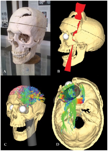
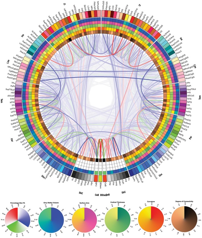
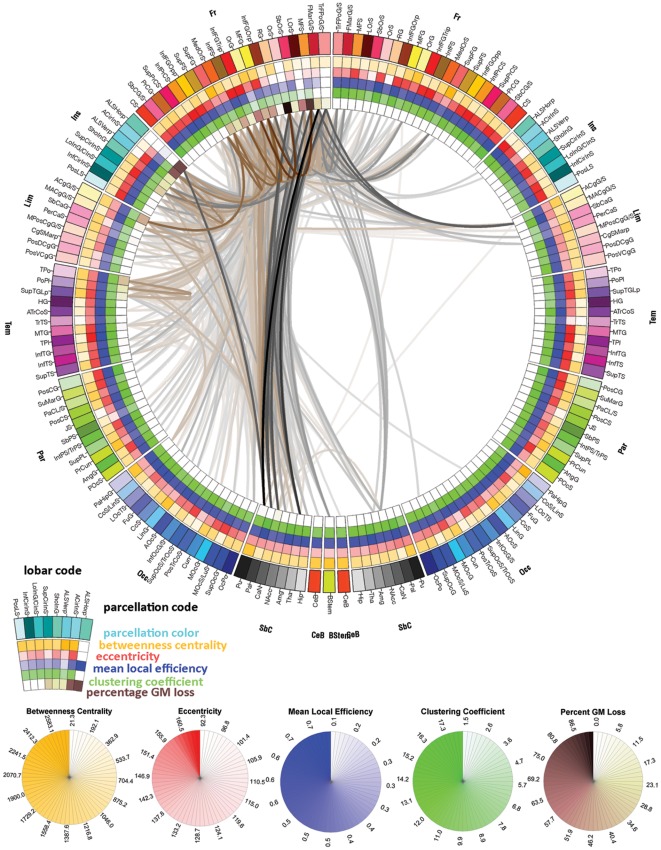
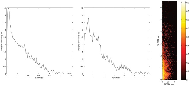
Verification
Claims the paper makes
The claims and statistics of this paper haven’t been checked yet.
Run the check — free, no account needed. It reads the open-access text and checks the sources it cites.
Claims other papers make about this one
Each is checked against this paper’s own text: does it say what the citing paper says it does?
The cited source says what the claim attributes to it.
In the citing paper
“While this indeed confirmed notable local injury, it also identified patterns of widespread and longrange connectivity loss that could also have contributed to the characteristic postinjury behavioral patterns described.”
From Graph theory analysis of complex brain networks: new concepts in brain mapping applied to neurosurgery (2016) · cites this paper as [99]
Self-check
Percentages, sums and reported differences re-computed from the paper’s own numbers.
The self-check needs the paper’s full text.
Citations
Cites
Collective dynamics of ‘small-world’ networks
Watts, D. J., & Strogatz, S. H. (1998) · Nature · doi:10.1038/30918 · Cited by 43,060
Complex brain networks: graph theoretical analysis of structural and functional systems
Bullmore, E., & Sporns, O. (2009) · Nature Reviews Neuroscience · doi:10.1038/nrn2575 · Cited by 11,820
Complex network measures of brain connectivity: Uses and interpretations
Rubinov, M., & Sporns, O. (2010) · NeuroImage · doi:10.1016/j.neuroimage.2009.10.003 · Cited by 11,522
Circos: An information aesthetic for comparative genomicsPreprint
Krzywinski, M. I., Schein, J. E., Birol, İ., Connors, J. M., Gascoyne, R. D., Horsman, D. E., Jones, S. J. M., & Marra, M. A. (2009) · Genome Research · doi:10.1101/gr.092759.109 · Cited by 11,073
Cortical Surface-Based Analysis
Dale, A. M., Fischl, B., & Sereno, M. I. (1999) · NeuroImage · doi:10.1006/nimg.1998.0395 · Cited by 10,910
Fischl, B., Salat, D. H., Busa, E., Albert, M., Dieterich, M., Haselgrove, C., van der Kouwe, A., Killiany, R., Kennedy, D., Klaveness, S., et al. (2002) · Neuron · doi:10.1016/s0896-6273(02)00569-x · Cited by 8,610
Cortical Surface-Based Analysis
Fischl, B., Sereno, M. I., & Dale, A. M. (1999) · NeuroImage · doi:10.1006/nimg.1998.0396 · Cited by 6,147
Measuring the thickness of the human cerebral cortex from magnetic resonance images
Fischl, B. R., & Dale, A. M. (2000) · Proceedings of the National Academy of Sciences · doi:10.1073/pnas.200033797 · Cited by 5,995
Automatically Parcellating the Human Cerebral Cortex
Fischl, B. R. (2003) · Cerebral Cortex · doi:10.1093/cercor/bhg087 · Cited by 4,208
Mapping the structural core of human cerebral cortex
Hagmann, P., Cammoun, L., Gigandet, X., Meuli, R., Honey, C. J., Wedeen, V. J., & Sporns, O. (2008) · PLoS Biology · doi:10.1371/journal.pbio.0060159 · Cited by 4,173
Research works printed but not linked: 7
- E Dupuy, 1877, A critical review of the prevailing theories concerning the physiology and pathology of the brain., Medical Times and Gazette, ii, 11
- AM Dale, 1999, Cortical surface-based analysis - I. Segmentation and surface reconstruction., Neuro Image, 9, 179
- B Fischl, 2002, Whole Brain Segmentation: Automated Labeling of Neuroanatomical Structures in the Human Brain., Neurotechnique, 33, 341
- B Fischl, 1999, Cortical surface-based analysis. II: Inflation, flattening, and a surface-based coordinate system., Neuroimage, 9, 195
- J Alstott, 2009, Modeling the impact of lesions in the human brain., Plos Computational Biology, 5, e1000408, 10.1371/journal.pcbi.1000408 · DOI · Paper page
- I Dinov, 2010, Neuroimaging Study Designs, Computational Analyses and Data Provenance Using the LONI Pipeline., PLoS ONE
- I Dinov, 2010, Efficient, distributed and interactive neuroimaging data analysis using the LONI pipeline., Frontiers in Neuroinformatics, 3, 1
Cited by
Sorted by citations
Network hubs in the human brain
van den Heuvel, M. P., & Sporns, O. (2013) · Trends in Cognitive Sciences · doi:10.1016/j.tics.2013.09.012 · Cited by 2,123
The social brain in psychiatric and neurological disorders
Kennedy, D. P., & Adolphs, R. (2012) · Trends in Cognitive Sciences · doi:10.1016/j.tics.2012.09.006 · Cited by 846
Network dysfunction after traumatic brain injury
Sharp, D. J., Scott, G., & Leech, R. (2014) · Nature Reviews Neurology · doi:10.1038/nrneurol.2014.15 · Cited by 657
Brain Networks in Schizophrenia
van den Heuvel, M. P., & Fornito, A. (2014) · Neuropsychology Review · doi:10.1007/s11065-014-9248-7 · Cited by 531
Understanding Emotions: Origins and Roles of the Amygdala
Šimić, G., Tkalčić, M., Vukić, V., Mulc, D., Španić, E., Šagud, M., Olucha-Bordonau, F. E., Vukšić, M., & Hof, P. R. (2021) · Biomolecules · doi:10.3390/biom11060823 · Cited by 631
Insights into Human Behavior from Lesions to the Prefrontal Cortex
Szczepanski, S. M., & Knight, R. T. (2014) · Neuron · doi:10.1016/j.neuron.2014.08.011 · Cited by 529
Imaging human connectomes at the macroscale
Craddock, R. C., Jbabdi, S., Yan, C.-G., Vogelstein, J. T., Castellanos, F. X., Di Martino, A., Kelly, C., Heberlein, K., Colcombe, S., & Milham, M. P. (2013) · Nature Methods · doi:10.1038/nmeth.2482 · Cited by 419
Zhang, F., Daducci, A., He, Y., Schiavi, S., Seguin, C., Smith, R. E., Yeh, C.-H., Zhao, T., & O’Donnell, L. J. (2022) · NeuroImage · doi:10.1016/j.neuroimage.2021.118870 · Cited by 302
From Phineas Gage and Monsieur Leborgne to H.M.: Revisiting Disconnection Syndromes
de Schotten, M. T., Dell’Acqua, F., Ratiu, P., Leslie, A., Howells, H., Cabanis, E. A., Iba-Zizen, M. T., Plaisant, O., Simmons, A., Dronkers, N. F., et al. (2015) · Cerebral Cortex · doi:10.1093/cercor/bhv173 · Cited by 223
Monoamine-sensitive developmental periods impacting adult emotional and cognitive behaviors
Suri, D., Teixeira, C. M., Cagliostro, M. K. C., Mahadevia, D., & Ansorge, M. S. (2015) · Neuropsychopharmacology · doi:10.1038/npp.2014.231 · Cited by 167
McCormick, C., Ciaramelli, E., Luca, F. D., & Maguire, E. A. (2017) · Neuroscience · doi:10.1016/j.neuroscience.2017.07.066 · Cited by 174
The DTI connectivity of the human claustrum
Torgerson, C. M., Andrei Irimia, A., Goh, S. Y. M., & Horn, J. D. V. (2014) · Human Brain Mapping · doi:10.1002/hbm.22667 · Cited by 154
Self-citationThe dynamic functional core network of the human brain at rest
Kabbara, A., Falou, W. E., Khalil, M. A., Wendling, F., & Hassan, M. (2017) · Scientific Reports · doi:10.1038/s41598-017-03420-6 · Cited by 147
Andrei Irimia, A., Wang, B., Aylward, S., Prastawa, M. W., Pace, D. F., Gerig, G., Hovda, D. A., Kikinis, R., Vespa, P. M., & Horn, J. D. V. (2012) · NeuroImage Clinical · doi:10.1016/j.nicl.2012.08.002 · Cited by 122
Self-citationThe Original Social Network: White Matter and Social Cognition
Wang, Y., & Olson, I. R. (2018) · Trends in Cognitive Sciences · doi:10.1016/j.tics.2018.03.005 · Cited by 121
Cortico-Limbic Interactions Mediate Adaptive and Maladaptive Responses Relevant to Psychopathology
Kovner, R., Oler, J. A., & Kalin, N. H. (2019) · American Journal of Psychiatry · doi:10.1176/appi.ajp.2019.19101064 · Cited by 111
Visualizing the human connectome
Margulies, D. S., Böttger, J., Watanabe, A., & Gorgolewski, K. J. (2013) · NeuroImage · doi:10.1016/j.neuroimage.2013.04.111 · Cited by 102
Executive function in Down syndrome: A meta-analysis
Tungate, A. S., & Conners, F. A. (2020) · Research in Developmental Disabilities · doi:10.1016/j.ridd.2020.103802 · Cited by 116
Hart, M. G., Ypma, R. J. F., Romero-Garcia, R., Price, S. J., & Suckling, J. (2016) · Journal of Neurosurgery · doi:10.3171/2015.4.jns142683 · Cited by 74
Mitra, J., Shen, K., Ghose, S., Bourgeat, P., Fripp, J., Salvado, O., Pannek, K., Taylor, D. J., Mathias, J. L., & Rose, S. (2016) · NeuroImage · doi:10.1016/j.neuroimage.2016.01.056 · Cited by 72
Bianciardi, M., Toschi, N., Eichner, C., Polimeni, J. R., Setsompop, K., Brown, E. N., Hämäläinen, M., Rosen, B. R., & Wald, L. L. (2016) · Magnetic Resonance Materials in Physics Biology and Medicine · doi:10.1007/s10334-016-0546-3 · Cited by 71
Daianu, M., Mezher, A. F., Mendez, M. F., Jahanshad, N., Jimenez, E. E., & Thompson, P. M. (2015) · Human Brain Mapping · doi:10.1002/hbm.23069 · Cited by 64
Traumatic brain injury and reserve
Bigler, E. D., & Stern, Y. (2015) · Handbook of clinical neurology · doi:10.1016/b978-0-444-63521-1.00043-1 · Cited by 72
Connectome analysis for pre-operative brain mapping in neurosurgery
Hart, M. G., Price, S. J., & Suckling, J. (2016) · British Journal of Neurosurgery · doi:10.1080/02688697.2016.1208809 · Cited by 60
A framework For brain atlases: Lessons from seizure dynamics
Revell, A. Y., Silva, A. B., Arnold, T. C., Stein, J. M., Das, S. R., Shinohara, R. T., Bassett, D. S., Litt, B., & Davis, K. A. (2022) · NeuroImage · doi:10.1016/j.neuroimage.2022.118986 · Cited by 53
Systematic network lesioning reveals the core white matter scaffold of the human brain
Andrei Irimia, A., & Horn, J. D. V. (2014) · Frontiers in Human Neuroscience · doi:10.3389/fnhum.2014.00051 · Cited by 51
Prefrontal Computation as Active Inference
Parr, T., Rikhye, R. V., Halassa, M. M., & Friston, K. J. (2019) · Cerebral Cortex · doi:10.1093/cercor/bhz118 · Cited by 52
Orbitofrontal cortical thinning and aggression in mild traumatic brain injury patients
Epstein, D. J., Legarreta, M., Bueler, E., King, J., McGlade, E., & Yurgelun‐Todd, D. (2016) · Brain and Behavior · doi:10.1002/brb3.581 · Cited by 47
Andrei Irimia, A., Sy, G., Torgerson, C. M., Vespa, P. M., & Horn, J. D. V. (2014) · PubMed · Cited by 42
Self-citationFunctional neuroimaging of traumatic brain injury: advances and clinical utility
Andrei Irimia, A., & Horn, J. D. V. (2015) · Neuropsychiatric Disease and Treatment · doi:10.2147/ndt.s79174 · Cited by 44
Ethical Challenges in Digital Psychology and Cyberpsychology
Parsons, T. D. (2019) · Cambridge University Press eBooks · doi:10.1017/9781108553384 · Cited by 33
Effects of spatial smoothing on group-level differences in functional brain networks
Triana, A. M., Glerean, E., Saramäki, J., & Korhonen, O. (2020) · Network Neuroscience · doi:10.1162/netn_a_00132 · Cited by 33
Neuroimaging Biomarkers of New-Onset Psychiatric Disorders Following Traumatic Brain Injury
Mayer, A. R., & Quinn, D. K. (2021) · Biological Psychiatry · doi:10.1016/j.biopsych.2021.06.005 · Cited by 32
The promotion of recovery through rehabilitation after acquired brain injury in children
Forsyth, R. J., & Basu, A. P. (2014) · Developmental Medicine & Child Neurology · doi:10.1111/dmcn.12575 · Cited by 31
Experimental Methods in Cognitive Neuroscience
Ruff, C. C., & Huettel, S. A. (2013) · Elsevier eBooks · doi:10.1016/b978-0-12-416008-8.00006-1 · Cited by 35
Yu, L. Q., Kan, I. P., & Kable, J. W. (2019) · Cognitive Neuropsychology · doi:10.1080/02643294.2019.1690981 · Cited by 35
The neurobiology of psychopathy: recent developments and new directions in research and treatment
Cummings, M. A. (2015) · CNS Spectrums · doi:10.1017/s1092852914000741 · Cited by 27
Big data sharing and analysis to advance research in post-traumatic epilepsy
Duncan, D., Vespa, P. M., Pitkänen, A., Braimah, A., Lapinlampi, N., & Toga, A. W. (2018) · Neurobiology of Disease · doi:10.1016/j.nbd.2018.05.026 · Cited by 26
Self-citationFunctional differentiation in the human ventromedial frontal lobe: A data‐driven parcellation
Chase, H. W., Grace, A. A., Fox, P. T., Phillips, M. L., & Eickhoff, S. B. (2020) · Human Brain Mapping · doi:10.1002/hbm.25014 · Cited by 21
Huepe, D., & Salas, N. (2013) · Frontiers in Human Neuroscience · doi:10.3389/fnhum.2013.00287 · Cited by 23
Social Cognition and White Matter: Connectivity and Cooperation
Filley, C. M. (2020) · Cognitive and Behavioral Neurology · doi:10.1097/wnn.0000000000000223 · Cited by 21
Deciding with (or without) the Future in Mind: Individual Differences in Decision-Making
Cosenza, M., Matarazzo, O., Baldassarre, I., & Nigro, G. (2013) · Smart innovation, systems and technologies · doi:10.1007/978-3-319-04129-2_44 · Cited by 20
Elimari, N., & Lafargue, G. (2020) · Frontiers in Psychology · doi:10.3389/fpsyg.2020.545632 · Cited by 21
Filho, R. V. T. (2020) · Dementia & Neuropsychologia · doi:10.1590/1980-57642020dn14-040013 · Cited by 20
The ever-changing OFC landscape: What neural signals in OFC can tell us about inhibitory control.
Brockett, A. T., & Roesch, M. R. (2020) · Behavioral Neuroscience · doi:10.1037/bne0000412 · Cited by 20
Goh, S. Y. M., Andrei Irimia, A., Torgerson, C. M., & Horn, J. D. V. (2014) · Frontiers in Neuroinformatics · doi:10.3389/fninf.2014.00019 · Cited by 19
Self-citationConnectivity Matrices and Brain Graphs
(2016) · Elsevier eBooks · doi:10.1016/b978-0-12-407908-3.00003-0 · Cited by 21
Shelley, B. P. (2016) · Archives of Medicine and Health Sciences · doi:10.4103/2321-4848.196182 · Cited by 18
Protzko, J., & Colom, R. (2021) · Intelligence · doi:10.1016/j.intell.2021.101581 · Cited by 18
Salas, N., Escobar, M. J., & Huepe, D. (2021) · Frontiers in Neurology · doi:10.3389/fneur.2021.599378 · Cited by 18
Human Genetics and Clinical Aspects of Neurodevelopmental Disorders
Lyon, G. J., & O'Rawe, J. (2015) · Wiley eBooks · doi:10.1002/9781118524947.ch13 · Cited by 16
Integrating precision medicine in the study and clinical treatment of a severely mentally ill person
O'Rawe, J., Fang, H., Rynearson, S. G., Robison, R. J., Kiruluta, E. S., Higgins, G. A., Eilbeck, K. L., Reese, M. G., & Lyon, G. J. (2013) · PeerJ · doi:10.7717/peerj.177 · Cited by 16
Prediction errors and valence: From single units to multidimensional encoding in the amygdala
Brockett, A. T., Vázquez, D., & Roesch, M. R. (2021) · Behavioural Brain Research · doi:10.1016/j.bbr.2021.113176 · Cited by 14
Langen, C. D., Muetzel, R. L., Blanken, L. M. E., van der Lugt, A., Tiemeier, H. W., Verhulst, F. C., Niessen, W. J., & White, T. J. H. (2018) · Brain and Behavior · doi:10.1002/brb3.1031 · Cited by 18
Dávid, S., Heesink, L., Geuze, E., Gladwin, T. E., van Honk, J., Kleber, R. J., & Leemans, A. (2019) · Brain Structure and Function · doi:10.1007/s00429-019-02016-2 · Cited by 17
Fritz, R. G., Zimmermann, E., Meier, M., Mestre‐Francés, N., Radespiel, U., & Schmidtke, D. (2020) · Brain and Behavior · doi:10.1002/brb3.1752 · Cited by 13
Schleim, S. (2022) · Frontiers in Human Neuroscience · doi:10.3389/fnhum.2022.734174 · Cited by 14
Craver, C. F., Graham, B., & Rosenbaum, R. S. (2013) · Cortex · doi:10.1016/j.cortex.2013.11.001 · Cited by 13
Mapping the rest of the human connectome: Atlasing the spinal cord and peripheral nervous system
Andrei Irimia, A., & Horn, J. D. V. (2020) · NeuroImage · doi:10.1016/j.neuroimage.2020.117478 · Cited by 14
Coverage of the Phineas Gage Story in Introductory Psychology Textbooks
Griggs, R. A. (2015) · Teaching of Psychology · doi:10.1177/0098628315587614 · Cited by 12
Gender and Hemispheric Asymmetries in Acquired Sociopathy
de Oliveira‐Souza, R., Paranhos, T., Moll, J., & Grafman, J. H. (2019) · Frontiers in Psychology · doi:10.3389/fpsyg.2019.00346 · Cited by 13
Patients with multiple sclerosis present low levels of empathy
de Almeida, M. B., Going, L. C., & Fragoso, Y. D. (2016) · Arquivos de Neuro-Psiquiatria · doi:10.1590/0004-282x20160154 · Cited by 12
Taylor, P. N., & Forsyth, R. J. (2016) · NeuroImage Clinical · doi:10.1016/j.nicl.2016.07.010 · Cited by 10
Neuromodulation of cursing in American English: A combined tDCS and pupillometry study
Reilly, J. J., Zuckerman, B. M., Kelly, A. E., Flurie, M., & Rao, S. (2020) · Brain and Language · doi:10.1016/j.bandl.2020.104791 · Cited by 10
Morality and the Brain: The Right Hemisphere and Doing Right
Filley, C. M., Kletenik, I., & Churchland, P. S. (2020) · Cognitive and Behavioral Neurology · doi:10.1097/wnn.0000000000000253 · Cited by 9
Fleischhacker, W. W., Brooks, D. J., Dr, P., Fleischhacker, W. W., Dr, P., & Brooks, D. J. (2005) · Springer eBooks · doi:10.1007/3-211-31222-6 · Cited by 9
Oler, J. A., & Fudge, J. L. (2019) · eLife · doi:10.7554/elife.51990 · Cited by 8
Hart, M. G., Romero-Garcia, R., Price, S. J., Santarius, T., & Suckling, J. (2020) · World Neurosurgery · doi:10.1016/j.wneu.2020.03.116 · Cited by 7
Coolidge, F. L. (2020) · doi:10.1093/oso/9780190940942.001.0001 · Cited by 11
Dissecting white matter pathways: A neuroanatomical approach
Forkel, S. J., Bortolami, C., Dulyan, L., Barrett, R. L. C., & Beyh, A. (2025) · Elsevier eBooks · doi:10.1016/b978-0-12-818894-1.00012-4 · Cited by 6
Langen, C. D., White, T. J. H., Ikram, M. A., Vernooij, M. W., & Niessen, W. J. (2015) · PLoS ONE · doi:10.1371/journal.pone.0137484 · Cited by 6
Introduction to Social Cognition
Dulau, C. (2015) · Neuropsychiatric symptoms of neurological disease · doi:10.1007/978-3-319-18464-7_13 · Cited by 8
Explaining Violence ‐ Towards a Critical Friendship with Neuroscience?
Ray, L. (2016) · Journal for the Theory of Social Behaviour · doi:10.1111/jtsb.12102 · Cited by 8
Giacomo, R. D., Didato, G., Belcastro, V., Deleo, F., Pastori, C., Arnaldi, D., Rosa, G. J., & Villani, F. S. (2021) · Epileptic Disorders · doi:10.1684/epd.2021.1277 · Cited by 5
Maratos, F. A., Chu, K.-C. K., Lipka, S., Stupple, E. J. N., & Parente, F. (2022) · Cognitive Processing · doi:10.1007/s10339-022-01111-3 · Cited by 6
Integrated Visualization of Human Brain Connectome Data
Li, H., Fang, S., Goñi, J., Contreras, J. A., Liang, Y., Cai, C., West, J. D., Risacher, S. L., Wang, Y., Sporns, O., et al. (2015) · Lecture notes in computer science · doi:10.1007/978-3-319-23344-4_29 · Cited by 5
Rasheed, W., Yik, N. Y., Hamid, N. H., Reza, F., Idris, Z., & Tang, T. B. (2017) · Computers in Biology and Medicine · doi:10.1016/j.compbiomed.2017.05.005 · Cited by 5
Brockett, A. T., Kumar, N., Sharalla, P., & Roesch, M. R. (2024) · eNeuro · doi:10.1523/eneuro.0015-24.2024 · Cited by 5
White Matter and Social CognitionPreprint
Wang, Y., Metoki, A., Alm, K. H., & Olson, I. R. (2017) · bioRxiv (Cold Spring Harbor Laboratory) · doi:10.1101/179473 · Cited by 5
Shaw, C. A. (2017) · Wiley eBooks · doi:10.1002/9781118634523.refs · Cited by 9
Cartocci, G., Boccia, M., Pompili, P. M., Ferracuti, S., Frati, P., Fineschi, V., Fiorelli, M., & Caramia, F. (2021) · Psychiatry Research Neuroimaging · doi:10.1016/j.pscychresns.2021.111259 · Cited by 4
Stjepanović, D., Hall, W., & Leung, J. (2023) · Handbook of clinical neurology · doi:10.1016/b978-0-12-821375-9.00010-4 · Cited by 7
Pediatric Traumatic Brain Injury and Attention Deficit Hyperactivity Disorder
Eme, R. F. (2015) · The ADHD Report · doi:10.1521/adhd.2015.23.5.1 · Cited by 4
Kayser, A. S. (2019) · Handbook of clinical neurology · doi:10.1016/b978-0-12-804281-6.00004-5 · Cited by 4
Chronic Traumatic Encephalopathy
Kanthasamy, A. G., Anantharam, V., Jin, H., Ghaisas, S., Zenitsky, G. D., & Kanthasamy, A. (2016) · Springer eBooks · doi:10.1007/978-3-319-44022-4_38 · Cited by 5
Brain explorer for connectomic analysis
Initiative, A. D. N., Li, H., Fang, S., Contreras, J. A., West, J. D., Risacher, S. L., Wang, Y., Sporns, O., Saykin, A. J., Goñi, J., et al. (2017) · Brain Informatics · doi:10.1007/s40708-017-0071-9 · Cited by 4
Nishimura, M., Okanishi, T., Fujimoto, A., Kanai, S., Sasaki, Y., Homma, Y., Otsubo, H., & Enoki, H. (2018) · Epilepsy Research · doi:10.1016/j.eplepsyres.2018.02.006 · Cited by 4
Phineas Gage: The brain and the behavior
Damásio, H. (2018) · Revue Neurologique · doi:10.1016/j.neurol.2018.09.005 · Cited by 4
Human genetics and clinical aspects of neurodevelopmental disordersPreprint
Lyon, G. J., & O'Rawe, J. (2013) · bioRxiv (Cold Spring Harbor Laboratory) · doi:10.1101/000687 · Cited by 6
Lesion-Symptom Mapping: From Single Cases to the Human Disconnectome
Forkel, S. J. (2021) · Elsevier eBooks · doi:10.1016/b978-0-12-819641-0.00056-6 · Cited by 5
Brain network estimation from dense EEG signals : application to neurological disorders
Kabbara, A. (2018) · theses.fr (ABES) · doi:10.70675/d3d377dbze714z4938z995cz0e931172cb78 · Cited by 4
Social Media Ethics Section 2: Ethical Research with Social Media
Parsons, T. D. (2019) · Cambridge University Press eBooks · doi:10.1017/9781108553384.011 · Cited by 3
Sigmund Freud—early network theories of the brain
Surbeck, W., Killeen, T., Vetter, J., & Hildebrandt, G. (2018) · Acta Neurochirurgica · doi:10.1007/s00701-018-3519-7 · Cited by 3
A Study of Acupuncture or Moxibustion Contraindications inTaipingshenghuifangandTongrenzhenjiujing
Kwon, S., & Kim, S. (2018) · Korean Journal of Acupuncture · doi:10.14406/acu.2018.001 · Cited by 3
Case Study: Frontal Lobe Epilepsy and Links with Aggression
Giacomo, R. D., & Villani, F. S. (2023) · Springer eBooks · doi:10.1007/978-3-031-31547-3_159 · Cited by 2
López-Ibor, J. J., & López‐Ibor, M. (2013) · PubMed · Cited by 2
Underlying biological substrates associated with cognitive dysfunction in major depressive disorder
(2015) · Cambridge University Press eBooks · doi:10.1017/cbo9781139860567.012 · Cited by 2
Willmott, C. (2016) · SpringerBriefs in ethics · doi:10.1007/978-3-319-30391-8_3 · Cited by 2
E.L., a modern-day Phineas Gage: Revisiting frontal lobe injury
Freitas, P. H., Monteiro, R., Bertani, R., Perret, C. M., Rodrigues, P., Vicentini, J. C., de Morais, T. M. G., Rozental, S. F. A., Galvão, G. D. F., de Mattos, F., et al. (2022) · The Lancet Regional Health - Americas · doi:10.1016/j.lana.2022.100340 · Cited by 2
Human Communication and the Brain
Stolk, A. (2017) · Frontiers for Young Minds · doi:10.3389/frym.2017.00012 · Cited by 2
Metadata
- Authors
- Van Horn, J. D., Irimia, A., Torgerson, C. M., Chambers, M. C., Kikinis, R., & Toga, A. W.
- Affiliations
- University of California, Los Angeles (US)
- Brigham and Women's Hospital (US)
- Venue
- PLoS ONE
- Publisher
- Public Library of Science (PLoS)
- Published
- 2012-05-16
- Volume
- 7
- Issue
- 5
- Pages
- e37454
- ISSN
- 1932-6203 (ISSN-L)
- PMC
- PMC3353935
- OpenAlex
- W2043275179
- Cited by
- 196
- References
- 95
- Citations by year
- 2026: 6 · 2025: 3 · 2024: 5 · 2023: 6 · 2022: 7 · 2021: 16 · 2020: 34 · 2019: 29 · 2018: 18 · 2017: 8 · 2016: 15 · 2015: 19 · 2014: 9 · 2013: 15 · 2012: 4
- FWCI
- 11.35
- Type
- journal article
- Language
- en
- Open access
- gold
- doi.org
- Locations
- PLoS ONE (publishedVersion, cc-by)
- Europe PMC (PubMed Central) (publishedVersion, cc-by)
- PubMed Central (publishedVersion, cc-by)
- PubMed (publishedVersion)
- Digital Access to Scholarship at Harvard (DASH) (Harvard University) (submittedVersion, cc-by)
- DOAJ (DOAJ: Directory of Open Access Journals) (submittedVersion, cc-by-sa)
- Figshare (submittedVersion, cc-by)
- Funders
- University of California, Los Angeles
- National Institute of Mental Health (RC1MH088194)
- National Institute of Biomedical Imaging and Bioengineering (U54EB005149)
- National Center for Research Resources (P41RR013642, P41RR013218)
- Topics
- Health Sciences › Medicine › Radiology, Nuclear Medicine and Imaging › Advanced Neuroimaging Techniques and Applications
- Life Sciences › Neuroscience › Cognitive Neuroscience › Functional Brain Connectivity Studies
- Health Sciences › Medicine › Clinical Neurology › Traumatic Brain Injury Research
- Keywords
- Phineas Gage, traumatic brain injury, white matter injury, connectomics, diffusion MRI, brain network connectivity, frontal lobe lesions, network hubs, history of neuroscience
- MeSH terms
- Adult, Brain Injuries, Frontal Lobe, Humans, Magnetic Resonance Imaging, Male, Nerve Fibers, Myelinated, Personality Disorders, Tomography, X-Ray Computed, Vermont, History, 19th Century, Neuroimaging
About these checks
Registry, retraction, Cites and statistics results are computed, not judged, with one exception: a printed reference that no index lists is read once by a model before it counts as a citation error. Claims are extracted by a model and kept only when their quote appears verbatim in the text; each cited source is then checked, with a model’s help, for whether it says what the claim relies on. Those verdicts can be wrong — read the claim’s quote and the cited source before relying on one.
Statistics and arithmetic are recomputed, not judged.
Confidence bands are shown only for fields calibrated on our annotated test set. None is calibrated yet, so no band is shown.
What counts as a claim ›How we measure confidence ›How we check this ›
Are you an author, or do you see a problem with this page? Request a review
