

The DOI matches its Crossref record; 93 of the 95 references it cites were found in a registry or an index.
Mapping Connectivity Damage in the Case of Phineas Gage
Open accessAbstract
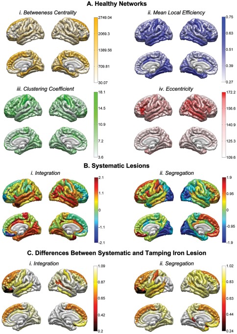
White matter (WM) mapping of the human brain using neuroimaging techniques has gained considerable interest in the neuroscience community. Using diffusion weighted (DWI) and magnetic resonance imaging (MRI), WM fiber pathways between brain regions may be systematically assessed to make inferences concerning their role in normal brain function, influence on behavior, as well as concerning the consequences of network-level brain damage. In this paper, we investigate the detailed connectomics in a noted example of severe traumatic brain injury (TBI) which has proved important to and controversial in the history of neuroscience. We model the WM damage in the notable case of Phineas P. Gage, in whom a "tamping iron" was accidentally shot through his skull and brain, resulting in profound behavioral changes. The specific effects of this injury on Mr. Gage's WM connectivity have not previously been considered in detail. Using computed tomography (CT) image data of the Gage skull in conjunction with modern anatomical MRI and diffusion imaging data obtained in contemporary right handed male subjects (aged 25-36), we computationally simulate the passage of the iron through the skull on the basis of reported and observed skull fiducial landmarks and assess the extent of cortical gray matter (GM) and WM damage. Specifically, we find that while considerable damage was, indeed, localized to the left frontal cortex, the impact on measures of network connectedness between directly affected and other brain areas was profound, widespread, and a probable contributor to both the reported acute as well as long-term behavioral changes. Yet, while significantly affecting several likely network hubs, damage to Mr. Gage's WM network may not have been more severe than expected from that of a similarly sized "average" brain lesion. These results provide new insight into the remarkable brain injury experienced by this noteworthy patient.
Overview
The claims and statistics of this paper haven’t been checked yet.
Run the check — free, no account needed. It reads the open-access text and checks the sources it cites.
Integrity signals
Registry
Cites
Statistics
Figures
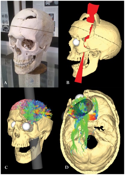
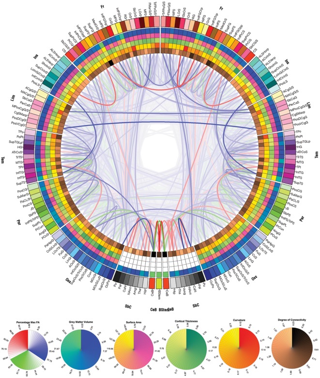
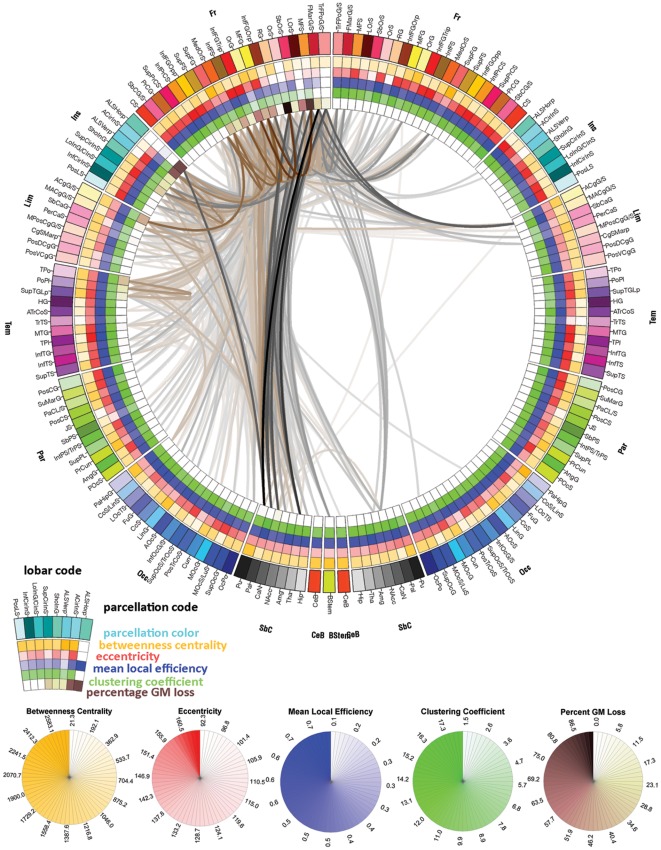
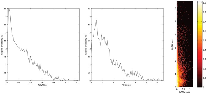
Verification
Claims the paper makes
The claims and statistics of this paper haven’t been checked yet.
Run the check — free, no account needed. It reads the open-access text and checks the sources it cites.
Claims other papers make about this one
Each is checked against this paper’s own text: does it say what the citing paper says it does?
The cited source says what the claim attributes to it.
In the citing paper
“While this indeed confirmed notable local injury, it also identified patterns of widespread and longrange connectivity loss that could also have contributed to the characteristic postinjury behavioral patterns described.”
From Graph theory analysis of complex brain networks: new concepts in brain mapping applied to neurosurgery (2016) · cites this paper as [99]
Self-check
Percentages, sums and reported differences re-computed from the paper’s own numbers.
The self-check needs the paper’s full text.
Citations
Cites
Collective dynamics of ‘small-world’ networks
Watts, D. J., & Strogatz, S. H. (1998) · Nature · doi:10.1038/30918 · Cited by 43,060
Complex brain networks: graph theoretical analysis of structural and functional systems
Bullmore, E., & Sporns, O. (2009) · Nature Reviews Neuroscience · doi:10.1038/nrn2575 · Cited by 11,820
Complex network measures of brain connectivity: Uses and interpretations
Rubinov, M., & Sporns, O. (2010) · NeuroImage · doi:10.1016/j.neuroimage.2009.10.003 · Cited by 11,522
Circos: An information aesthetic for comparative genomicsPreprint
Krzywinski, M. I., Schein, J. E., Birol, İ., Connors, J. M., Gascoyne, R. D., Horsman, D. E., Jones, S. J. M., & Marra, M. A. (2009) · Genome Research · doi:10.1101/gr.092759.109 · Cited by 11,073
Cortical Surface-Based Analysis
Dale, A. M., Fischl, B., & Sereno, M. I. (1999) · NeuroImage · doi:10.1006/nimg.1998.0395 · Cited by 10,910
Fischl, B., Salat, D. H., Busa, E., Albert, M., Dieterich, M., Haselgrove, C., van der Kouwe, A., Killiany, R., Kennedy, D., Klaveness, S., et al. (2002) · Neuron · doi:10.1016/s0896-6273(02)00569-x · Cited by 8,610
Cortical Surface-Based Analysis
Fischl, B., Sereno, M. I., & Dale, A. M. (1999) · NeuroImage · doi:10.1006/nimg.1998.0396 · Cited by 6,147
Measuring the thickness of the human cerebral cortex from magnetic resonance images
Fischl, B. R., & Dale, A. M. (2000) · Proceedings of the National Academy of Sciences · doi:10.1073/pnas.200033797 · Cited by 5,995
Automatically Parcellating the Human Cerebral Cortex
Fischl, B. R. (2003) · Cerebral Cortex · doi:10.1093/cercor/bhg087 · Cited by 4,208
Mapping the structural core of human cerebral cortex
Hagmann, P., Cammoun, L., Gigandet, X., Meuli, R., Honey, C. J., Wedeen, V. J., & Sporns, O. (2008) · PLoS Biology · doi:10.1371/journal.pbio.0060159 · Cited by 4,173
Automatic parcellation of human cortical gyri and sulci using standard anatomical nomenclature
Destrieux, C., Fischl, B., Dale, A., & Halgren, E. (2010) · NeuroImage · doi:10.1016/j.neuroimage.2010.06.010 · Cited by 2,956
The Human Connectome Project: A data acquisition perspective
Essen, D. C. V., Uǧurbil, K., Auerbach, E. J., Barch, D. M., Behrens, T. E. J., Bucholz, R. D., Chang, A., Chen, L., Corbetta, M., Curtiss, S. W., et al. (2012) · NeuroImage · doi:10.1016/j.neuroimage.2012.02.018 · Cited by 2,638
Achard, S., Salvador, R., Whitcher, B., Suckling, J., & Bullmore, E. (2006) · The Journal of Neuroscience · doi:10.1523/jneurosci.3874-05.2006 · Cited by 2,393
Organization, development and function of complex brain networks
SPORNS, O., CHIALVO, D., KAISER, M., & HILGETAG, C. (2004) · Trends in Cognitive Sciences · doi:10.1016/j.tics.2004.07.008 · Cited by 2,055
Networks of the Brain: Quantitative Analysis and Modeling
Sporns, O. (2010) · The MIT Press eBooks · doi:10.7551/mitpress/8476.001.0001 · Cited by 1,750
The Return of Phineas Gage: Clues About the Brain from the Skull of a Famous Patient
Damásio, H., Grabowski, T. J., Frank, R. J., Galaburda, A. M., & Damásio, A. R. (1994) · Science · doi:10.1126/science.8178168 · Cited by 1,520
The human connectome: a complex network
Sporns, O. (2011) · Annals of the New York Academy of Sciences · doi:10.1111/j.1749-6632.2010.05888.x · Cited by 1,440
Zhou, J., Greicius, M. D., Gennatas, E. D., Growdon, M. E., Jang, J. Y., Rabinovici, G. D., Kramer, J. H., Weiner, M., Miller, B. L., & Seeley, W. W. (2010) · Brain · doi:10.1093/brain/awq075 · Cited by 1,016
Measures of degeneracy and redundancy in biological networks
Tononi, G., Sporns, O., & Edelman, G. M. (1999) · Proceedings of the National Academy of Sciences · doi:10.1073/pnas.96.6.3257 · Cited by 632
Modeling the impact of lesions in the human brain
Alstott, J., Breakspear, M., Hagmann, P., Cammoun, L., & Sporns, O. (2009) · PLoS Computational Biology · doi:10.1371/journal.pcbi.1000408 · Cited by 561
Mixture models and exploratory analysis in networks
Newman, M. E. J., & Leicht, E. A. (2007) · Proceedings of the National Academy of Sciences · doi:10.1073/pnas.0610537104 · Cited by 556
Differential Working Memory Load Effects after Mild Traumatic Brain Injury
McAllister, T. W., Sparling, M. B., Flashman, L. A., Guerin, S. J., Mamourian, A. C., & Saykin, A. J. (2001) · NeuroImage · doi:10.1006/nimg.2001.0899 · Cited by 533
Sidaros, A., Engberg, A. W., Sidaros, K., Liptrot, M. G., Herning, M., Petersen, P., Paulson, O. B., Jernigan, T. L., & Rostrup, E. (2007) · Brain · doi:10.1093/brain/awm294 · Cited by 526
Episodic memory and the self in a case of isolated retrograde amnesia
Levine, B. (1998) · Brain · doi:10.1093/brain/121.10.1951 · Cited by 472
Uncinate Fasciculus Findings in Schizophrenia: A Magnetic Resonance Diffusion Tensor Imaging Study
Kubicki, M. R., Westin, C., Maier, S. E., Frumin, M., Nestor, P. G., Salisbury, D. F., Kikinis, R., Jolesz, F. A., McCarley, R. W., & Shenton, M. E. (2002) · American Journal of Psychiatry · doi:10.1176/appi.ajp.159.5.813 · Cited by 454
Self-citationDisrupted axonal fiber connectivity in schizophrenia
Zalesky, A., Fornito, A., Seal, M. L., Cocchi, L., Westin, C.-F., Bullmore, E. T., Egan, G. F., & Pantelis, C. (2011) · Biological Psychiatry · doi:10.1016/j.biopsych.2010.08.022 · Cited by 439
de Haan, W., Pijnenburg, Y. A. L., STRIJERS, R. L. M., van der Made, Y., van der Flier, W. M., Scheltens, P., & Stam, C. J. (2009) · BMC Neuroscience · doi:10.1186/1471-2202-10-101 · Cited by 415
Hines, M. L. (2009) · Frontiers in Neuroinformatics · doi:10.3389/neuro.11.001.2009 · Cited by 414
Recovery from the passage of an iron bar through the head
Harlow, J. M. (1993) · History of Psychiatry · doi:10.1177/0957154x9300401407 · Cited by 403
Effects of focal frontal lesions on response inhibition
Picton, T. W., Stuss, D. T., Alexander, M. P., Shallice, T., Binns, M. A., & Gillingham, S. (2006) · Cerebral Cortex · doi:10.1093/cercor/bhk031 · Cited by 387
Dynamical consequences of lesions in cortical networks
Honey, C. J., & Sporns, O. (2008) · Human Brain Mapping · doi:10.1002/hbm.20579 · Cited by 353
Passage of an Iron Rod through the Head
(1848) · New England Journal of Medicine · doi:10.1056/nejm184812130392001 · Cited by 353
Passage of an Iron Rod Through the Head
Harlow, J. M. (1999) · Journal of Neuropsychiatry · doi:10.1176/jnp.11.2.281 · Cited by 338
Executive dysfunction following traumatic brain injury: Neural substrates and treatment strategies
McDonald, B. C., Flashman, L. A., & Saykin, A. J. (2002) · NeuroRehabilitation · doi:10.3233/nre-2002-17407 · Cited by 302
Lipton, M. L., Gulko, E., Zimmerman, M. E., Friedman, B. W., Kim, M., Gellella, E., Gold, T., Shifteh, K., Ardekani, B. A., & Branch, C. A. (2009) · Radiology · doi:10.1148/radiol.2523081584 · Cited by 291
The Brain of LB1, Homo floresiensis
Falk, D., Hildebolt, C. F., Smith, K., Morwood, M. J., Sutikna, T., Brown, P., Jatmiko, J., Saptomo, E. W., Brunsden, B. S., & Prior, F. (2005) · Science · doi:10.1126/science.1109727 · Cited by 279
MR connectomics: Principles and challenges
Hagmann, P., Cammoun, L., Gigandet, X., Gerhard, S., Grant, P. E., Wedeen, V. J., Meuli, R. A., Thiran, J., Honey, C. J., & Sporns, O. (2010) · Journal of Neuroscience Methods · doi:10.1016/j.jneumeth.2010.01.014 · Cited by 276
Diffusion Tensor Imaging of Mild to Moderate Blast-Related Traumatic Brain Injury and Its Sequelae
Levin, H. S., Wilde, E. A., Troyanskaya, M., Petersen, N. J., Scheibel, R. S., Newsome, M. R., Radaideh, M., Wu, T. C., Yallampalli, R., Chu, Z. D., et al. (2010) · Journal of Neurotrauma · doi:10.1089/neu.2009.1073 · Cited by 263
White matter tract integrity in aging and Alzheimer's disease
Damoiseaux, J. S., Smith, S. M., Witter, M. P., Sanz‐Arigita, E., Barkhof, F., Scheltens, P., Stam, C. J., Zarei, M., & Rombouts, S. A. R. B. (2008) · Human Brain Mapping · doi:10.1002/hbm.20563 · Cited by 259
Genetics of white matter development: A DTI study of 705 twins and their siblings aged 12 to 29
Chiang, M.-C., McMahon, K. L., de Zubicaray, G. I., Martin, N. G., Hickie, I. B., Toga, A. W., Wright, M. J., & Thompson, P. M. (2010) · NeuroImage · doi:10.1016/j.neuroimage.2010.10.015 · Cited by 255
Self-citationSimulation of robustness against lesions of cortical networks
Kaiser, M., Martin, R., Andras, P., & Young, M. P. (2007) · European Journal of Neuroscience · doi:10.1111/j.1460-9568.2007.05574.x · Cited by 244
Stuss, D. T. (2011) · Current Opinion in Neurology · doi:10.1097/wco.0b013e32834c7eb9 · Cited by 234
The fractions of short- and long-range connections in the visual cortex
Stepanyants, A., Martinez, L. M., Ferecskó, A. S., & Kisvárday, Z. F. (2009) · Proceedings of the National Academy of Sciences · doi:10.1073/pnas.0810390106 · Cited by 208
Hasan, K. M., Iftikhar, A., Kamali, A., Kramer, L. A., Ashtari, M., Cirino, P. T., Papanicolaou, A. C., Fletcher, J. M., & Ewing‐Cobbs, L. (2009) · Brain Research · doi:10.1016/j.brainres.2009.04.025 · Cited by 187
Early hominid brain evolution: a new look at old endocasts
Falk, D., Redmond, J. C., Guyer, J., Conroy, C., Recheis, W. A., Weber, G. W., & Seidler, H. (2000) · Journal of Human Evolution · doi:10.1006/jhev.1999.0378 · Cited by 174
The inferior longitudinal fasciculus: A reexamination in humans and monkeys
Tusa, R. J., & Ungerleider, L. G. (1985) · Annals of Neurology · doi:10.1002/ana.410180512 · Cited by 172
Reduced white matter integrity in attention-deficit hyperactivity disorder
Hamilton, L. S., Levitt, J. G., O'Neill, J., Alger, J. R., Luders, E., Phillips, O. R., Caplan, R., Toga, A. W., McCracken, J., & Narr, K. L. (2008) · NeuroReport · doi:10.1097/wnr.0b013e3283174415 · Cited by 167
Self-citationNeuroplasticity following non-penetrating traumatic brain injury
Levin, H. S. (2003) · Brain Injury · doi:10.1080/0269905031000107151 · Cited by 162
An odd kind of fame: stories of Phineas Gage
(2001) · Choice Reviews Online · doi:10.5860/choice.38-2778 · Cited by 157
Efficient, Distributed and Interactive Neuroimaging Data Analysis using the LONI Pipeline
Dinov, I. D. (2009) · Frontiers in Neuroinformatics · doi:10.3389/neuro.11.022.2009 · Cited by 149
Andrei Irimia, A., Chambers, M. C., Torgerson, C. M., & Horn, J. D. V. (2012) · NeuroImage · doi:10.1016/j.neuroimage.2012.01.107 · Cited by 142
Self-citationNeuroimaging Study Designs, Computational Analyses and Data Provenance Using the LONI Pipeline
Dinov, I. D., Lozev, K., Petrosyan, P., Liu, Z., Eggert, P., Pierce, J., Zamanyan, A., Chakrapani, S., Horn, J. D. V., Parker, D. S., et al. (2010) · PLoS ONE · doi:10.1371/journal.pone.0013070 · Cited by 136
Self-citationMood Change following Bilateral Hemisphere Brain Injury
Lipsey, J. R., Robinson, R. G., Pearlson, G. D., Rao, K., & Price, T. R. (1983) · The British Journal of Psychiatry · doi:10.1192/bjp.143.3.266 · Cited by 119
The Tale of Phineas Gage, Digitally Remastered
Ratiu, P., Talos, I., Haker, S. J., Lieberman, D. E., & Everett, P. C. (2004) · Journal of Neurotrauma · doi:10.1089/089771504774129964 · Cited by 109
Social and Emotional Decision-making Following Frontal Lobe Injury
Clark, L., & Manes, F. F. (2004) · Neurocase · doi:10.1080/13554790490882799 · Cited by 106
An odd kind of fame. Stories of Phineas Gage
Hodges, J. R. (2001) · Journal of Neurology Neurosurgery & Psychiatry · doi:10.1136/jnnp.71.1.136c · Cited by 102
Traumatic brain injury, major depression, and diffusion tensor imaging: Making connections
Maller, J. J., Thomson, R. H., Lewis, P., Rose, S., Pannek, K., & Fitzgerald, P. B. (2010) · Brain Research Reviews · doi:10.1016/j.brainresrev.2010.04.003 · Cited by 100
Shaw, P., Bramham, J., Lawrence, E. J., Morris, R., Baron-Cohen, S., & David, A. S. (2005) · Journal of Cognitive Neuroscience · doi:10.1162/0898929054985491 · Cited by 96
Principles of recovery from traumatic brain injury: Reorganization of functional networks
Castellanos, N. P., Leyva, I., Buldú, J. M., Bajo, R., Paúl, N., Cuesta, P., Ordóñez, V. E., Pascua, C. L., Boccaletti, S., Maestú, F., et al. (2011) · NeuroImage · doi:10.1016/j.neuroimage.2010.12.046 · Cited by 96
Aarabi, B., Taghipour, M., Haghnegahdar, A., Farokhi, M. R., & Mobley, L. W. (2000) · Neurosurgical FOCUS · doi:10.3171/foc.2000.8.1.155 · Cited by 86
Art. I.—Dr. Harlowʼs Case of Recovery from the passage of an Iron Bar through the Head.
BIGELOW, H. J. (1850) · The American Journal of the Medical Sciences · doi:10.1097/00000441-185016390-00001 · Cited by 80
Fiber tractography reveals disruption of temporal lobe white matter tracts in schizophrenia
Phillips, O. R., Nuechterlein, K. H., Clark, K. A., Hamilton, L. S., Asarnow, R. F., Hageman, N. S., Toga, A. W., & Narr, K. L. (2009) · Schizophrenia Research · doi:10.1016/j.schres.2008.10.019 · Cited by 80
Self-citationAn Examination of the Degrees of Freedom of Human Jaw Motion in Speech and Mastication
Ostry, D. J., Vatikiotis‐Bateson, E., & Gribble, P. L. (1997) · Journal of Speech Language and Hearing Research · doi:10.1044/jslhr.4006.1341 · Cited by 78
Baird, A. A., Colvin, M. K., Horn, J. D. V., Inati, S., & Gazzaniga, M. S. (2005) · Journal of Cognitive Neuroscience · doi:10.1162/0898929053467569 · Cited by 76
Traumatic brain injury alters the functional brain network mediating working memory
Kasahara, M., Menon, D. K., Salmond, C. H., Outtrim, J. G., Tavares, J. V. T., Carpenter, T. A., Pickard, J. D., Sahakian, B. J., & Stamatakis, E. A. (2011) · Brain Injury · doi:10.3109/02699052.2011.608210 · Cited by 75
MacKenzie‐Graham, A. J., Horn, J. D. V., Woods, R. P., Crawford, K. L., & Toga, A. W. (2008) · NeuroImage · doi:10.1016/j.neuroimage.2008.04.186 · Cited by 66
Self-citationInvestigation of resting-state EEG functional connectivity in frontotemporal lobar degeneration
Pijnenburg, Y. A. L., STRIJERS, R. L. M., van der Made, Y., van der Flier, W. M., Scheltens, P., & Stam, C. J. (2008) · Clinical Neurophysiology · doi:10.1016/j.clinph.2008.02.024 · Cited by 63
Andrei Irimia, A., Chambers, M. C., Torgerson, C. M., Filippou-Frye, M., Hovda, D. A., Alger, J. R., Gerig, G., Toga, A. W., Vespa, P. M., Kikinis, R., et al. (2012) · Frontiers in Neurology · doi:10.3389/fneur.2012.00010 · Cited by 62
Self-citationJohnson, C. P., Juranek, J. J., Kramer, L. A., Prasad, M. R., Swank, P. R., & Ewing‐Cobbs, L. (2011) · Journal of the International Neuropsychological Society · doi:10.1017/s1355617711000464 · Cited by 56
Damage to the lateral prefrontal cortex impairs familiarity but not recollection
Aly, M., Yonelinas, A. P., Kishiyama, M. M., & Knight, R. T. (2011) · Behavioural Brain Research · doi:10.1016/j.bbr.2011.07.043 · Cited by 49
White Matter Changes Associated With Psychotic Symptoms in Alzheimer's Disease Patients
Lee, D. Y., Choo, I. H., Kim, K. W., Jhoo, J. H., Youn, J. C., Lee, U.-J., & Woo, J. I. (2006) · Journal of Neuropsychiatry · doi:10.1176/jnp.2006.18.2.191 · Cited by 49
The Tale of Phineas Gage, Digitally Remastered
Ratiu, P., & Talos, I. (2004) · New England Journal of Medicine · doi:10.1056/nejmicm031024 · Cited by 49
The Goulstonian Lectures on the Localisation of Cerebral Disease
Ferrier, D. (1878) · BMJ · doi:10.1136/bmj.1.904.591 · Cited by 45
Penetrating craniocerebral injuries from nail-gun use
Selvanathan, S. K., Goldschlager, T., McMillen, J., & Campbell, S. (2007) · Journal of Clinical Neuroscience · doi:10.1016/j.jocn.2006.02.017 · Cited by 43
Neurophysiological Alterations During Strategy-Based Verbal Learning in Traumatic Brain Injury
Strangman, G. E., Goldstein, R., O’Neil-Pirozzi, T. M., Kelkar, K., Supelana, C. J., Burke, D. T., Katz, D. I., Rauch, S. L., Savage, C. R., & Glenn, M. B. (2008) · Neurorehabilitation and neural repair · doi:10.1177/1545968308324225 · Cited by 41
Changes in white matter microstructure in patients with TLE and hippocampal sclerosis
Knake, S., Salat, D. H., Halgren, E., Halko, M. A., Greve, D. N., & Grant, P. E. (2009) · Epileptic Disorders · doi:10.1684/epd.2009.0272 · Cited by 36
Inhibition and the control of behavior
Macmillan, M. (1992) · Brain and Cognition · doi:10.1016/0278-2626(92)90038-n · Cited by 34
Validation of plaster endocast morphology through 3D CT image analysis
Schoenemann, P. T., Gee, J. C., Avants, B., Holloway, R. L., Monge, J., & Lewis, J. (2006) · American Journal of Physical Anthropology · doi:10.1002/ajpa.20499 · Cited by 32
Penetrating ballistic-like frontal brain injury caused by a metallic rod
Pascual, J. M., Navas, M., & Carrasco, R. (2009) · Acta Neurochirurgica · doi:10.1007/s00701-009-0222-8 · Cited by 30
Pal, D., Gupta, R. K., Agarwal, S., Yadav, A., Ojha, B. K., Awasthi, A., Rathore, R. K. S., Pandey, C. M., & Narayana, P. A. (2011) · Clinical Neurology and Neurosurgery · doi:10.1016/j.clineuro.2011.12.002 · Cited by 29
Face to Face with Phineas Gage
Wilgus, J., & Wilgus, B. (2009) · Journal of the History of the Neurosciences · doi:10.1080/09647040903018402 · Cited by 19
The Goulstonian Lectures on the Localisation of Cerebral Disease
Ferrier, D. (1878) · BMJ · doi:10.1136/bmj.1.900.443 · Cited by 17
Darwin and the ghost of Phineas Gage: Neuro-evolution and the social brain
Dunbar, R. (2009) · Cortex · doi:10.1016/j.cortex.2009.05.005 · Cited by 13
Randolph, J. (1837) · The American Journal of the Medical Sciences · doi:10.1097/00000441-183720410-00001 · Cited by 13
Essen, D. C. V. (2011) · Journal of Vision · doi:10.1167/11.15.8 · Cited by 13
Gunshot wound to the head with full recovery
Rothschild, M. A., & Schneider, V. (2000) · International Journal of Legal Medicine · doi:10.1007/s004149900120 · Cited by 13
Attentional Inhibition in Patients with Focal Frontal Lobe Lesions
McDonald, C. R., Bauer, R. M., Filoteo, J. V., Grande, L. J., Roper, S. N., & Gilmore, R. L. (2005) · Journal of Clinical and Experimental Neuropsychology · doi:10.1080/13803390490520454 · Cited by 13
Brain impalement by an angle metal bar
Ordia, J. I. (2008) · Clinical Neurology and Neurosurgery · doi:10.1016/j.clineuro.2008.10.016 · Cited by 9
REVIEW OF NEUROPSYCHIATRY FOR 1940
COBB, S. S. (1940) · Archives of Internal Medicine · doi:10.1001/archinte.1940.00190180153011 · Cited by 8
Fernie, H. M. (1889) · BMJ · doi:10.1136/bmj.1.1469.443-b · Cited by 6
Postmortem Diagnosis of Leukodystrophies
Shields, L., Handy, T., Parker, J. C., & Burns, C. (1998) · Journal of Forensic Sciences · doi:10.1520/jfs14359j · Cited by 3
Deep Penetrating Orbitocerebral Steel Spring Injury With Minimal Sequelae: A Case Report
Nobe, M. Y., Yoon, S. J., Wachter, B., & Tao, J. (2010) · Ophthalmic Plastic and Reconstructive Surgery · doi:10.1097/iop.0b013e3181cff655 · Cited by 1
High-resolution intersubject averaging and a coordinate system for the cortical surface
Fischl, B., Sereno, M. I., Tootell, R. B. H., & Dale, A. M. (1999) · Human Brain Mapping · doi:10.1002/(sici)1097-0193(1999)8:4
Research works printed but not linked: 7
- E Dupuy, 1877, A critical review of the prevailing theories concerning the physiology and pathology of the brain., Medical Times and Gazette, ii, 11
- AM Dale, 1999, Cortical surface-based analysis - I. Segmentation and surface reconstruction., Neuro Image, 9, 179
- B Fischl, 2002, Whole Brain Segmentation: Automated Labeling of Neuroanatomical Structures in the Human Brain., Neurotechnique, 33, 341
- B Fischl, 1999, Cortical surface-based analysis. II: Inflation, flattening, and a surface-based coordinate system., Neuroimage, 9, 195
- J Alstott, 2009, Modeling the impact of lesions in the human brain., Plos Computational Biology, 5, e1000408, 10.1371/journal.pcbi.1000408 · DOI · Paper page
- I Dinov, 2010, Neuroimaging Study Designs, Computational Analyses and Data Provenance Using the LONI Pipeline., PLoS ONE
- I Dinov, 2010, Efficient, distributed and interactive neuroimaging data analysis using the LONI pipeline., Frontiers in Neuroinformatics, 3, 1
Cited by
Sorted by citations
Network hubs in the human brain
van den Heuvel, M. P., & Sporns, O. (2013) · Trends in Cognitive Sciences · doi:10.1016/j.tics.2013.09.012 · Cited by 2,123
The social brain in psychiatric and neurological disorders
Kennedy, D. P., & Adolphs, R. (2012) · Trends in Cognitive Sciences · doi:10.1016/j.tics.2012.09.006 · Cited by 846
Network dysfunction after traumatic brain injury
Sharp, D. J., Scott, G., & Leech, R. (2014) · Nature Reviews Neurology · doi:10.1038/nrneurol.2014.15 · Cited by 657
Brain Networks in Schizophrenia
van den Heuvel, M. P., & Fornito, A. (2014) · Neuropsychology Review · doi:10.1007/s11065-014-9248-7 · Cited by 531
Understanding Emotions: Origins and Roles of the Amygdala
Šimić, G., Tkalčić, M., Vukić, V., Mulc, D., Španić, E., Šagud, M., Olucha-Bordonau, F. E., Vukšić, M., & Hof, P. R. (2021) · Biomolecules · doi:10.3390/biom11060823 · Cited by 631
Metadata
- Authors
- Van Horn, J. D., Irimia, A., Torgerson, C. M., Chambers, M. C., Kikinis, R., & Toga, A. W.
- Affiliations
- University of California, Los Angeles (US)
- Brigham and Women's Hospital (US)
- Venue
- PLoS ONE
- Publisher
- Public Library of Science (PLoS)
- Published
- 2012-05-16
- Volume
- 7
- Issue
- 5
- Pages
- e37454
- ISSN
- 1932-6203 (ISSN-L)
- PMC
- PMC3353935
- OpenAlex
- W2043275179
- Cited by
- 196
- References
- 95
- Citations by year
- 2026: 6 · 2025: 3 · 2024: 5 · 2023: 6 · 2022: 7 · 2021: 16 · 2020: 34 · 2019: 29 · 2018: 18 · 2017: 8 · 2016: 15 · 2015: 19 · 2014: 9 · 2013: 15 · 2012: 4
- FWCI
- 11.35
- Type
- journal article
- Language
- en
- Open access
- gold
- doi.org
- Locations
- PLoS ONE (publishedVersion, cc-by)
- Europe PMC (PubMed Central) (publishedVersion, cc-by)
- PubMed Central (publishedVersion, cc-by)
- PubMed (publishedVersion)
- Digital Access to Scholarship at Harvard (DASH) (Harvard University) (submittedVersion, cc-by)
- DOAJ (DOAJ: Directory of Open Access Journals) (submittedVersion, cc-by-sa)
- Figshare (submittedVersion, cc-by)
- Funders
- University of California, Los Angeles
- National Institute of Mental Health (RC1MH088194)
- National Institute of Biomedical Imaging and Bioengineering (U54EB005149)
- National Center for Research Resources (P41RR013642, P41RR013218)
- Topics
- Health Sciences › Medicine › Radiology, Nuclear Medicine and Imaging › Advanced Neuroimaging Techniques and Applications
- Life Sciences › Neuroscience › Cognitive Neuroscience › Functional Brain Connectivity Studies
- Health Sciences › Medicine › Clinical Neurology › Traumatic Brain Injury Research
- Keywords
- Phineas Gage, traumatic brain injury, white matter injury, connectomics, diffusion MRI, brain network connectivity, frontal lobe lesions, network hubs, history of neuroscience
- MeSH terms
- Adult, Brain Injuries, Frontal Lobe, Humans, Magnetic Resonance Imaging, Male, Nerve Fibers, Myelinated, Personality Disorders, Tomography, X-Ray Computed, Vermont, History, 19th Century, Neuroimaging
About these checks
Registry, retraction, Cites and statistics results are computed, not judged, with one exception: a printed reference that no index lists is read once by a model before it counts as a citation error. Claims are extracted by a model and kept only when their quote appears verbatim in the text; each cited source is then checked, with a model’s help, for whether it says what the claim relies on. Those verdicts can be wrong — read the claim’s quote and the cited source before relying on one.
Statistics and arithmetic are recomputed, not judged.
Confidence bands are shown only for fields calibrated on our annotated test set. None is calibrated yet, so no band is shown.
What counts as a claim ›How we measure confidence ›How we check this ›
Are you an author, or do you see a problem with this page? Request a review
