

The DOI matches its Crossref record; 33 of the 33 references it cites were found in a registry or an index.
Glycerol monolaurate antibacterial activity in broth and biofilm cultures.
Open accessAbstract
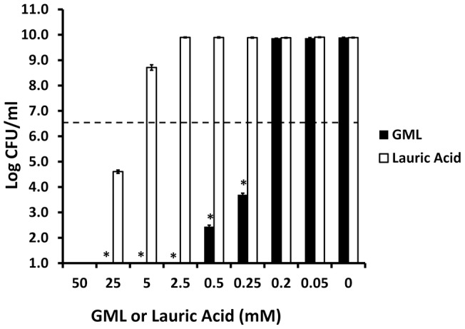
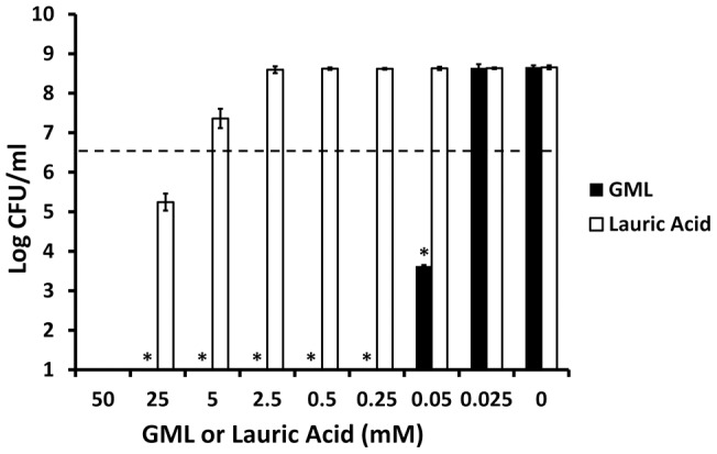
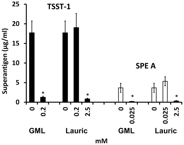
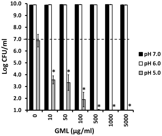
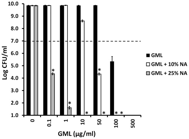
BACKGROUND: Glycerol monolaurate (GML) is an antimicrobial agent that has potent activity against gram-positive bacteria. This study examines GML antibacterial activity in comparison to lauric acid, in broth cultures compared to biofilm cultures, and against a wide range of gram-positive, gram-negative, and non-gram staining bacteria. METHODOLOGY/PRINCIPAL FINDINGS: GML is ≥ 200 times more effective than lauric acid in bactericidal activity, defined as a ≥ 3 log reduction in colony-forming units (CFU)/ml, against Staphylococcus aureus and Streptococcus pyogenes in broth cultures. Both molecules inhibit superantigen production by these organisms at concentrations that are not bactericidal. GML prevents biofilm formation by Staphylococcus aureus and Haemophilus influenzae, as representative gram-positive and gram-negative organisms, tested in 96 well microtiter plates, and simultaneously is bactericidal for both organisms in mature biofilms. GML is bactericidal for a wide range of potential bacterial pathogens, except for Pseudomonas aeruginosa and Enterobacteriaceae. In the presence of acidic pH and the cation chelator ethylene diamine tetraacetic acid, GML has greatly enhanced bactericidal activity for Pseudomonas aeruginosa and Enterobacteriaceae. Solubilization of GML in a nonaqueous delivery vehicle (related to K-Y Warming®) enhances its bactericidal activity against S. aureus. Both R and S, and 1 and 2 position lauric acid derivatives of GML exhibit bactericidal activity. Despite year-long passage of Staphylococcus aureus on sub-growth inhibitory concentrations of GML (0.5 x minimum bactericidal concentration), resistance to GML did not develop. CONCLUSIONS/SIGNIFICANCE: GML may be useful as a broad-spectrum human or animal topical microbicide and may be useful as an environmental surface microbicide for management of bacterial infections and contamination.
Overview
The claims and statistics of this paper haven’t been checked yet.
Run the check — free, no account needed. It reads the open-access text and checks the sources it cites.
Integrity signals
Registry
Cites
Statistics
Figures
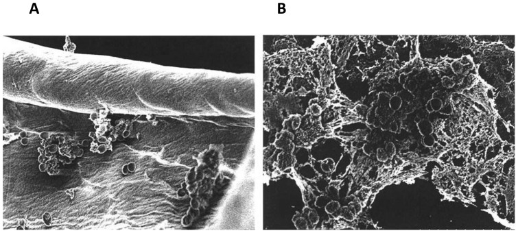
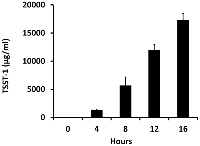
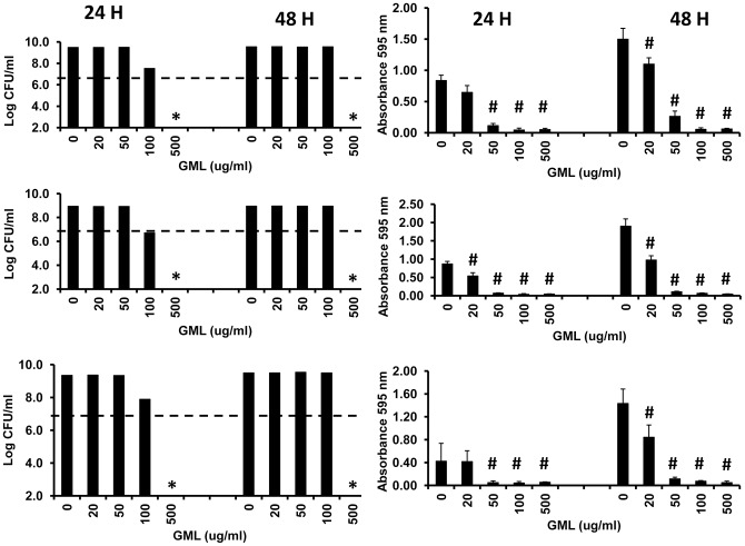
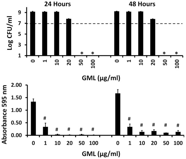
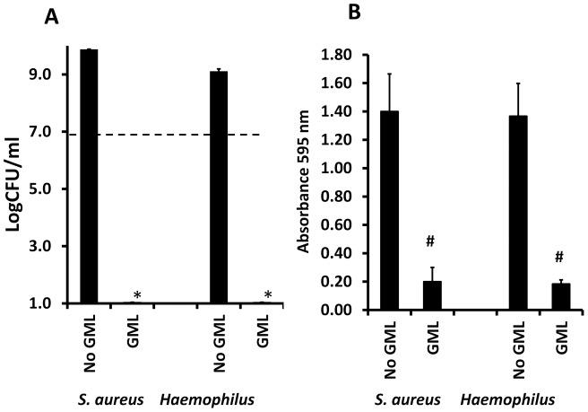
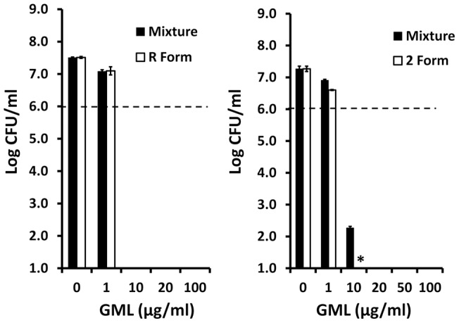
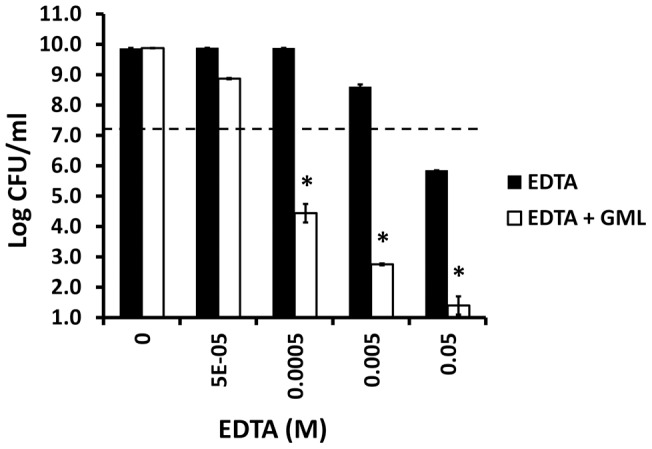
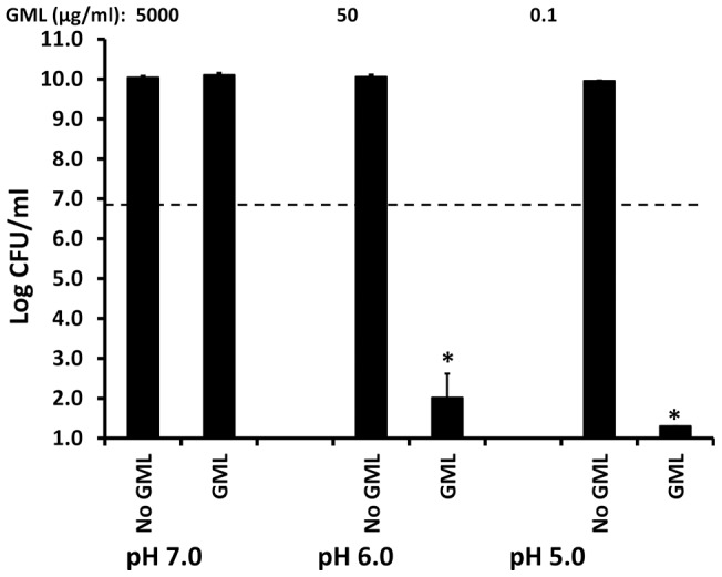
Verification
Claims the paper makes
The claims and statistics of this paper haven’t been checked yet.
Run the check — free, no account needed. It reads the open-access text and checks the sources it cites.
Claims other papers make about this one
Each is checked against this paper’s own text: does it say what the citing paper says it does?
The source backs part of the claim, not all of it — or backs it for a different population, condition or comparison.
In the citing paper
“because these bacteria are also unlikely to acquire resistance to these compounds (Petschow et al., 1996; Schlievert and Peterson, 2012; Jackman et al., 2020).”
From Insect Derived Lauric Acid as Promising Alternative Strategy to Antibiotics in the Antimicrobial Resistance Scenario (2021) · cites this paper as Schlievert and Peterson, 2012
Experimental — accuracy not yet measuredThe source backs part of the claim, not all of it — or backs it for a different population, condition or comparison.
In the citing paper
“Their ability to destabilize the bacterial cell membrane makes them promising candidates among novel antimicrobials”
From Insect Derived Lauric Acid as Promising Alternative Strategy to Antibiotics in the Antimicrobial Resistance Scenario (2021) · cites this paper as Schlievert and Peterson, 2012
Experimental — accuracy not yet measured
Self-check
Percentages, sums and reported differences re-computed from the paper’s own numbers.
The self-check needs the paper’s full text.
Citations
Cites
Bacterial biofilms: from the Natural environment to infectious diseases
Hall‐Stoodley, L., Costerton, J. W., & Stoodley, P. (2004) · Nature Reviews Microbiology · doi:10.1038/nrmicro821 · Cited by 7,133
Microtiter Plate Assay for Assessment of Listeria monocytogenes Biofilm Formation
Djordjevic, D., Wiedmann, M., & McLandsborough, L. (2002) · Applied and Environmental Microbiology · doi:10.1128/aem.68.6.2950-2958.2002 · Cited by 960
Schlievert, P. M., Shands, K. N., Dan, B. B., Schmid, G. P., & Nishimura, R. D. (1981) · The Journal of Infectious Diseases · doi:10.1093/infdis/143.4.509 · Cited by 666
Self-citationGlycerol monolaurate prevents mucosal SIV transmission
Li, Q., Estes, J. D., Schlievert, P. M., Duan, L., Brosnahan, A. J., Southern, P. J., Reilly, C., Peterson, M. L., SCHULTZ‐DARKEN, N. J., Brunner, K. G., et al. (2009) · Nature · doi:10.1038/nature07831 · Cited by 638
Self-citationBergdoll, M. S. (1981) · The Lancet · doi:10.1016/s0140-6736(81)92186-3 · Cited by 630
Regulation of exoprotein gene expression in Staphylococcus aureus by agr
Recsei, P. A., Kreiswirth, B. N., O’Reilly, M., Schlievert, P. M., Gruss, A., & Novick, R. P. (1986) · Molecular and General Genetics MGG · doi:10.1007/bf00330517 · Cited by 619
Self-citationABSENCE OF CROSS-REACTIVITY BETWEEN CAPTOPRIL AND ENALAPRIL
Navis, G. J., de Jong, P. E., Kallenberg, C. G. M., de Monchy, J., & de Zeeuw, D. (1984) · The Lancet · doi:10.1016/s0140-6736(84)92353-5 · Cited by 485
Inactivation of enveloped viruses and killing of cells by fatty acids and monoglycerides
Thormar, H., Isaacs, C. E., Brown, H. R., Barshatzky, M., & Pessolano, T. L. (1987) · Antimicrobial Agents and Chemotherapy · doi:10.1128/aac.31.1.27 · Cited by 377
Kaufmann, G. F., Sartorio, R., Lee, S., Rogers, C. J., Meijler, M. M., Moss, J. A., Clapham, B., Brogan, A. P., Dickerson, T. J., & Janda, K. D. (2004) · Proceedings of the National Academy of Sciences · doi:10.1073/pnas.0408639102 · Cited by 336
The Methyl-Branched Fortifications of Mycobacterium tuberculosis
Minnikin, D. E., Kremer, L., Dover, L. G., & Besra, G. S. (2002) · Chemistry & Biology · doi:10.1016/s1074-5521(02)00142-4 · Cited by 268
Clindamycin-Induced Suppression of Toxic-Shock Syndrome-Associated Exotoxin Production
Schlievert, P. M., & Kelly, J. A. (1984) · The Journal of Infectious Diseases · doi:10.1093/infdis/149.3.471 · Cited by 162
Self-citationProjan, S. J., Brown-Skrobot, S., Schlievert, P. M., Vandenesch, F., & Novick, R. P. (1994) · Journal of Bacteriology · doi:10.1128/jb.176.14.4204-4209.1994 · Cited by 151
Self-citationDeLeo, F. R., Kennedy, A. D., Chen, L., Wardenburg, J. B., Kobayashi, S. D., Mathema, B., Braughton, K. R., Whitney, A. R., Villaruz, A. E., Martens, C., et al. (2011) · Proceedings of the National Academy of Sciences · doi:10.1073/pnas.1111084108 · Cited by 148
Effect of glycerol monolaurate on bacterial growth and toxin production
Schlievert, P. M., Deringer, J. R., Kim, M. H., Projan, S. J., & Novick, R. P. (1992) · Antimicrobial Agents and Chemotherapy · doi:10.1128/aac.36.3.626 · Cited by 127
Self-citationTHE ROLE OF TEMPERATE BACTERIOPHAGE IN THE PRODUCTION OF ERYTHROGENIC TOXIN BY GROUP A STREPTOCOCCI
Zabriskie, J. B. (1964) · The Journal of Experimental Medicine · doi:10.1084/jem.119.5.761 · Cited by 122
Ruzin, A., & Novick, R. P. (2000) · Journal of Bacteriology · doi:10.1128/jb.182.9.2668-2671.2000 · Cited by 105
Glycerol Monolaurate Inhibits the Effects of Gram-Positive Select Agents on Eukaryotic Cells
Peterson, M. L., & Schlievert, P. M. (2006) · Biochemistry · doi:10.1021/bi051992u · Cited by 91
Self-citationDelauné, A., Poupel, O., Mallet, A., Coïc, Y., Msadek, T., & Dubrac, S. (2011) · PLoS ONE · doi:10.1371/journal.pone.0017054 · Cited by 88
Lowery, C. A., Park, J., Gloeckner, C., Meijler, M. M., Mueller, R., Boshoff, H. I., Ulrich, R. L., Clifton E. Barry, Bartlett, D. H., Kravchenko, V. V., et al. (2009) · Journal of the American Chemical Society · doi:10.1021/ja9056079 · Cited by 85
Giantonio, B. J., Alpaugh, R. K., Schultz, J., McAleer, C., Newton, D. W., Shannon, B. J., Guédez, Y., Kotb, M., Vı́tek, L., Persson, R., et al. (1997) · Journal of Clinical Oncology · doi:10.1200/jco.1997.15.5.1994 · Cited by 83
Studies on the Mode of Action of Reutericyclin
Gänzle, M. G., & Vogel, R. F. (2003) · Applied and Environmental Microbiology · doi:10.1128/aem.69.2.1305-1307.2003 · Cited by 80
Inactivation of Visna Virus and Other Enveloped Viruses by Free Fatty Acids and Monoglycerides
Thormar, H., Isaacs, C. E., KIM, K. S., & Brown, H. R. (1994) · Annals of the New York Academy of Sciences · doi:10.1111/j.1749-6632.1994.tb38948.x · Cited by 74
Strandberg, K. L., Peterson, M. L., Lin, Y., Pack, M. C., Chase, D. J., & Schlievert, P. M. (2009) · Antimicrobial Agents and Chemotherapy · doi:10.1128/aac.01151-09 · Cited by 69
Self-citationSchlievert, P. M., Strandberg, K. L., Brosnahan, A. J., Peterson, M. L., Pambuccian, S. E., Nephew, K. R., Brunner, K. G., SCHULTZ‐DARKEN, N. J., & Haase, A. T. (2008) · Antimicrobial Agents and Chemotherapy · doi:10.1128/aac.00989-08 · Cited by 68
Self-citationα and β Chains of Hemoglobin Inhibit Production of Staphylococcus aureus Exotoxins
Schlievert, P. M., Case, L. C., Nemeth, K. A., Davis, C., Sun, Y., Qin, W., Wang, F., Brosnahan, A. J., Mleziva, J. A., Peterson, M. L., et al. (2007) · Biochemistry · doi:10.1021/bi701202w · Cited by 59
Self-citationFood-Grade Chemicals for Use in Designing Food Preservative Systems
Kabara, J. J. (1981) · Journal of Food Protection · doi:10.4315/0362-028x-44.8.633 · Cited by 52
Lin, Y., Schlievert, P. M., Anderson, M. J., Fair, C., Schaefers, M. M., Muthyala, R., & Peterson, M. L. (2009) · PLoS ONE · doi:10.1371/journal.pone.0007499 · Cited by 50
Self-citationGlycerol Monolaurate Inhibits Virulence Factor Production in Bacillus anthracis
Vetter, S. M., & Schlievert, P. M. (2005) · Antimicrobial Agents and Chemotherapy · doi:10.1128/aac.49.4.1302-1305.2005 · Cited by 49
Self-citationPechous, R. D., Ledala, N., Wilkinson, B. J., & Jayaswal, R. K. (2004) · Antimicrobial Agents and Chemotherapy · doi:10.1128/aac.48.8.3057-3063.2004 · Cited by 41
Staphylococcus aureus Exotoxins Are Present In Vivo in Tampons
Schlievert, P. M., Nemeth, K. A., Davis, C., Peterson, M. L., & Jones, B. E. (2010) · Clinical and Vaccine Immunology · doi:10.1128/cvi.00483-09 · Cited by 41
Self-citationStrandberg, K. L., Peterson, M. L., Schaefers, M. M., Case, L. C., Pack, M. C., Chase, D. J., & Schlievert, P. M. (2009) · Clinical Infectious Diseases · doi:10.1086/644614 · Cited by 34
Self-citationEvidence for Two Molecular Forms of Streptococcal Erythrogenic Toxin
Nauciel, C., Blass, J., Mangalo, R., & Raynaud, M. (1969) · European Journal of Biochemistry · doi:10.1111/j.1432-1033.1969.tb00754.x · Cited by 25
Reiser, R. F., Hinzman, S. J., & Bergdoll, M. S. (1987) · Journal of Clinical Microbiology · doi:10.1128/jcm.25.8.1450-1452.1987 · Cited by 23
Research works printed but not linked: 1
- JJ Kabara, 1980, GRAS antimicrobial agents for cosmetic products., J Soc Cosmet Chem, 31, 1
Cited by
Sorted by citations
Yoon, B., Jackman, J., Valle-González, E., & Cho, N.-J. (2018) · International Journal of Molecular Sciences · doi:10.3390/ijms19041114 · Cited by 637
Bacterial Biofilm Eradication Agents: A Current Review
Verderosa, A. D., Totsika, M., & Fairfull‐Smith, K. E. (2019) · Frontiers in Chemistry · doi:10.3389/fchem.2019.00824 · Cited by 577
Staphylococcal and Streptococcal Superantigen Exotoxins
Spaulding, A. R., Salgado‐Pabón, W., Kohler, P. L., Horswill, A. R., Leung, D. Y. M., & Schlievert, P. M. (2013) · Clinical Microbiology Reviews · doi:10.1128/cmr.00104-12 · Cited by 566
Self-citationProspects of using nanotechnology for food preservation, safety, and security
Bajpai, V. K., Kamle, M., Shukla, S., Mahato, D. K., Chandra, P., Hwang, S. K., Kumar, P., Huh, Y. S., & Han, Y. (2018) · Journal of Food and Drug Analysis · doi:10.1016/j.jfda.2018.06.011 · Cited by 464
Ramanathan, S., Santhakumari, S., Poonguzhali, P., Mani, G., Dyavaiah, M., & Xiangmin, L. (2021) · Frontiers in Microbiology · doi:10.3389/fmicb.2021.676458 · Cited by 363
Metadata
- Authors
- Schlievert, P. M., & Peterson, M. L.
- Affiliations
- University of Iowa (US)
- University of Minnesota (US)
- Corresponding author
- Schlievert, Patrick M.
- Venue
- PLoS ONE
- Publisher
- Public Library of Science (PLoS)
- Published
- 2012-07-11
- Volume
- 7
- Issue
- 7
- Pages
- e40350
- ISSN
- 1932-6203 (ISSN-L)
- PMC
- PMC3394780
- OpenAlex
- W2035186660
- Cited by
- 198
- References
- 33
- Citations by year
- 2026: 6 · 2025: 13 · 2024: 32 · 2023: 15 · 2022: 15 · 2021: 23 · 2020: 18 · 2019: 20 · 2018: 19 · 2017: 10 · 2016: 11 · 2015: 7 · 2014: 3 · 2013: 5
- FWCI
- 2.21
- Type
- journal article
- Language
- en
- Open access
- gold
- doi.org
- Locations
- PLoS ONE (publishedVersion, cc-by)
- Europe PMC (PubMed Central) (publishedVersion, cc-by)
- PubMed Central (publishedVersion, cc-by)
- PubMed (publishedVersion)
- DOAJ (DOAJ: Directory of Open Access Journals) (submittedVersion, cc-by-sa)
- Funders
- National Institutes of Health
- National Institute of Allergy and Infectious Diseases (U54 AI 57153, R01AI073366, R01 AI73366, U54AI057153, R01AI074283)
- Topics
- Life Sciences › Immunology and Microbiology › Applied Microbiology and Biotechnology › Antimicrobial agents and applications
- Life Sciences › Agricultural and Biological Sciences › Food Science › Essential Oils and Antimicrobial Activity
- Life Sciences › Biochemistry, Genetics and Molecular Biology › Molecular Biology › Bacterial biofilms and quorum sensing
- Keywords
- glycerol monolaurate, lauric acid, antibacterial activity, biofilm inhibition, Staphylococcus aureus, Streptococcus pyogenes, Haemophilus influenzae, Pseudomonas aeruginosa, Enterobacteriaceae, bactericidal activity, microbicides, topical antimicrobial agents, surface disinfectants, antimicrobial resistance
- MeSH terms
- Anti-Bacterial Agents, Bacteria, Glycerol, Humans, Hydrogen-Ion Concentration, Laurates, Lauric Acids, Microbial Sensitivity Tests, Community-Acquired Infections, Superantigens, Biofilms, Monoglycerides
About these checks
Registry, retraction, Cites and statistics results are computed, not judged, with one exception: a printed reference that no index lists is read once by a model before it counts as a citation error. Claims are extracted by a model and kept only when their quote appears verbatim in the text; each cited source is then checked, with a model’s help, for whether it says what the claim relies on. Those verdicts can be wrong — read the claim’s quote and the cited source before relying on one.
Statistics and arithmetic are recomputed, not judged.
Confidence bands are shown only for fields calibrated on our annotated test set. None is calibrated yet, so no band is shown.
What counts as a claim ›How we measure confidence ›How we check this ›
Are you an author, or do you see a problem with this page? Request a review
