

The DOI matches its Crossref record; 41 of the 42 references it cites were found in a registry or an index.
Molecular Imaging of Microglial Activation in Amyotrophic Lateral Sclerosis
Open accessAbstract
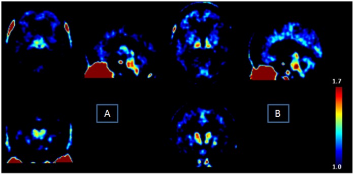
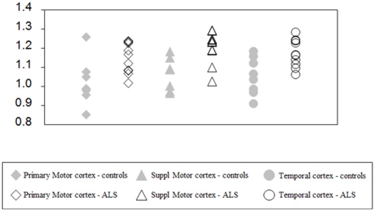
There is growing evidence of activated microglia and inflammatory processes in the cerebral cortex in amyotrophic lateral sclerosis (ALS). Activated microglia is characterized by increased expression of the 18 kDa translocator protein (TSPO) in the brain and may be a useful biomarker of inflammation. In this study, we evaluated neuroinflammation in ALS patients using a radioligand of TSPO, (18)F-DPA-714. Ten patients with probable or definite ALS (all right-handed, without dementia, and untreated by riluzole or other medication that might bias the binding on the TSPO), were enrolled prospectively and eight healthy controls matched for age underwent a PET study. Comparison of the distribution volume ratios between both groups were performed using a Mann-Whitney's test. Significant increase of distribution of volume ratios values corresponding to microglial activation was found in the ALS sample in primary motor, supplementary motor and temporal cortex (p = 0.009, p = 0.001 and p = 0.004, respectively). These results suggested that the cortical uptake of (18)F-DPA-714 was increased in ALS patients during the "time of diagnosis" phase of the disease. This finding might improve our understanding of the pathophysiology of ALS and might be a surrogate marker of efficacy of treatment on microglial activation.
Overview
The claims and statistics of this paper haven’t been checked yet.
Run the check — free, no account needed. It reads the open-access text and checks the sources it cites.
Integrity signals
Registry
Cites
Statistics
Figures
Verification
Claims the paper makes
The claims and statistics of this paper haven’t been checked yet.
Run the check — free, no account needed. It reads the open-access text and checks the sources it cites.
Claims other papers make about this one
Each is checked against this paper’s own text: does it say what the citing paper says it does?
In the citing paper
“presumably due to increased CNS microglia or their activation, as suggested by positron emission tomography (PET) imaging using radiotracers specific for activated microglia [423,424].”
From Autophagy and ALS: mechanistic insights and therapeutic implications (2022) · cites this paper as [423]
Self-check
Percentages, sums and reported differences re-computed from the paper’s own numbers.
The self-check needs the paper’s full text.
Citations
Cites
Folstein, M. F., Folstein, S. E., & McHugh, P. R. (1975) · Journal of Psychiatric Research · doi:10.1016/0022-3956(75)90026-6 · Cited by 88,282
Rosen, D. R., Siddique, T., Patterson, D., Figlewicz, D. A., Sapp, P., Hentati, A., Donaldson, D., Goto, J., O'Regan, J. P., Deng, H.-X., et al. (1993) · Nature · doi:10.1038/362059a0 · Cited by 7,062
El Escorial revisited: revised criteria for the diagnosis of amyotrophic lateral sclerosis
Brooks, B. R., Miller, R. G., Swash, M., & Munsat, T. L. (2000) · Amyotrophic Lateral Sclerosis and Other Motor Neuron Disorders · doi:10.1080/146608200300079536 · Cited by 5,433
The FAB: A frontal assessment battery at bedside
Dubois, B., Slachevsky, A., Litvan, I., & Pillon, B. (2000) · Neurology · doi:10.1212/wnl.55.11.1621 · Cited by 3,913
Cedarbaum, J. M., Stambler, N., Malta, E., Fuller, C., Hilt, D., Thurmond, B., & Nakanishi, A. (1999) · Journal of the Neurological Sciences · doi:10.1016/s0022-510x(99)00210-5 · Cited by 3,307
Kiernan, M. C., Vucic, S., Cheah, B. C., Turner, M. R., Eisen, A., Hardiman, O., Burrell, J. R., & Zoing, M. C. (2011) · The Lancet · doi:10.1016/s0140-6736(10)61156-7 · Cited by 2,603
Pochet, P. (2006) · Griffith Research Online · Cited by 1,550
Papadopoulos, V., Baraldi, M., Guilarte, T. R., Knudsen, T. B., Lacapère, J.-J., Lindemann, P., Norenberg, M. D., Nutt, D., Weizman, A., Zhang, M.-R., et al. (2006) · Trends in Pharmacological Sciences · doi:10.1016/j.tips.2006.06.005 · Cited by 1,389
UNRAVELING THE MECHANISMS INVOLVED IN MOTOR NEURON DEGENERATION IN ALS
Bruijn, L. I., Miller, T. M., & Cleveland, D. W. (2004) · Annual Review of Neuroscience · doi:10.1146/annurev.neuro.27.070203.144244 · Cited by 1,185
Prevalence and patterns of cognitive impairment in sporadic ALS
Ringholz, G. M., Appel, S. H., Bradshaw, M. R., Cooke, N., Mosnik, D. M., & Schulz, P. E. (2005) · Neurology · doi:10.1212/01.wnl.0000172911.39167.b6 · Cited by 843
Turner, M. R., Cagnin, A., Turkheimer, F. E., Miller, C. C. J., Shaw, C. E., Brooks, D. J., Leigh, P. N., & Banati, R. B. (2004) · Neurobiology of Disease · doi:10.1016/j.nbd.2003.12.012 · Cited by 737
Clinical genetics of amyotrophic lateral sclerosis: what do we really know?
Andersen, P. M., & Al-Chalabi, A. (2011) · Nature Reviews Neurology · doi:10.1038/nrneurol.2011.150 · Cited by 687
Neuroinflammation in amyotrophic lateral sclerosis: role of glial activation in motor neuron disease
Philips, T., & Robberecht, W. (2011) · The Lancet Neurology · doi:10.1016/s1474-4422(11)70015-1 · Cited by 646
Cognitive impairment in amyotrophic lateral sclerosis
Phukan, J., Pender, N. P., & Hardiman, O. M. (2007) · The Lancet Neurology · doi:10.1016/s1474-4422(07)70265-x · Cited by 635
Mitchell, J. D., & Borasio, G. D. (2007) · The Lancet · doi:10.1016/s0140-6736(07)60944-1 · Cited by 606
Inflammatory processes in amyotrophic lateral sclerosis
McGeer, P. L., & McGeer, E. G. (2002) · Muscle & Nerve · doi:10.1002/mus.10191 · Cited by 470
Visualising microglial activation in vivo
Banati, R. B. (2002) · Glia · doi:10.1002/glia.10144 · Cited by 428
Mixed-Affinity Binding in Humans with 18-kDa Translocator Protein Ligands
Owen, D. R., Gunn, R. N., Rabiner, E. A., Bennacef, I., Fujita, M., Kreisl, W. C., Innis, R. B., Pike, V. W., Reynolds, R., Matthews, P. M., et al. (2010) · Journal of Nuclear Medicine · doi:10.2967/jnumed.110.079459 · Cited by 361
Microglia in ALS: The Good, The Bad, and The Resting
Henkel, J. S., Beers, D. R., Zhao, W., & Appel, S. H. (2009) · Journal of Neuroimmune Pharmacology · doi:10.1007/s11481-009-9171-5 · Cited by 353
Geser, F., Brandmeir, N. J., Kwong, L. K., Martinez‐Lage, M., Elman, L. B., McCluskey, L. F., Xie, S. X., Lee, V. M. ‐., & Trojanowski, J. Q. (2008) · Archives of Neurology · doi:10.1001/archneur.65.5.636 · Cited by 308
James, M. L., Fulton, R., Vercouillie, J., Henderson, D., Garreau, L., Chalon, S., Dollé, F., Selleri, S., Guilloteau, D., & Kassiou, M. (2008) · Journal of Nuclear Medicine · doi:10.2967/jnumed.107.046151 · Cited by 283
Self-citationCorpus callosum involvement is a consistent feature of amyotrophic lateral sclerosis
Filippini, N., Douaud, G., Mackay, C. E., Knight, S., Talbot, K., & Turner, M. R. (2010) · Neurology · doi:10.1212/wnl.0b013e3181fb84d1 · Cited by 280
Kreisl, W. C., Fujita, M., Fujimura, Y., Kimura, N., Jenko, K. J., Kannan, P., Hong, J. S., Morse, C. L., Zoghbi, S. S., Gladding, R. L., et al. (2009) · NeuroImage · doi:10.1016/j.neuroimage.2009.11.056 · Cited by 275
Hsiung, G. R., DeJesus-Hernandez, M., Feldman, H., Sengdy, P., Bouchard‐Kerr, P., Dwosh, E., Butler, R. K., Leung, B., Fok, A., Rutherford, N. J., et al. (2012) · Brain · doi:10.1093/brain/awr354 · Cited by 229
Piao, Y., Wakabayashi, K., Kakita, A., Yamada, M., Hayashi, S., Morita, T., Ikuta, F., Oyanagi, K., & Takahashi, H. (2003) · Brain Pathology · doi:10.1111/j.1750-3639.2003.tb00002.x · Cited by 210
Quantitative Functional Brain Imaging with Positron Emission Tomography
(1998) · Elsevier eBooks · doi:10.1016/b978-0-12-161340-2.x5000-8 · Cited by 186
The microglial-motoneuron dialogue in ALS.
Appel, S. H., Zhao, W., Beers, D. R., & Henkel-Tigges, J. (2011) · Europe PMC (PubMed Central) · Cited by 179
Agosta, F., Pagani, E., Rocca, M. A., Caputo, D., Perini, M., Salvi, F., Prelle, A. C., & Filippi, M. (2007) · Human Brain Mapping · doi:10.1002/hbm.20364 · Cited by 169
Arlicot, N., Vercouillie, J., Ribeiro, M. J., Tauber, C., Venel, Y., Baulieu, J., Maïa, S., Corcia, P., Stabin, M. G., Reynolds, A. J., et al. (2011) · Nuclear Medicine and Biology · doi:10.1016/j.nucmedbio.2011.10.012 · Cited by 149
Self-citationAmyotrophic lateral sclerosis and frontotemporal dementia: A behavioural and cognitive continuum
Lillo, P., Savage, S., Mioshi, E., Kiernan, M., & Hodges, J. (2012) · Amyotrophic Lateral Sclerosis · doi:10.3109/17482968.2011.639376 · Cited by 143
Abourbeh, G., Thezé, B., Maroy, R., Dubois, A., Brulon, V., Fontyn, Y., Dollé, F., Tavitian, B., & Boisgard, R. (2012) · Journal of Neuroscience · doi:10.1523/jneurosci.2900-11.2012 · Cited by 112
Petit-Taboué, M.-C., Baron, J., Barré, L., Travère, J.-M., Speckel, D., Camsonne, R., & MacKenzie, E. T. (1991) · European Journal of Pharmacology · doi:10.1016/0014-2999(91)90594-g · Cited by 82
Veenman, L. S., & Gavish, M. (2012) · Current Molecular Medicine · doi:10.2174/1566524011207040398 · Cited by 81
Tang, D., Hight, M. R., McKinley, E. T., Fu, A., Buck, J. R., Smith, R. A., Tantawy, M. N., Peterson, T. E., Colvin, D. C., Ansari, M. S., et al. (2012) · Journal of Nuclear Medicine · doi:10.2967/jnumed.111.095653 · Cited by 63
Hirvonen, J., Roivainen, A., Virta, J. R., Helin, S., Någren, K., & Rinne, J. O. (2009) · European Journal of Nuclear Medicine and Molecular Imaging · doi:10.1007/s00259-009-1298-3 · Cited by 46
Quantitative analysis of [18F]FDDNP PET using subcortical white matter as reference region
Wong, K. P., Wardak, M., Shao, W., Dahlbom, M. L., Kepe, V., Liu, J., Satyamurthy, N., Small, G. W., Barrio, J. R., & Huang, S. (2009) · European Journal of Nuclear Medicine and Molecular Imaging · doi:10.1007/s00259-009-1293-8 · Cited by 37
Frontal and temporal lobe involvement on verbal fluency measures in amyotrophic lateral sclerosis
Lepow, L., Sweringen, J. V., Strutt, A. M., Jawaid, A., MacAdam, C., Harati, Y., Schulz, P. E., & York, M. K. (2010) · Journal of Clinical and Experimental Neuropsychology · doi:10.1080/13803391003596439 · Cited by 29
Gunn, R. N., Lammertsma, A. A., & Cunningham, V. J. (1998) · Elsevier eBooks · doi:10.1016/b978-012161340-2/50062-7 · Cited by 26
Veenman, L., & Gavish, M. (2012) · Current Molecular Medicine · doi:10.2174/156652412800163343 · Cited by 22
Kiernan and others 2011 Amyotrophic Lateral Sclerosis - Review Article
Kiernan, M. C., Vucic, S., Cheah, B. C., Turner, M. R., Eisen, A., Hardiman, O. M., & Burrell, J. R. (2011) · Cited by 18
Peptidyl epoxides as selective protease inactivators
Albeck, A. (2000) · Drug Development Research · doi:10.1002/1098-2299(200007/08)50:3/4 · Cited by 12
Research works printed but not linked: 1
- I Bertrand, 1925, La sclérose latérale amyotrophique (Anatomie pathologique), RevNeurol, 1, 779
Cited by
Sorted by citations
Hardiman, O. M., Al‐Chalabi, A., Chiò, A., Corr, E. M., Logroscino, G., Robberecht, W., Shaw, P. J., Simmons, Z., & van den Berg, L. H. (2017) · Nature Reviews Disease Primers · doi:10.1038/nrdp.2017.71 · Cited by 2,262
ALS Genetics, Mechanisms, and Therapeutics: Where Are We Now?
Mejzini, R., Flynn, L. L., Pitout, I. L., Fletcher, S., Wilton, S. D., & Akkari, P. A. (2019) · Frontiers in Neuroscience · doi:10.3389/fnins.2019.01310 · Cited by 803
Liu, J., & Wang, F. (2017) · Frontiers in Immunology · doi:10.3389/fimmu.2017.01005 · Cited by 376
Inflammation in ALS/FTD pathogenesis
McCauley, M. E., & Baloh, R. H. (2019) · Acta Neuropathologica · doi:10.1007/s00401-018-1933-9 · Cited by 341
RETRACTED: Neuroinflammatory Markers: Key Indicators in the Pathology of Neurodegenerative Diseases
Rauf, A., Badoni, H., Abu‐Izneid, T., Olatunde, A., Rahman, M. M., Painuli, S., Semwal, P., Wilairatana, P., & Mubarak, M. S. (2022) · Molecules · doi:10.3390/molecules27103194 · Cited by 316
Retracted
Metadata
- Authors
- Corcia, P., Tauber, C., Vercoullie, J., Arlicot, N., Prunier, C., Praline, J., Nicolas, G., Venel, Y., Hommet, C., Baulieu, J.-L., Cottier, J.-P., Roussel, C., Kassiou, M., Guilloteau, D., & Ribeiro, M.-J.
- Affiliations
- Université de Tours (FR)
- Centre Hospitalier Universitaire de Tours (FR)
- Centre Hospitalier Universitaire d'Angers (FR)
- Corresponding author
- Corcia, Philippe
- ORCID
- Venue
- PLoS ONE
- Publisher
- Public Library of Science (PLoS)
- Published
- 2012-12-31
- Volume
- 7
- Issue
- 12
- Pages
- e52941
- ISSN
- 1932-6203 (ISSN-L)
- PMC
- PMC3534121
- OpenAlex
- W2051387942
- Cited by
- 249
- References
- 42
- Citations by year
- 2026: 7 · 2025: 14 · 2024: 8 · 2023: 13 · 2022: 19 · 2021: 28 · 2020: 28 · 2019: 20 · 2018: 13 · 2017: 21 · 2016: 23 · 2015: 23 · 2014: 25 · 2013: 6
- FWCI
- 11.46
- Type
- journal article
- Language
- en
- Open access
- gold
- doi.org
- Locations
- PLoS ONE (publishedVersion, cc-by)
- PubMed Central (publishedVersion, cc-by)
- PubMed (publishedVersion)
- DOAJ (DOAJ: Directory of Open Access Journals) (submittedVersion, cc-by-sa)
- Zenodo (CERN European Organization for Nuclear Research) (submittedVersion, cc-by)
- Funders
- FP7 Health (FP7/2007-2013)
- Topics
- Health Sciences › Medicine › Clinical Neurology › Amyotrophic Lateral Sclerosis Research
- Life Sciences › Neuroscience › Neurology › Neuroinflammation and Neurodegeneration Mechanisms
- Life Sciences › Neuroscience › Cellular and Molecular Neuroscience › Alzheimer's disease research and treatments
- Keywords
- amyotrophic lateral sclerosis, microglial activation, neuroinflammation, translocator protein (TSPO), [18F]DPA-714, positron emission tomography, distribution volume ratio, cerebral cortex, primary motor cortex, supplementary motor area, temporal cortex, biomarkers
- MeSH terms
- Aged, Amyotrophic Lateral Sclerosis, Brain, Female, Humans, Inflammation, Male, Middle Aged, Radionuclide Imaging, Microglia, Receptors, GABA, Molecular Imaging, Neuroimaging
About these checks
Registry, retraction, Cites and statistics results are computed, not judged, with one exception: a printed reference that no index lists is read once by a model before it counts as a citation error. Claims are extracted by a model and kept only when their quote appears verbatim in the text; each cited source is then checked, with a model’s help, for whether it says what the claim relies on. Those verdicts can be wrong — read the claim’s quote and the cited source before relying on one.
Statistics and arithmetic are recomputed, not judged.
Confidence bands are shown only for fields calibrated on our annotated test set. None is calibrated yet, so no band is shown.
What counts as a claim ›How we measure confidence ›How we check this ›
Are you an author, or do you see a problem with this page? Request a review
