

The DOI matches its Crossref record.
Metal and Silicate Particles Including Nanoparticles Are Present in Electronic Cigarette Cartomizer Fluid and Aerosol
Overview
The claims and statistics of this paper haven’t been checked yet.
Run the check — free, no account needed. It reads the open-access text and checks the sources it cites.
Integrity signals
Registry
Cites
Statistics
Figures
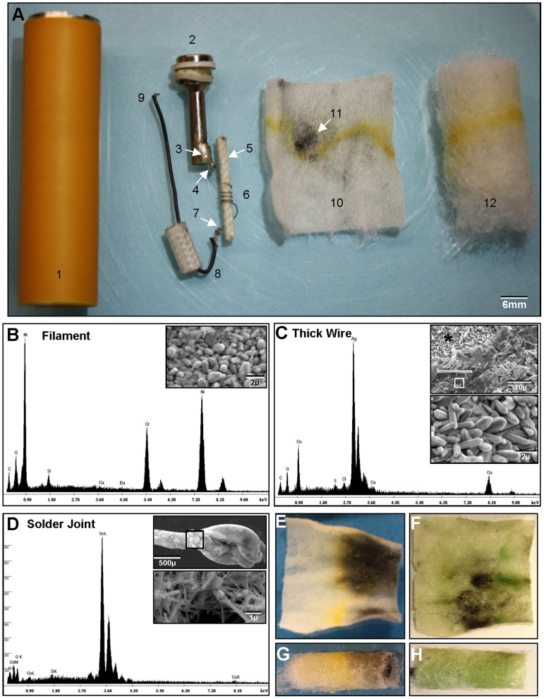
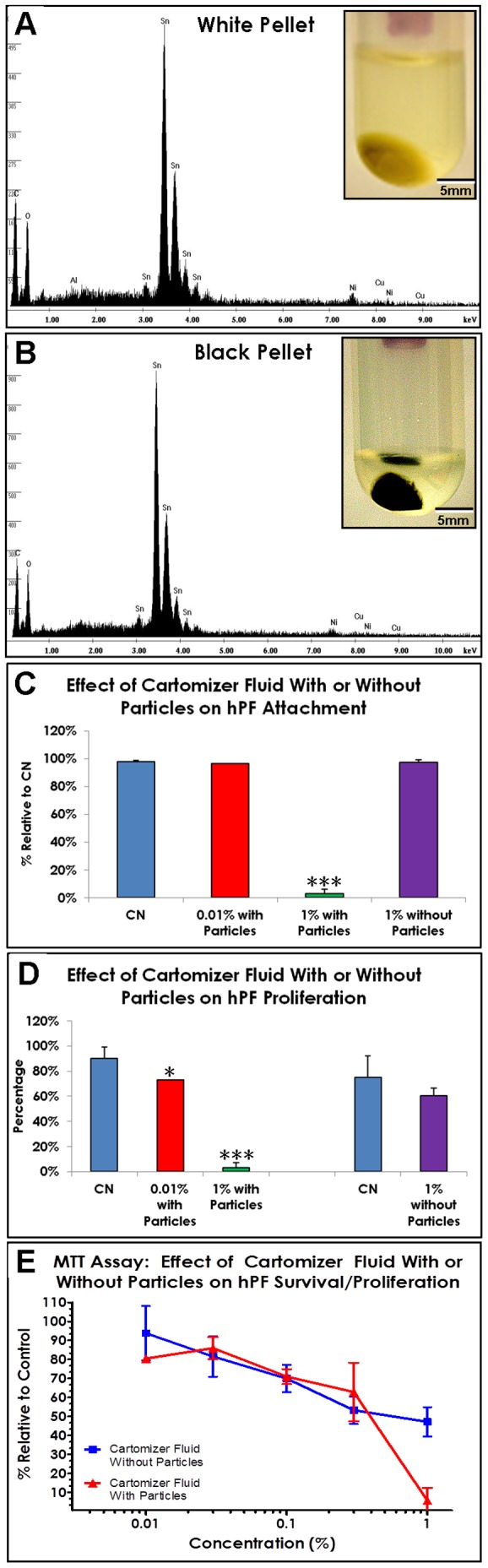
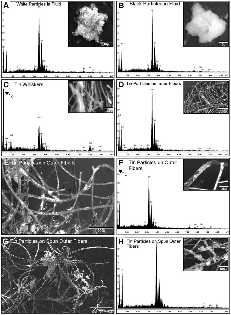
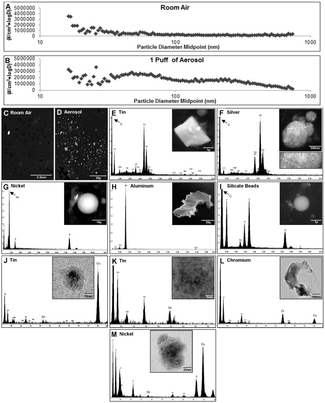
Verification
Claims the paper makes
The claims and statistics of this paper haven’t been checked yet.
Run the check — free, no account needed. It reads the open-access text and checks the sources it cites.
Claims other papers make about this one
Each is checked against this paper’s own text: does it say what the citing paper says it does?
In the citing paper
“While the mechanisms of fetal harm from EC use during pregnancy are not known, they may be mediated via exposure to volatile organic compounds, heavy metals, carcinogens, nicotine, and carrier agents, among others [17].”
In the citing paper
“Ingredients other than nicotine have also been shown to hinder the function of trophoblasts in the placenta and lead to complications in placental structuring [17, 18].”
Self-check
Percentages, sums and reported differences re-computed from the paper’s own numbers.
The self-check needs the paper’s full text.
Citations
Cites
No references could be resolved yet.
Research works printed but not linked: 40
- JW Ayers, 2011, Tracking the rise in popularity of electronic nicotine delivery systems (electronic cigarettes) using search query surveillance, Am J Prev Med, 40, 448, 10.1016/j.amepre.2010.12.007 · DOI
- JK Noel, 2011, Electronic cigarettes: a new ‘tobacco’ industry?, Tob Control, 20, 81, 10.1136/tc.2010.038562 · DOI
- A Trtchounian, 2010, Electronic nicotine delivery systems - Is there a need for regulation?, Tobacco Control, 20, 47, 10.1136/tc.2010.037259 · DOI
- AD Flouris, 2010, Electronic cigarettes: miracle or menace?, BMJ, 340, c311, 10.1136/bmj.c311 · DOI
- M Williams, 2011, Variability Among Electronic Cigarettes in the Pressure Drop, Airflow Rate, and Aerosol Production, Nicotine & Tobacco Research, 13, 1276, 10.1093/ntr/ntr164 · DOI
- JF Etter, 2011, Saliva cotinine levels in users of electronic cigarettes, Eur Respir J, 38, 1219, 10.1183/09031936.00066011 · DOI
- AR Vansickel, 2012, Electronic Cigarettes: Effective Nicotine Delivery After Acute Administration, Nicotine Tob Res, 15, 267, 10.1093/ntr/ntr316 · DOI
- P Caponnetto, 2012, The emerging phenomenon of electronic cigarettes, Expert Rev Respir Med, 6, 63, 10.1586/ers.11.92 · DOI
- LE Odum, 2012, Electronic Cigarettes: Do They Have a Role in Smoking Cessation?, J Pharm Pract, 25, 611, 10.1177/0897190012451909 · DOI
- P Caponnetto, 2011, Successful smoking cessation with electronic cigarettes in smokers with a documented history of recurring relapses: a case series, J Med Case Reports, 5, 585, 10.1186/1752-1947-5-585 · DOI
- JF Etter, 2010, Electronic cigarettes: a survey of users, BMC Public Health, 10, 231, 10.1186/1471-2458-10-231 · DOI
- IL Chen, 2012, FDA Summary of Adverse Events on Electronic Cigarettes, Nicotine Tob Res, 15, 615, 10.1093/ntr/nts145 · DOI
- L McCauley, 2012, An unexpected consequence of electronic cigarette use, Chest, 141, 1110, 10.1378/chest.11-1334 · DOI
- ML Trehy, 2011, Analysis of electronic cigarette cartridges, refill solutions, and smoke for nicotine and nicotine related impurities, Journal of Liquid Chromatography & Related Technologies, 34, 1442, 10.1080/10826076.2011.572213 · DOI
- ML Goniewicz, 2012, Nicotine Levels in Electronic Cigarettes, Nicotine Tob Res, 15, 158, 10.1093/ntr/nts103 · DOI
- A Trtchounian, 2010, Conventional and Electronic Cigarettes (E-cigarettes) Have Different Smoking Properties, Nicotine & Tobacco Research, 12, 905, 10.1093/ntr/ntq114 · DOI
- JA Young, 2004, Tin, Journal of Chemical Education, 81, 1562, 10.1021/ed081p1562 · DOI
- JA Young, 2008, Tin(II) Oxide, Journal of Chemical Education, 85, 193, 10.1021/ed085p193 · DOI
- R Behar, 2012, A method for rapid dose-response screening of environmental chemicals using human embryonic stem cells, J Pharmacol Toxicol Methods, 66, 238, 10.1016/j.vascn.2012.07.003 · DOI
- Lee NC (2009) Lead-free soldering; Lee NC, editor. New York: Springer. · DOI
- SH Liu, 2004, Tin whisker growth driven by electrical currents, Journal of Applied Physics, 95, 7742, 10.1063/1.1712019 · DOI
- RA Smith, 1986, Boron in glass and glass making, Journal of non-crystalline solids, 84, 421, 10.1016/0022-3093(86)90805-7 · DOI
- WD van Dijk, 2011, Nanoparticles in cigarette smoke; real-time undiluted measurements by a scanning mobility particle sizer, Anal Bioanal Chem, 399, 3573, 10.1007/s00216-011-4701-4 · DOI
- S Bakand, 2012, Nanoparticles: a review of particle toxicology following inhalation exposure, Inhal Toxicol, 24, 125, 10.3109/08958378.2010.642021 · DOI
- W Yang, 2008, Inhaled nanoparticles–a current review, Int J Pharm, 356, 239, 10.1016/j.ijpharm.2008.02.011 · DOI
- AM Schrand, 2010, Metal-based nanoparticles and their toxicity assessment, Wiley Interdiscip Rev Nanomed Nanobiotechnol, 2, 544, 10.1002/wnan.103 · DOI
- G Oberdorster, 2005, Nanotoxicology: an emerging discipline evolving from studies of ultrafine particles, Environ Health Perspect, 113, 823, 10.1289/ehp.7339 · DOI · Paper page
- S Takenaka, 2001, Pulmonary and systemic distribution of inhaled ultrafine silver particles in rats, Environ Health Perspect, 109 Suppl 4, 547, 10.1289/ehp.01109s4547 · DOI
- C Terzano, 2010, Air pollution ultrafine particles: toxicity beyond the lung, Eur Rev Med Pharmacol Sci, 14, 809
- G Bhabra, 2009, Nanoparticles can cause DNA damage across a cellular barrier, Nat Nanotechnol, 4, 876, 10.1038/nnano.2009.313 · DOI
- M Horie, 2011, Chromium(III) oxide nanoparticles induced remarkable oxidative stress and apoptosis on culture cells, Environ Toxicol, 28, 61, 10.1002/tox.20695 · DOI
- GS Kang, 2011, Long-term inhalation exposure to nickel nanoparticles exacerbated atherosclerosis in a susceptible mouse model, Environ Health Perspect, 119, 176, 10.1289/ehp.1002508 · DOI
- WH Chen, 2005, [Biological responses of tin mine particles and their association with adverse effects on health in tin mine], Zhonghua Lao Dong Wei Sheng Zhi Ye Bing Za Zhi, 23, 366
- L Bian, 2011, Induction of lung epithelial cell transformation and fibroblast activation by Yunnan tin mine dust and their interaction, Med Oncol, 28 Suppl 1, S560, 10.1007/s12032-010-9655-4 · DOI
- P Schuler, 1958, Stannosis: benign pneumoconiosis owing to inhalation of tin dust and fume. II. Clinical study, Ind Med Surg, 27, 432
- M Hua, 2011, Mining data on usage of electronic nicotine delivery systems (ENDS) from YouTube videos, Tobacco Control
- SJ Stohs, 1997, Toxicity of trace elements in tobacco smoke, Inhalation Toxicology, 9, 867, 10.1080/089583797197926 · DOI
- M Chiba, 1992, Toxic and trace elements in tobacco and tobacco smoke, Bull World Health Organ, 70, 269
- D Bernhard, 2005, Metals in cigarette smoke, IUBMB Life, 57, 805, 10.1080/15216540500459667 · DOI
- C Smith, 1997, An international literature survey of “IARC group 1 carcinogens” reported in mainstream cigarette smoke, Food and Chemical Toxicology, 35, 1107, 10.1016/S0278-6915(97)00063-X · DOI
Cited by
Sorted by citations
E-Cigarettes: A Scientific Review
Grana, R., Benowitz, N., & Glantz, S. A. (2014) · Circulation · doi:10.1161/circulationaha.114.007667 · Cited by 1,194
Lerner, C. A., Sundar, I. K., Yao, H., Gerloff, J., Ossip, D. J., McIntosh, S., Robinson, R., & Rahman, I. (2015) · PLOS ONE · doi:10.1371/journal.pone.0116732 · Cited by 666
Hajek, P., Etter, J., Benowitz, N., Eissenberg, T., & McRobbie, H. (2014) · Addiction · doi:10.1111/add.12659 · Cited by 546
Sussan, T. E., Gajghate, S., Thimmulappa, R. K., Ma, J., Kim, J.-H., Sudini, K., Consolini, N., Cormier, S. A., Lomnicki, S., Hasan, F., et al. (2015) · PLOS ONE · doi:10.1371/journal.pone.0116861 · Cited by 429
Garcia-Arcos, I., Geraghty, P., Baumlin, N., Campos, M., Dabo, A. J., Jundi, B., Cummins, N., Eden, E., Grosche, A., Salathe, M., et al. (2016) · Thorax · doi:10.1136/thoraxjnl-2015-208039 · Cited by 339
Metadata
- Authors
- Williams, M., Villarreal, A., Bozhilov, K., Lin, S., & Talbot, P.
- Venue
- PLoS ONE
- Publisher
- Public Library of Science (PLoS)
- Published
- 2013-03-20
- Volume
- 8
- Issue
- 3
- Pages
- e57987
- ISSN
- 1932-6203
- PMC
- PMC3603976
- Cited by
- 530
- References
- 0
- Language
- en
About these checks
Registry, retraction, Cites and statistics results are computed, not judged, with one exception: a printed reference that no index lists is read once by a model before it counts as a citation error. Claims are extracted by a model and kept only when their quote appears verbatim in the text; each cited source is then checked, with a model’s help, for whether it says what the claim relies on. Those verdicts can be wrong — read the claim’s quote and the cited source before relying on one.
Statistics and arithmetic are recomputed, not judged.
Confidence bands are shown only for fields calibrated on our annotated test set. None is calibrated yet, so no band is shown.
What counts as a claim ›How we measure confidence ›How we check this ›
Are you an author, or do you see a problem with this page? Request a review
How to cite
Cite CoChat for the checks on this page, and the paper for its own findings.
- This page
- CoChat. (2026). Citation check: Metal and Silicate Particles Including Nanoparticles Are Present in Electronic Cigarette Cartomizer Fluid and Aerosol. https://app.cochat.ai/papers/10.1371/journal.pone.0057987
- The paper
- Williams, M., Villarreal, A., Bozhilov, K., Lin, S., & Talbot, P. (2013). Metal and Silicate Particles Including Nanoparticles Are Present in Electronic Cigarette Cartomizer Fluid and Aerosol. PLoS ONE. https://doi.org/10.1371/journal.pone.0057987
