

The DOI matches its Crossref record; 41 of the 41 references it cites were found in a registry or an index.
Functional Impairment of Microglia Coincides with Beta-Amyloid Deposition in Mice with Alzheimer-Like Pathology
Open accessAbstract
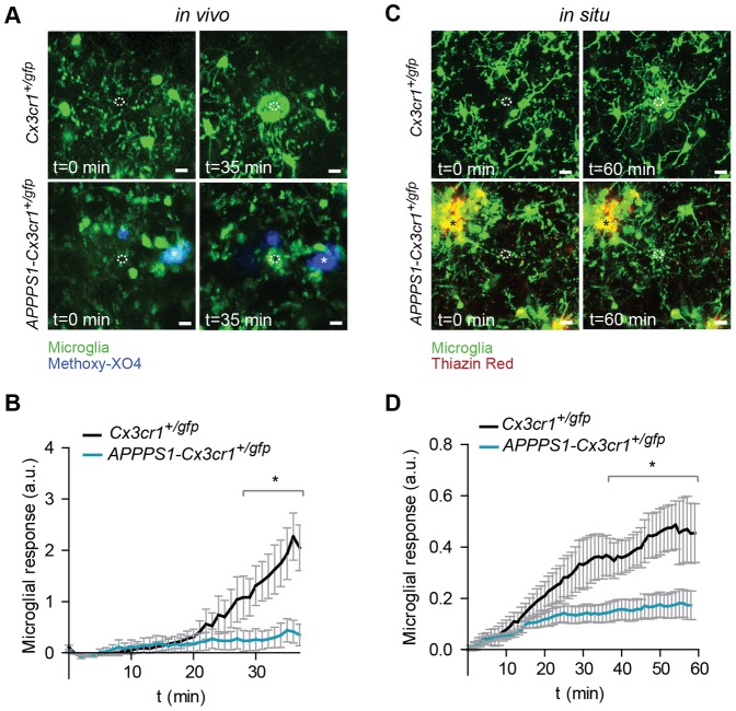
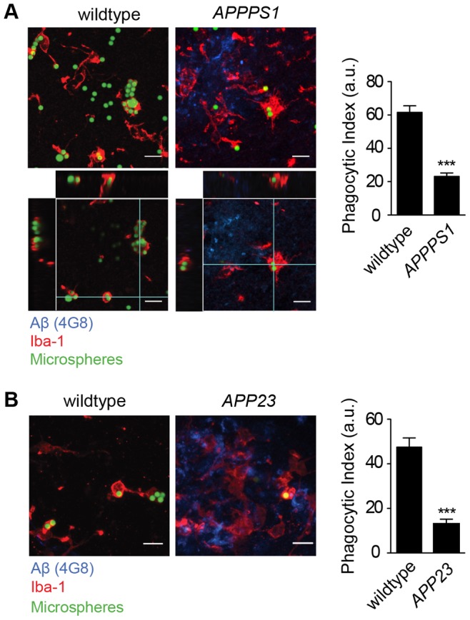
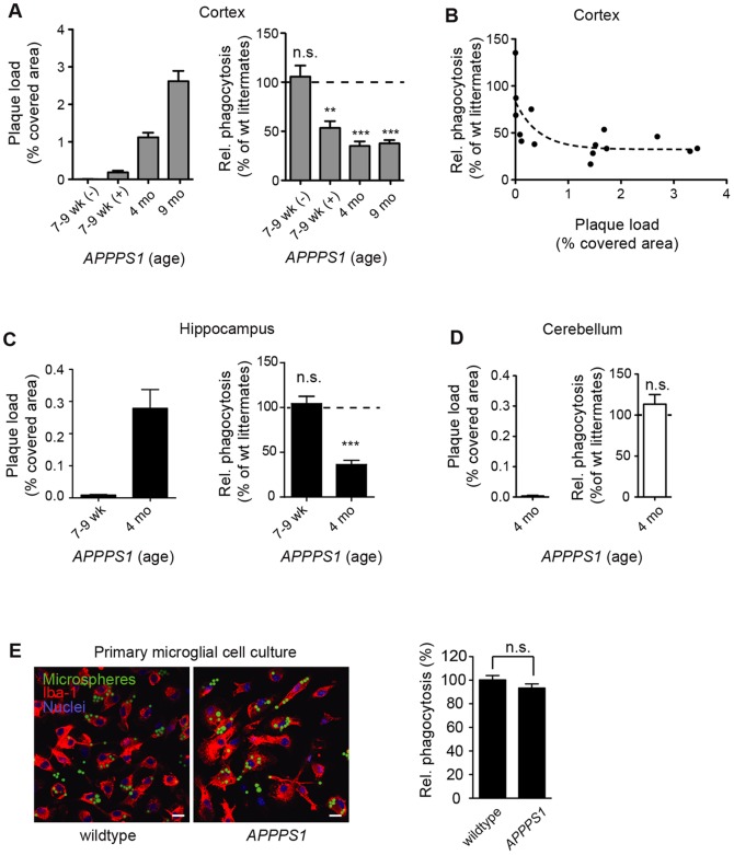
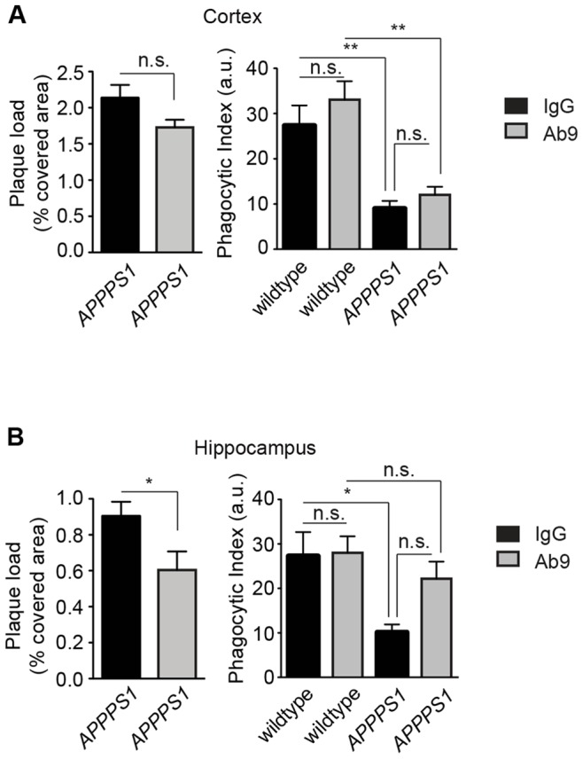
Microglial cells closely interact with senile plaques in Alzheimer's disease and acquire the morphological appearance of an activated phenotype. The significance of this microglial phenotype and the impact of microglia for disease progression have remained controversial. To uncover and characterize putative changes in the functionality of microglia during Alzheimer's disease, we directly assessed microglial behavior in two mouse models of Alzheimer's disease. Using in vivo two-photon microscopy and acute brain slice preparations, we found that important microglial functions - directed process motility and phagocytic activity - were strongly impaired in mice with Alzheimer's disease-like pathology compared to age-matched non-transgenic animals. Notably, impairment of microglial function temporally and spatially correlated with Aβ plaque deposition, and phagocytic capacity of microglia could be restored by interventionally decreasing amyloid burden by Aβ vaccination. These data suggest that major microglial functions progressively decline in Alzheimer's disease with the appearance of Aβ plaques, and that this functional impairment is reversible by lowering Aβ burden, e.g. by means of Aβ vaccination.
Overview
The claims and statistics of this paper haven’t been checked yet.
Run the check — free, no account needed. It reads the open-access text and checks the sources it cites.
Integrity signals
Registry
Cites
Statistics
Figures
Verification
Claims the paper makes
The claims and statistics of this paper haven’t been checked yet.
Run the check — free, no account needed. It reads the open-access text and checks the sources it cites.
Claims other papers make about this one
Each is checked against this paper’s own text: does it say what the citing paper says it does?
The source backs part of the claim, not all of it — or backs it for a different population, condition or comparison.
In the citing paper
“Notably, previous studies have shown that microglial function may be adversely affected by Aβ, particularly as a result of chronic exposure to pro-inflammatory cytokines, resulting in increased Aβ accumulation and potentially underpinning the contradiction between our in vitro and in vivo results.39,40”
From Microglial INPP5D limits plaque formation and glial reactivity in the PSAPP mouse model of Alzheimer's disease (2023) · cites this paper as [39]
Experimental — accuracy not yet measuredThe source backs part of the claim, not all of it — or backs it for a different population, condition or comparison.
In the citing paper
“the ability of microglia to remove Αβ declines over time”
From Amyloid-β induces lipid droplet-mediated microglial dysfunction via the enzyme DGAT2 in Alzheimer’s disease (2025) · cites this paper as [17]
Experimental — accuracy not yet measuredThe source backs part of the claim, not all of it — or backs it for a different population, condition or comparison.
In the citing paper
“supporting that the progression of amyloid pathology correlates with impaired capacity of microglia to phagocytose Αβ17,16.”
From Amyloid-β induces lipid droplet-mediated microglial dysfunction via the enzyme DGAT2 in Alzheimer’s disease (2025) · cites this paper as [17]
Experimental — accuracy not yet measuredThe source backs part of the claim, not all of it — or backs it for a different population, condition or comparison.
In the citing paper
“their phagocytic capacity declines with aging, chronic exposure to Αβ, and during sustained inflammation in the AD brain76,16,17.”
From Amyloid-β induces lipid droplet-mediated microglial dysfunction via the enzyme DGAT2 in Alzheimer’s disease (2025) · cites this paper as [17]
Experimental — accuracy not yet measuredThe source backs part of the claim, not all of it — or backs it for a different population, condition or comparison.
In the citing paper
“However, perturbations in microglial activity, often associated with aging, can affect the effective clearance of these neuron-derived Aβ fibrils, leading to increased plaque formation and the progression of cognitive and behavioral impairments (17–20).”
From Reducing microglial lipid load enhances β amyloid phagocytosis in an Alzheimer’s disease mouse model (2025) · cites this paper as [19]
Experimental — accuracy not yet measured
The cited source says what the claim attributes to it.
In the citing paper
“Microglial phagocytosis is believed to be dysfunctional in AD [17–19].”
From IL-1β-driven amyloid plaque clearance is associated with an expansion of transcriptionally reprogrammed microglia (2019) · cites this paper as [18]
The cited source says what the claim attributes to it.
In the citing paper
“However, several studies have shown that defects in the phagocytic function of microglia can be restored with Aβ immunization [18, 23] and anti-inflammatory drug treatment [24, 25].”
From IL-1β-driven amyloid plaque clearance is associated with an expansion of transcriptionally reprogrammed microglia (2019) · cites this paper as [18]
The cited source says what the claim attributes to it.
In the citing paper
“Notably, microglial process motility directed toward site of damage was significantly impaired in mice with A β plaques 48 .”
From Transcriptomic analysis of purified human cortical microglia reveals age-associated changes (2017) · cites this paper as [48]
Self-check
Percentages, sums and reported differences re-computed from the paper’s own numbers.
The self-check needs the paper’s full text.
Citations
Cites
Resting microglial cells are highly dynamic surveillants of brain parenchyma in vivo
Nimmerjahn, A., Kirchhoff, F., & Helmchen, F. (2005) · Science · doi:10.1126/science.1110647 · Cited by 5,827 · cited 1×
“To investigate a second biologically relevant microglial function we studied process motility by assessing process extensions towards an acute injury induced by a laser lesion [1,2].”
BackgroundATP mediates rapid microglial response to local brain injury in vivo
Davalos, D., Grutzendler, J., Yang, G., Kim, J. V., Zuo, Y., Jung, S., Littman, D. R., Dustin, M. L., & Gan, W.-B. (2005) · Nature Neuroscience · doi:10.1038/nn1472 · Cited by 3,913 · cited 3×
“Microglia respond to defined tissue injuries by extending their processes towards the lesion in an ATP-dependent fashion [2].”
BackgroundMethodMicroglia: Active sensor and versatile effector cells in the normal and pathologic brain
Hanisch, U.-K., & Kettenmann, H. (2007) · Nature Neuroscience · doi:10.1038/nn1997 · Cited by 3,676 · cited 1×
“process and the morphology of microglial cells does not necessarily match their functional state [14,15].”
BackgroundSelf-citationJung, S., Aliberti, J., Graemmel, P., Sunshine, M. J., Kreutzberg, G. W., Sher, A., & Littman, D. R. (2000) · Molecular and Cellular Biology · doi:10.1128/mcb.20.11.4106-4114.2000 · Cited by 2,632 · cited 2×
“1A and B and Videos S1 and S2), which were obtained by crossbreeding APPPS1 mice, a mouse model of cerebral amyloidosis [21] with Cx3cr1 mice [23], a mouse model that allows visualization of microglia.”
MethodMicroglia Shape Adult Hippocampal Neurogenesis through Apoptosis-Coupled Phagocytosis
Sierra, A., Encinas, J. M., Deudero, J. J. P., Chancey, J. H., Enikolopov, G., Overstreet-Wadiche, L. S., Tsirka, S. E., & Maletic-Savatic, M. (2010) · Cell Stem Cell · doi:10.1016/j.stem.2010.08.014 · Cited by 1,622 · cited 1×
“newborn neuroblasts [5].”
Wake, H., Moorhouse, A. J., Jinno, S., Kohsaka, S., & Nabekura, J. (2009) · The Journal of Neuroscience · doi:10.1523/jneurosci.4363-08.2009 · Cited by 1,619 · cited 1×
“neuronal structure [3,4].”
Two amyloid precursor protein transgenic mouse models with Alzheimer disease-like pathology
Sturchler-Pierrat, C., Abramowski, D., Duke, M., Wiederhold, K.-H., Mistl, C., Rothacher, S., Ledermann, B., Bürki, K., Frey, P., Paganetti, P. A., et al. (1997) · Proceedings of the National Academy of Sciences · doi:10.1073/pnas.94.24.13287 · Cited by 1,507 · cited 2×
“APP23 mice, which express APP with the Swedish mutation [22] were provided by Novartis, Basel, Switzerland.”
BackgroundMethodMicroglial Dysfunction and Defective β-Amyloid Clearance Pathways in Aging Alzheimer's Disease Mice
Hickman, S. E., Allison, E. K., & El Khoury, J. (2008) · The Journal of Neuroscience · doi:10.1523/jneurosci.0616-08.2008 · Cited by 1,329 · cited 4×
“Interestingly, expression of receptors and enzymes involved in microglial Ab-uptake and degradation is progressively downregulated in a transgenic mouse model of Alzheimer’s disease [20].”
BackgroundResultInfluentialMCGEER, P., & MCGEER, E. (1995) · Brain Research Reviews · doi:10.1016/0165-0173(95)00011-9 · Cited by 1,287 · cited 1×
“A number of in vitro studies have not only shown that administration of Ab stimulates the production of inflammatory cytokines and activation markers in microglial cells [8] but also triggers its uptake by receptor-mediated phagocytosis [16].”
BackgroundLee, J.-H., Yu, W. H., Kumar, A., Lee, S., Mohan, P. S., Peterhoff, C. M., Wolfe, D. M., Martinez-Vicente, M., Massey, A. C., Sovak, G., et al. (2010) · Cell · doi:10.1016/j.cell.2010.05.008 · Cited by 1,212
Cited by
Sorted by citations
Neuroinflammation in Alzheimer's disease
Heneka, M. T., Carson, M. J., Khoury, J. E., Landreth, G. E., Brosseron, F., Feinstein, D. L., Jacobs, A. H., Wyss-Coray, T., Vitorica, J., Ransohoff, R. M., et al. (2015) · The Lancet Neurology · doi:10.1016/s1474-4422(15)70016-5 · Cited by 6,226
“The sustained exposure to A β itself, chemokines, cytokines, and other inflammatory mediators seems responsible for the persistent functional impairment of microglial cells observed at plaque sites 40,41 .”
Self-citationInflammation as a central mechanism in Alzheimer's disease
Kinney, J. W., Bemiller, S. M., Murtishaw, A. S., Leisgang, A. M., Salazar, A. M., & Lamb, B. T. (2018) · Alzheimer's & Dementia: Translational Research & Clinical Interventions · doi:10.1016/j.trci.2018.06.014 · Cited by 2,253
Immune attack: the role of inflammation in Alzheimer disease
Heppner, F. L., Ransohoff, R. M., & Becher, B. (2015) · Nature Reviews Neuroscience · doi:10.1038/nrn3880 · Cited by 2,134
Self-citationMicroglia in physiology and disease
Wolf, S. A., Boddeke, H. W. G. M., & Kettenmann, H. (2017) · Annual Review of Physiology · doi:10.1146/annurev-physiol-022516-034406 · Cited by 1,426
Self-citationMicroglia and brain macrophages in the molecular age: from origin to neuropsychiatric disease
Prinz, M., & Priller, J. (2014) · Nature Reviews Neuroscience · doi:10.1038/nrn3722 · Cited by 1,329
Metadata
- Authors
- Krabbe, G., Halle, A., Matyash, V., Rinnenthal, J. L., Eom, G. D., Bernhardt, U., Miller, K. R., Prokop, S., Kettenmann, H., & Heppner, F. L.
- Affiliations
- Max Delbrück Center (DE)
- Center of Advanced European Studies and Research (DE)
- Charité – Universitätsmedizin Berlin (DE)
- Corresponding author
- Kettenmann, Helmut
- Heppner, Frank L.
- Venue
- PLoS ONE
- Publisher
- Public Library of Science (PLoS)
- Published
- 2013-04-08
- Volume
- 8
- Issue
- 4
- Pages
- e60921
- ISSN
- 1932-6203 (ISSN-L)
- PMC
- PMC3620049
- OpenAlex
- W2044090829
- Cited by
- 478
- References
- 41
- Citations by year
- 2026: 15 · 2025: 29 · 2024: 31 · 2023: 25 · 2022: 44 · 2021: 56 · 2020: 40 · 2019: 41 · 2018: 38 · 2017: 38 · 2016: 33 · 2015: 39 · 2014: 34 · 2013: 13
- FWCI
- 22.45
- Type
- journal article
- Language
- en
- Open access
- gold
- doi.org
- Locations
- PLoS ONE (publishedVersion, cc-by)
- Europe PMC (PubMed Central) (publishedVersion, other-oa)
- PubMed Central (publishedVersion, cc-by)
- PubMed (publishedVersion)
- MDC Repository (Max-Delbrueck-Center for Molecular Medicine) (acceptedVersion)
- MPG.PuRe (Max Planck Society) (submittedVersion)
- DOAJ (DOAJ: Directory of Open Access Journals) (submittedVersion, cc-by-sa)
- Max Planck Digital Library (submittedVersion)
- Figshare (submittedVersion, cc-by)
- Funders
- European Commission (FP7 HEALTH)
- Deutsche Forschungsgemeinschaft (SFB TRR 43, NeuroCure EXC257, GRK1258, EXC257)
- Jung-Stiftung für Wissenschaft und Forschung
- National Institutes of Health
- National Institute of Neurological Disorders and Stroke (R01NS046006, NS046006)
- FP7 Health
- Topics
- Life Sciences › Neuroscience › Neurology › Neuroinflammation and Neurodegeneration Mechanisms
- Life Sciences › Neuroscience › Cellular and Molecular Neuroscience › Alzheimer's disease research and treatments
- Life Sciences › Neuroscience › Neurology › Neurological Disease Mechanisms and Treatments
- Keywords
- microglia, Alzheimer's disease, amyloid plaques, microglial activation, microglial phagocytosis, mouse models of Alzheimer's disease
- MeSH terms
- Alzheimer Disease, Animals, Cell Movement, Disease Models, Animal, Female, Humans, Male, Mice, Transgenic, Mutation, Phagocytosis, Amyloid beta-Peptides, Protein Structure, Secondary, Microglia, Mice, Protein Multimerization, Plaque, Amyloid, Spatio-Temporal Analysis
About these checks
Registry, retraction, Cites and statistics results are computed, not judged, with one exception: a printed reference that no index lists is read once by a model before it counts as a citation error. Claims are extracted by a model and kept only when their quote appears verbatim in the text; each cited source is then checked, with a model’s help, for whether it says what the claim relies on. Those verdicts can be wrong — read the claim’s quote and the cited source before relying on one.
Statistics and arithmetic are recomputed, not judged.
Confidence bands are shown only for fields calibrated on our annotated test set. None is calibrated yet, so no band is shown.
What counts as a claim ›How we measure confidence ›How we check this ›
Are you an author, or do you see a problem with this page? Request a review
