

The DOI matches its Crossref record.
HPV16 Oncoproteins Promote Cervical Cancer Invasiveness by Upregulating Specific Matrix Metalloproteinases
Overview
The claims and statistics of this paper haven’t been checked yet.
Run the check — free, no account needed. It reads the open-access text and checks the sources it cites.
Integrity signals
Registry
Cites
Statistics
Figures
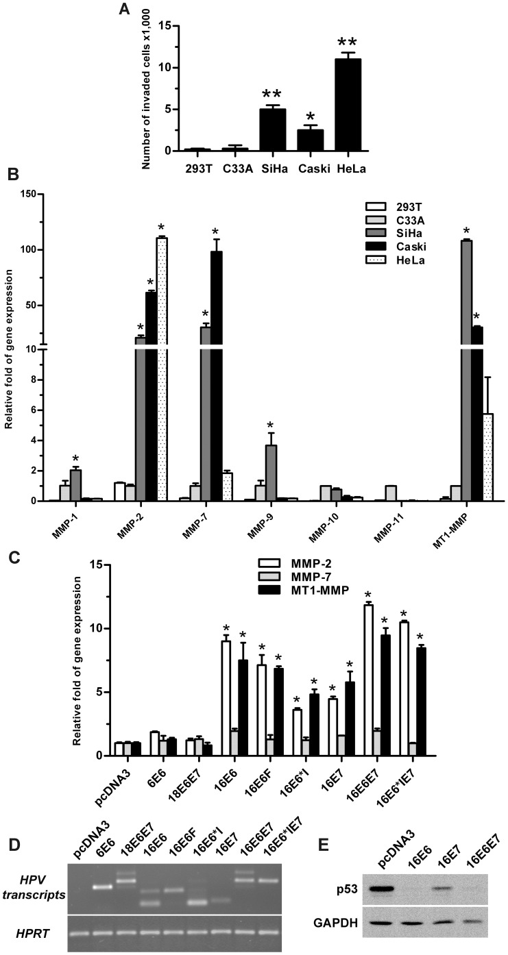
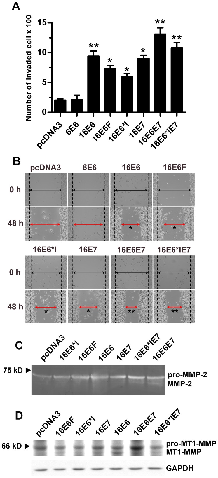
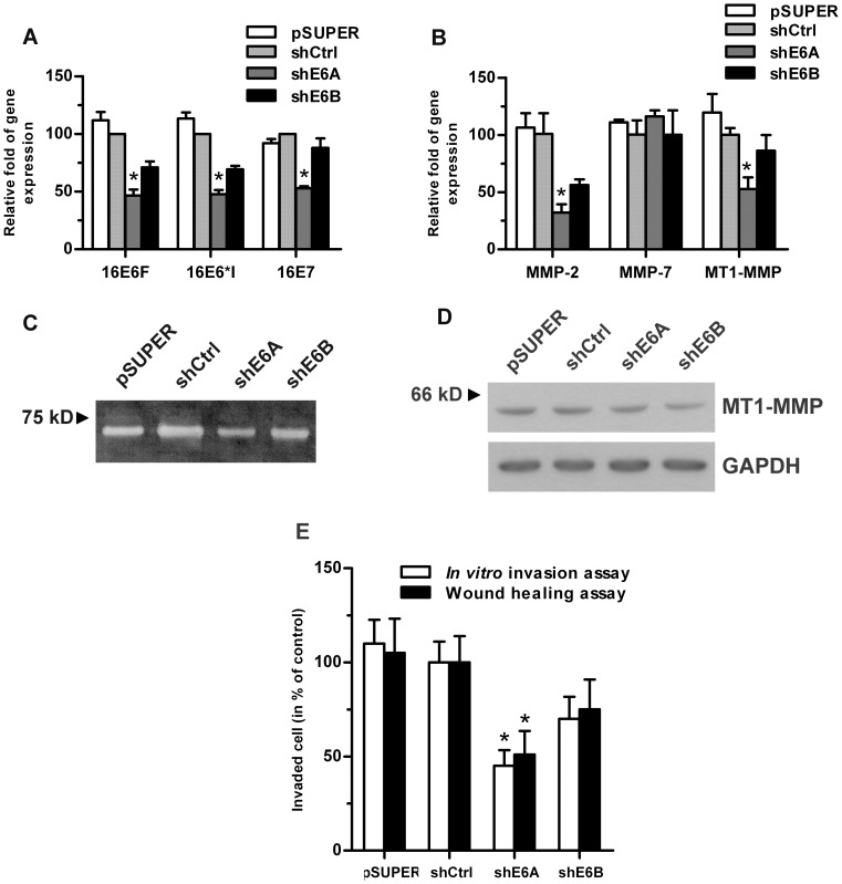
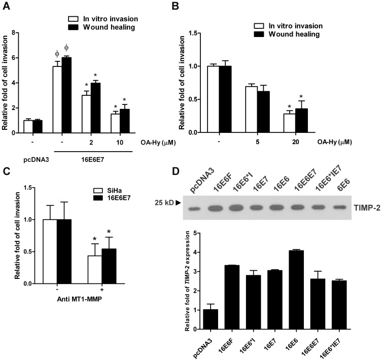
Verification
Claims the paper makes
The claims and statistics of this paper haven’t been checked yet.
Run the check — free, no account needed. It reads the open-access text and checks the sources it cites.
Claims other papers make about this one
Each is checked against this paper’s own text: does it say what the citing paper says it does?
In the citing paper
“the upregulation of MMP1, MT1-MMP, MMP2 and MMP9 has been shown to be due to the expression of HPV E6 and E7 oncoproteins in various cellular models of cervical cancer [260,261,262,263,264]”
From The Not-So-Good, the Bad and the Ugly: HPV E5, E6 and E7 Oncoproteins in the Orchestration of Carcinogenesis (2021) · cites this paper as [263]
Self-check
Percentages, sums and reported differences re-computed from the paper’s own numbers.
The self-check needs the paper’s full text.
Citations
Cites
No references could be resolved yet.
Research works printed but not linked: 45
- S Sukvirach, 2003, Population-based human papillomavirus prevalence in Lampang and Songkla, Thailand, J Infect Dis, 187(8), 1246, 10.1086/373901 · DOI
- H zur Hausen, 1996, Papillomavirus infections–a major cause of human cancers, Biochim Biophys Acta, 1288(2), F55
- K Munger, 1989, The E6 and E7 genes of the human papillomavirus type 16 together are necessary and sufficient for transformation of primary human keratinocytes, J Virol, 63(10), 4417, 10.1128/JVI.63.10.4417-4421.1989 · DOI · Paper page
- SN Boyer, 1996, E7 protein of human papilloma virus-16 induces degradation of retinoblastoma protein through the ubiquitin-proteasome pathway, Cancer Res, 56(20), 4620
- M Scheffner, 1990, The E6 oncoprotein encoded by human papillomavirus types 16 and 18 promotes the degradation of p53, Cell, 63(6), 1129, 10.1016/0092-8674(90)90409-8 · DOI · Paper page
- A Dey, 1997, HPV16 E6 oncoprotein stimulates the transforming growth factor-beta 1 promoter in fibroblasts through a specific GC-rich sequence, Virology, 228(2), 190, 10.1006/viro.1996.8363 · DOI
- S Oh, 2001, Telomerase activation by human papillomavirus type 16 E6 protein: induction of human telomerase reverse transcriptase expression through Myc and GC-rich Sp1 binding sites, J Virol, 75(12), 5559, 10.1128/JVI.75.12.5559-5566.2001 · DOI · Paper page
- TM Wise-Draper, 2005, The human DEK proto-oncogene is a senescence inhibitor and an upregulated target of high-risk human papillomavirus E7, J Virol, 79(22), 14309, 10.1128/JVI.79.22.14309-14317.2005 · DOI
- D Pim, 2009, The human papillomavirus (HPV) E6* proteins from high-risk, mucosal HPVs can direct degradation of cellular proteins in the absence of full-length E6 protein, J Virol, 83(19), 9863, 10.1128/JVI.00539-09 · DOI
- P Wanichwatanadecha, 2012, Transactivation activity of human papillomavirus type 16 E6*I on aldo-keto reductase genes enhances chemoresistance in cervical cancer cells, J Gen Virol, 93, 1081, 10.1099/vir.0.038265-0 · DOI
- E Lara-Pezzi, 2002, The hepatitis B virus X protein promotes tumor cell invasion by inducing membrane-type matrix metalloproteinase-1 and cyclooxygenase-2 expression, J Clin Invest, 110(12), 1831, 10.1172/JCI200215887 · DOI
- M Egeblad, 2002, New functions for the matrix metalloproteinases in cancer progression, Nat Rev Cancer, 2(3), 161, 10.1038/nrc745 · DOI · Paper page
- L Cardeal, 2006, Higher expression and activity of metalloproteinases in human cervical carcinoma cell lines is associated with HPV presence, Biochem Cell Biol, 84(5), 713, 10.1139/o06-084 · DOI
- S Smola-Hess, 2005, Expression of membrane type 1 matrix metalloproteinase in papillomavirus-positive cells: role of the human papillomavirus (HPV) 16 and HPV8 E7 gene products, J Gen Virol, 86(5), 1291, 10.1099/vir.0.80551-0 · DOI
- C Gilles, 1996, High level of MT-MMP expression is associated with invasiveness of cervical cancer cells, Int J Cancer, 65(2), 209, 10.1002/(SICI)1097-0215(19960117)65:2<209::AID-IJC14>3.0.CO;2-8 · DOI
- B-C Sheu, 2003, Increased expression and activation of gelatinolytic matrix metalloproteinases is associated with the progression and recurrence of human cervical cancer, Cancer Res, 63(19), 6537
- G Vazquez-Ortiz, 2005, Overexpression of cathepsin F, matrix metalloproteinases 11 and 12 in cervical cancer, BMC Cancer, 5, 68, 10.1186/1471-2407-5-68 · DOI
- LB Cardeal, 2012, HPV16 oncoproteins induce MMPs/RECK-TIMP-2 imbalance in primary keratinocytes: possible implications in cervical carcinogenesis, PLoS One, 7(3), e33585, 10.1371/journal.pone.0033585 · DOI
- Y Kato, 2002, Relationship between expression of matrix metalloproteinase-2 and matrix metalloproteinase-9 and invasion ability of cervical cancer cells, Oncol Rep, 9(3), 565
- H Sato, 1994, A matrix metalloproteinase expressed on the surface of invasive tumour cells, Nature, 370(6484), 61, 10.1038/370061a0 · DOI
- A Mitra, 2003, Membrane-associated MMP-2 in human cervical cancer, J Environ Pathol Toxicol Oncol, 22(2), 93
- I Clark, 2008, The regulation of matrix metalloproteinases and their inhibitors, Int J Biochem Cell Biol, 40(6–7), 1362, 10.1016/j.biocel.2007.12.006 · DOI · Paper page
- T Rampias, 2009, E6 and E7 gene silencing and transformed phenotype of human papillomavirus 16-positive oropharyngeal cancer cells, J Natl Cancer Inst, 101(6), 412, 10.1093/jnci/djp017 · DOI
- T Asano, 2008, Prognostic values of matrix metalloproteinase family expression in human colorectal carcinoma, J Surg Res, 146(1), 32, 10.1016/j.jss.2007.02.011 · DOI
- M Pesta, 2005, Quantitative estimation of matrix metalloproteinases 2 and 7 (MMP-2, MMP-7) and tissue inhibitors of matrix metalloproteinases 1 and 2 (TIMP-1, TIMP-2) in colorectal carcinoma tissue samples, Anticancer Res, 25(5), 3387
- P Pongcharoen, 2011, Silencing of CD44 by siRNA suppressed invasion, migration and adhesion to matrix, but not secretion of MMPs, of cholangiocarcinoma cells, Clin Exp Metastasis, 28(8), 827, 10.1007/s10585-011-9414-8 · DOI
- S Venturoli, 1998, Evaluation of immunoassays for the detection and typing of PCR amplified human papillomavirus DNA, J Clin Pathol, 51(2), 143, 10.1136/jcp.51.2.143 · DOI
- N Shirvaikar, 2012, Hematopoietic Stem Cell Mobilization and Homing after Transplantation: The Role of MMP-2, MMP-9, and MT1-MMP, Biochem Res Int, 2012, 685267, 10.1155/2012/685267 · DOI
- PA Eisenach, 2010, MT1-MMP regulates VEGF-A expression through a complex with VEGFR-2 and Src, J Cell Sci 123(Pt, 23), 4182, 10.1242/jcs.062711 · DOI
- A Thathiah, 2004, MT1-MMP mediates MUC1 shedding independent of TACE/ADAM17, Biochem J 382(Pt, 1), 363, 10.1042/BJ20040513 · DOI
- D Pim, 2010, Interaction of viral oncoproteins with cellular target molecules: infection with high-risk vs low-risk human papillomaviruses, APMIS, 118(6–7), 471, 10.1111/j.1600-0463.2010.02618.x · DOI
- Y Zhai, 2005, Expression of membrane type 1 matrix metalloproteinase is associated with cervical carcinoma progression and invasion, Cancer Res, 65(15), 6543, 10.1158/0008-5472.CAN-05-0231 · DOI · Paper page
- S Asha Nair, 2003, Changes in matrix metalloproteinases and their endogenous inhibitors during tumor progression in the uterine cervix, J Cancer Res Clin Oncol, 129(2), 123, 10.1007/s00432-002-0411-9 · DOI
- M Branca, 2006, Matrix metalloproteinase-2 (MMP-2) and its tissue inhibitor (TIMP-2) are prognostic factors in cervical cancer, related to invasive disease but not to high risk human papillomavirus (HPV) or virus persistence after treatment of CIN, Anticancer Res, 26, 1543
- M Nakada, 2001, Suppression of membrane-type 1 matrix metalloproteinase (MMP)-mediated MMP-2 activation and tumor invasion by testican3 and its splicing variant gene product, N-Tes, Cancer Res, 61, 8896
- T Uekita, 2004, Membrane type 1 matrix metalloproteinase cytoplasmic tail-binding protein 1 is a new member of the cupin superfamily, a possible multifunctional protein acting as an invasion suppressor down-regulated in tumors, J Biol Chem, 279, 12734, 10.1074/jbc.M309957200 · DOI
- KD Cowden Dahl, 2007, PEA3 is necessary for optimal epidermal growth factor receptor-stimulated matrix metalloproteinase expression and invasion of ovarian tumor cells, Mol Cancer Res, 5(5), 413, 10.1158/1541-7786.MCR-07-0019 · DOI
- J Lohi, 2000, Structural analysis and promoter characterization of the human membrane-type matrix metalloproteinase-1 (MT1-MMP) gene, Gene, 242(1–2), 75, 10.1016/S0378-1119(99)00549-1 · DOI
- H Qin, 1999, The transcription factors Sp1, Sp3, and AP-2 are required for constitutive matrix metalloproteinase-2 gene expression in astroglioma cells, J Biol Chem, 274(41), 29130, 10.1074/jbc.274.41.29130 · DOI
- L Qin, 2008, The AIB1 oncogene promotes breast cancer metastasis by activation of PEA3-mediated matrix metalloproteinase 2 (MMP2) and MMP9 expression, Mol Cell Biol, 28(19), 5937, 10.1128/MCB.00579-08 · DOI
- Z Zhang, 2009, Epidermal growth factor receptor regulates MT1-MMP and MMP-2 synthesis in SiHa cells via both PI3-K/AKT and MAPK/ERK pathways, Int J Gynecol Cancer, 19(6), 998, 10.1111/IGC.0b013e3181a83749 · DOI
- H Yamaguchi, 1999, A PEA3 site flanked by SP1, SP4, and GATA sites positively regulates the differentiation-dependent expression of Brachyury in embryonal carcinoma P19 cells, Biochem Biophys Res Commun, 254(3), 542, 10.1006/bbrc.1998.0102 · DOI
- O Lopez-Ocejo, 2000, Oncogenes and tumor angiogenesis: the HPV-16 E6 oncoprotein activates the vascular endothelial growth factor (VEGF) gene promoter in a p53 independent manner, Oncogene, 19(40), 4611, 10.1038/sj.onc.1203817 · DOI · Paper page
- K Munger, 2001, Biological activities and molecular targets of the human papillomavirus E7 oncoprotein, Oncogene, 20(54), 7888, 10.1038/sj.onc.1204860 · DOI
- A Takahashi, 2007, Adenovirus E1A negatively regulates E1AF, an ets family of the protein, Biochem Biophys Res Commun, 355(2), 438, 10.1016/j.bbrc.2007.01.165 · DOI
Cited by
Basukala, O., & Banks, L. (2021) · Viruses · doi:10.3390/v13101892 · Cited by 137
Metadata
- Authors
- Kaewprag, J., Umnajvijit, W., Ngamkham, J., & Ponglikitmongkol, M.
- Venue
- PLoS ONE
- Publisher
- Public Library of Science (PLoS)
- Published
- 2013-08-13
- Volume
- 8
- Issue
- 8
- Pages
- e71611
- ISSN
- 1932-6203
- PMC
- PMC3742460
- Cited by
- 35
- References
- 0
- Language
- en
About these checks
Registry, retraction, Cites and statistics results are computed, not judged, with one exception: a printed reference that no index lists is read once by a model before it counts as a citation error. Claims are extracted by a model and kept only when their quote appears verbatim in the text; each cited source is then checked, with a model’s help, for whether it says what the claim relies on. Those verdicts can be wrong — read the claim’s quote and the cited source before relying on one.
Statistics and arithmetic are recomputed, not judged.
Confidence bands are shown only for fields calibrated on our annotated test set. None is calibrated yet, so no band is shown.
What counts as a claim ›How we measure confidence ›How we check this ›
Are you an author, or do you see a problem with this page? Request a review
