Cited by 320Endoplasmic Reticulum Stress and DiseasePharmacological Receptor Mechanisms and EffectsIon Channels and Receptors

The DOI matches its Crossref record; 50 of the 51 references it cites were found in a registry or an index.

Sigma-1 receptor chaperone at the ER-mitochondrion interface mediates the mitochondrion-ER-nucleus signaling for cellular survival.

Open access

Abstract

The membrane of the endoplasmic reticulum (ER) of a cell forms contacts directly with mitochondria whereby the contact is referred to as the mitochondrion-associated ER membrane or the MAM. Here we found that the MAM regulates cellular survival via an MAM-residing ER chaperone the sigma-1 receptor (Sig-1R) in that the Sig-1R chaperones the ER stress sensor IRE1 to facilitate inter-organelle signaling for survival. IRE1 is found in this study to be enriched at the MAM in CHO cells. We found that IRE1 is stabilized at the MAM by Sig-1Rs when cells are under ER stress. Sig-1Rs stabilize IRE1 and thus allow for conformationally correct IRE1 to dimerize into the long-lasting, activated endonuclease. The IRE1 at the MAM also responds to reactive oxygen species derived from mitochondria. Therefore, the ER-mitochondrion interface serves as an important subcellular entity in the regulation of cellular survival by enhancing the stress-responding signaling between mitochondria, ER, and nucleus.

Overview

The claims and statistics of this paper haven’t been checked yet.

Run the check — free, no account needed. It reads the open-access text and checks the sources it cites.

A few free checks a day · papers already verified open instantly · results are public

Integrity signals

Registry

Crossref · 10.1371/journal.pone.0076941

Cites

50 of 50 references verified0 citation errors · 0 not checkable

Statistics

Figures

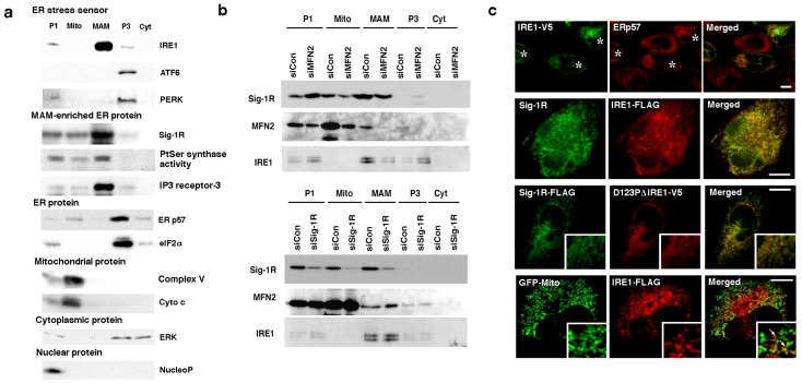
Figure 1 IRE1 localizes at the MAM. (a) The subcellular distribution of ER stress sensors. All endogenous proteins were prepared from wild-type non-stressed CHO cells. P1, nuclear; Mito, mitochondrial; P3, microsomal; Cyt, cytosolic fractions. NucleoP, nucleoporin p62; Complex V, complex V ATP synthase inhibitor; Cyto c, cytochrome c; ERp57, ER thiol-disulfide oxidoreductase p57; ERK, extracellular signal-regulated kinase. Phosphatidylserine (PtSer) synthase activity was measured by the autoradiographic measurement of 14C-PtSer as described [10]. All other proteins were measured by immunoblotting. (b) The subcellular distribution of IRE1 in CHO cells with reduced expression of mitofusin-2 (MFN2) or Sig-1Rs. Control (siCon) or specific siRNAs (siMFN2, siSig-1R) were transfected two days before the membrane fractionation. (c) Confocal microscopic observation of the subcellular distribution of Sig-1Rs and IRE1 in CHO cells. In top panels, endogenous ERp57 and transfected full-length IRE1-V5 were immunostained. Asterisks indicate CHO cells transfected with IRE1-V5 (Note: no V5 immunoreactivity in non-transfected cells, verifying the high selectivity of V5 immunostaining). In bottom panels, GFP targeting mitochondria was expressed by gene transfection. Arrows indicate clusters of IRE1-FLAG apposing mitochondria. Scale = 10 µm. Insets on a 5× magnification.
Figure 1 IRE1 localizes at the MAM. (a) The subcellular distribution of ER stress sensors. All endogenous proteins were prepared from wild-type non-stressed CHO cells. P1, nuclear; Mito, mitochondrial; P3, microsomal; Cyt, cytosolic fractions. NucleoP, nucleoporin p62; Complex V, complex V ATP synthase inhibitor; Cyto c, cytochrome c; ERp57, ER thiol-disulfide oxidoreductase p57; ERK, extracellular signal-regulated kinase. Phosphatidylserine (PtSer) synthase activity was measured by the autoradiographic measurement of 14C-PtSer as described [10]. All other proteins were measured by immunoblotting. (b) The subcellular distribution of IRE1 in CHO cells with reduced expression of mitofusin-2 (MFN2) or Sig-1Rs. Control (siCon) or specific siRNAs (siMFN2, siSig-1R) were transfected two days before the membrane fractionation. (c) Confocal microscopic observation of the subcellular distribution of Sig-1Rs and IRE1 in CHO cells. In top panels, endogenous ERp57 and transfected full-length IRE1-V5 were immunostained. Asterisks indicate CHO cells transfected with IRE1-V5 (Note: no V5 immunoreactivity in non-transfected cells, verifying the high selectivity of V5 immunostaining). In bottom panels, GFP targeting mitochondria was expressed by gene transfection. Arrows indicate clusters of IRE1-FLAG apposing mitochondria. Scale = 10 µm. Insets on a 5× magnification.CC0 · PubMed Central
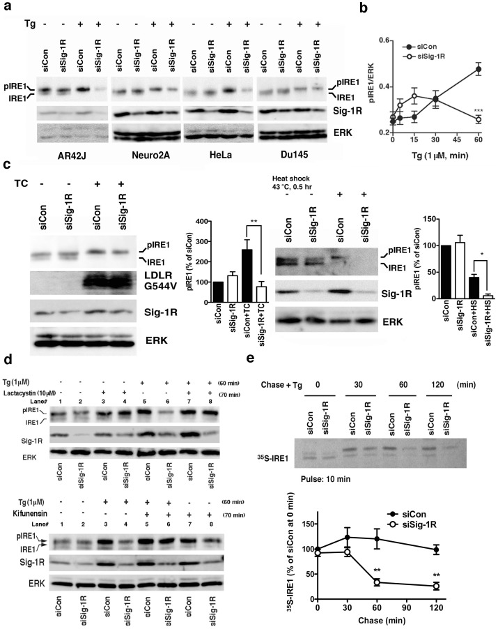
Figure 2 Sigma-1 receptors stabilize IRE1. (a) Sig-1R knockdown decreases phosphorylated IRE1 (pIRE1) in various types of cells when cells are under ER stress. Two days after the transfection of control siRNA (siCon) or Sig-1R siRNA (siSig-1R), cells were treated with thapsigargin (Tg) at 1 µM for 60 min. IRE1 were immunoprecipitated from 60–1000 µg of total protein lysates. (b) The temporal course of pIRE1 levels during ER stress. Control or Sig-1R siRNA was transfected to CHO cells two days before Tg. pIRE1 was measured by immunoprecipitation. The level of pIRE1 (partially phosphorylated IRE1 plus hyperphosphorylated IRE1) was normalized to ERK. The graph represents the means±S.E.M. ***p<0.001 (n = 6). (c) Effect of the Sig-1R knockdown on the level of pIRE1 in CHO cells either overexpressing LDL receptor G544V mutants or being under the heatshock treatment. LDL receptors G544V (LDLR G544V) were induced by the tetracycline treatment for 16 hrs (TC; see Methods). The level of pIRE1 was measured by immunoprecipitation and was normalized to ERK. Graphs represent means±S.E.M (n = 4). *p<0.05, **p<0.01. (d) Effects of lactacystin (10 µM; applied 10 min before Tg) or kifunensin (2 µg/ml; applied 10 min before Tg; lower panels) on Tg (1 µM for 1 hr)-induced decrease of pIRE1 in Sig-1R knockdown CHO cells. (e) Pulse-chase experiment measuring the effect of Sig-1R knockdown on the life-time of IRE1 when cells were under ER stress. After labeling with 35S-methionine, CHO cells transfected with control or Sig-1R siRNA were chased in the presence of Tg (1 µM) for indicated periods of time. IRE1 was immunoprecipitated and then detected by direct autoradiography. The graph represents mean±S.E.M (n = 4). **p<0.01 compared with siCon at the same time point.
Figure 2 Sigma-1 receptors stabilize IRE1. (a) Sig-1R knockdown decreases phosphorylated IRE1 (pIRE1) in various types of cells when cells are under ER stress. Two days after the transfection of control siRNA (siCon) or Sig-1R siRNA (siSig-1R), cells were treated with thapsigargin (Tg) at 1 µM for 60 min. IRE1 were immunoprecipitated from 60–1000 µg of total protein lysates. (b) The temporal course of pIRE1 levels during ER stress. Control or Sig-1R siRNA was transfected to CHO cells two days before Tg. pIRE1 was measured by immunoprecipitation. The level of pIRE1 (partially phosphorylated IRE1 plus hyperphosphorylated IRE1) was normalized to ERK. The graph represents the means±S.E.M. ***p<0.001 (n = 6). (c) Effect of the Sig-1R knockdown on the level of pIRE1 in CHO cells either overexpressing LDL receptor G544V mutants or being under the heatshock treatment. LDL receptors G544V (LDLR G544V) were induced by the tetracycline treatment for 16 hrs (TC; see Methods). The level of pIRE1 was measured by immunoprecipitation and was normalized to ERK. Graphs represent means±S.E.M (n = 4). *p<0.05, **p<0.01. (d) Effects of lactacystin (10 µM; applied 10 min before Tg) or kifunensin (2 µg/ml; applied 10 min before Tg; lower panels) on Tg (1 µM for 1 hr)-induced decrease of pIRE1 in Sig-1R knockdown CHO cells. (e) Pulse-chase experiment measuring the effect of Sig-1R knockdown on the life-time of IRE1 when cells were under ER stress. After labeling with 35S-methionine, CHO cells transfected with control or Sig-1R siRNA were chased in the presence of Tg (1 µM) for indicated periods of time. IRE1 was immunoprecipitated and then detected by direct autoradiography. The graph represents mean±S.E.M (n = 4). **p<0.01 compared with siCon at the same time point.CC0 · PubMed Central
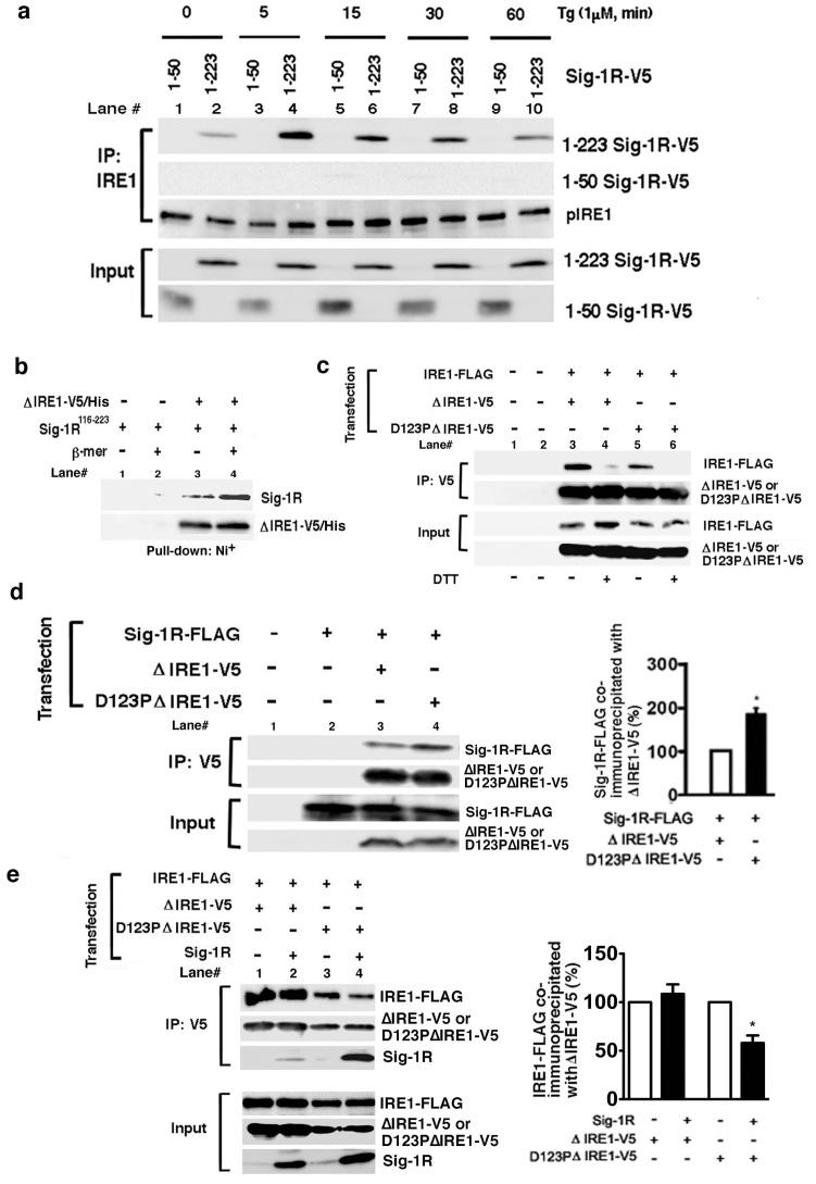
Figure 3 Sigma-1receptors preferentially associate with the monomeric form of IRE1 in the lumen of the ER. (a) Time-dependent association of V5-tagged Sig-1Rs with IRE1 during ER stress. IRE1 was immunoprecipitated in CHO cells expressing V5-tagged full-length Sig1R-V5 (1–223) or truncated Sig-1R-V5 (1–50). Thapsigargin (Tg) was applied to the medium for indicated periods of time. (b) Direct association between purified ΔIRE1-V5/His and the ER lumenal domain (116–223) of Sig-1Rs (GST-Sig-1R116–223) in vitro. Purified ΔIRE1 immobilized on a Ni+-column was incubated with purified GST-Sig-1R116–223 polypeptides. After extensive washing, the GST-Sig-1R116–223 associating with ΔIRE1-V5/His was measured by Western blotting. β-mer (+): the purified ΔIRE1-V5/His was pretreated with β-mercaptoethanol to prevent the dimerization of ΔIRE1-V5/His. The image represents three independent experiments. (c) Involvement of both the ionic bond (D123 site) and the disulfide bonds in the dimerization of IRE1. To assess the dimerization of IRE1, FLAG-tagged full-length IRE1 was co-immunoprecipitated with ΔIRE1-V5 with or without a point-mutation at D123 (e.g., ΔIRE1, D123PΔIRE1). Dimerization of IRE1, via disulfide bonds, was disrupted by DTT (20 mM for 30 min). (d) Association of Sig-1Rs with IRE1 depends on the dimerization status of IRE1. V5-tagged ΔIRE1 with or without a point-mutation at D123 were immunoprecipitated. The association of Sig-1R with Δ IRE1-V5 was assessed by measuring co-immunoprecipitated Sig-1R-FLAG. The graph represents means±S.E.M. *p<0.05 (n = 4). (e) Effects of overexpressed Sig-1Rs on the dimerization of IRE1. Dimerization of IRE1 was assessed by measuring IRE1-FLAG co-immunoprecipitated with V5-tagged IRE1 (ΔIRE1-V5 or D123PΔIRE1-V5). Note: overexpressed Sig-1Rs co-immunoprecipitated significantly more with IRE1-V5 when IRE1 was mutated at D123. The graph represents means±S.E.M (n = 4, *p<0.05).
Figure 3 Sigma-1receptors preferentially associate with the monomeric form of IRE1 in the lumen of the ER. (a) Time-dependent association of V5-tagged Sig-1Rs with IRE1 during ER stress. IRE1 was immunoprecipitated in CHO cells expressing V5-tagged full-length Sig1R-V5 (1–223) or truncated Sig-1R-V5 (1–50). Thapsigargin (Tg) was applied to the medium for indicated periods of time. (b) Direct association between purified ΔIRE1-V5/His and the ER lumenal domain (116–223) of Sig-1Rs (GST-Sig-1R116–223) in vitro. Purified ΔIRE1 immobilized on a Ni+-column was incubated with purified GST-Sig-1R116–223 polypeptides. After extensive washing, the GST-Sig-1R116–223 associating with ΔIRE1-V5/His was measured by Western blotting. β-mer (+): the purified ΔIRE1-V5/His was pretreated with β-mercaptoethanol to prevent the dimerization of ΔIRE1-V5/His. The image represents three independent experiments. (c) Involvement of both the ionic bond (D123 site) and the disulfide bonds in the dimerization of IRE1. To assess the dimerization of IRE1, FLAG-tagged full-length IRE1 was co-immunoprecipitated with ΔIRE1-V5 with or without a point-mutation at D123 (e.g., ΔIRE1, D123PΔIRE1). Dimerization of IRE1, via disulfide bonds, was disrupted by DTT (20 mM for 30 min). (d) Association of Sig-1Rs with IRE1 depends on the dimerization status of IRE1. V5-tagged ΔIRE1 with or without a point-mutation at D123 were immunoprecipitated. The association of Sig-1R with Δ IRE1-V5 was assessed by measuring co-immunoprecipitated Sig-1R-FLAG. The graph represents means±S.E.M. *p<0.05 (n = 4). (e) Effects of overexpressed Sig-1Rs on the dimerization of IRE1. Dimerization of IRE1 was assessed by measuring IRE1-FLAG co-immunoprecipitated with V5-tagged IRE1 (ΔIRE1-V5 or D123PΔIRE1-V5). Note: overexpressed Sig-1Rs co-immunoprecipitated significantly more with IRE1-V5 when IRE1 was mutated at D123. The graph represents means±S.E.M (n = 4, *p<0.05).CC0 · PubMed Central
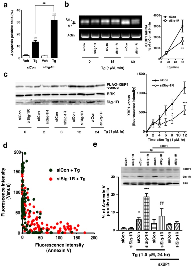
Figure 4 Effect of Sig-1R knockdown on the Tg-stimulated IRE1/XBP1 signaling pathway in CHO cells. (a) Effect of Sig-1R knockdown on Tg-induced apoptosis. Induction of apoptosis by Tg (1 µM, for 24 hr) was quantified by using Hoechst 33342 staining. The percentage of apoptotic cells was measured from 6 individual samples (more than 200 cells were counted in each sample) and is reported as means±S.E.M. ***P<0.001 compared with vehicle (Veh), ##P<0.01 compared with siCont with Tg. (b) Effects of Sig-1R knockdown on the splicing of XBP1 mRNA. CHO cells transfected with control or Sig-1R siRNA were treated with Tg for indicated periods of time. Total RNA was extracted and XBP-1 or actin transcripts were amplified by RT-PCR. Un, unspliced; S, spliced; sXBP1; spliced XBP1. The graph represents means±S.E.M. **p<0.01 compared with siCon at the same time point (n = 4). (c) Effects of Sig-1R siRNA on the expression of FLAG-XBP1-venus fusion proteins. CHO cells transfected with FLAG-XBP1-venus plasmids were treated with Tg, and the expression of FLAG-XBP1-venus was measured by Western blotting (left panels) or a fluorescence microplate reader (the right graph). *p<0.05, ***p<0.001 compared with siCon at the same time point (n = 8). Note that venus is expressed only when FLAG-XBP1 mRNA is spliced by active IRE1. (d) Sig-1R siRNA enhanced apoptosis in Tg-treated cells negatively correlates with the activity of XBP1 mRNA splicing. Fluorescence intensities (annexin V vs. venus) of individual CHO cells were plotted in the graph. Control siRNA (siCon) in green, Sig-1R siRNA (siSig-1R) in orange. (e) Overexpression of spliced XBP1 attenuates apoptosis enhanced by Sig-1R knockdown in Tg-treated cells. Each bar represents the means±S.E.M. (n = 6–7 samples; >50 cells/sample were counted). *P<0.05, ***P<0.001 compared with siCon alone. ##P<0.01 compared with siSig-1R with Tg.
Figure 4 Effect of Sig-1R knockdown on the Tg-stimulated IRE1/XBP1 signaling pathway in CHO cells. (a) Effect of Sig-1R knockdown on Tg-induced apoptosis. Induction of apoptosis by Tg (1 µM, for 24 hr) was quantified by using Hoechst 33342 staining. The percentage of apoptotic cells was measured from 6 individual samples (more than 200 cells were counted in each sample) and is reported as means±S.E.M. ***P<0.001 compared with vehicle (Veh), ##P<0.01 compared with siCont with Tg. (b) Effects of Sig-1R knockdown on the splicing of XBP1 mRNA. CHO cells transfected with control or Sig-1R siRNA were treated with Tg for indicated periods of time. Total RNA was extracted and XBP-1 or actin transcripts were amplified by RT-PCR. Un, unspliced; S, spliced; sXBP1; spliced XBP1. The graph represents means±S.E.M. **p<0.01 compared with siCon at the same time point (n = 4). (c) Effects of Sig-1R siRNA on the expression of FLAG-XBP1-venus fusion proteins. CHO cells transfected with FLAG-XBP1-venus plasmids were treated with Tg, and the expression of FLAG-XBP1-venus was measured by Western blotting (left panels) or a fluorescence microplate reader (the right graph). *p<0.05, ***p<0.001 compared with siCon at the same time point (n = 8). Note that venus is expressed only when FLAG-XBP1 mRNA is spliced by active IRE1. (d) Sig-1R siRNA enhanced apoptosis in Tg-treated cells negatively correlates with the activity of XBP1 mRNA splicing. Fluorescence intensities (annexin V vs. venus) of individual CHO cells were plotted in the graph. Control siRNA (siCon) in green, Sig-1R siRNA (siSig-1R) in orange. (e) Overexpression of spliced XBP1 attenuates apoptosis enhanced by Sig-1R knockdown in Tg-treated cells. Each bar represents the means±S.E.M. (n = 6–7 samples; >50 cells/sample were counted). *P<0.05, ***P<0.001 compared with siCon alone. ##P<0.01 compared with siSig-1R with Tg.CC0 · PubMed Central
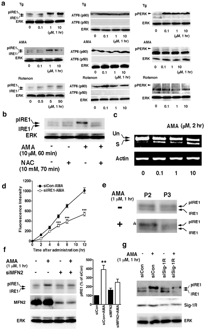
Figure 5 Mitochondria-derived oxidative factor activates IRE1. (a) Effects of Tg, antimycin A (AMA), or rotenone on the activation of ER stress sensors. IRE1 were immunoprecipitated before detection by western blottings. No active form of ATF6 (p50) or phosphorylated PERK was detected under the experimental condition with AMA or rotenone. (b) Effects of NAC on phosphorylation of IRE1 caused by AMA. (c) Effects of AMA on the splicing of the XBP1 mRNA in CHO cells. Un, unspliced; S, spliced. (d) AMA (1 µM) induced the expression of XBP1-venus in an IRE1-dependent manner. CHO cells were transfected with FLAG-XBP1-venus plasmids and siRNA (siCon, scrambled siRNA; siIRE1, IRE1 siRNA) two days before the AMA treatment. Induction of FLAG-XBP1-venus proteins was measured by a fluorescence microplate reader. ***P<0.001 (n = 12), compared with siCon. (e) Selective activation of IRE1 by AMA in the MAM-containing crude mitochondrial fraction. AMA-treated CHO cells were homogenized and subjected to differential centrifugation. The asterisk indicates the hyperphosphorylated form of IRE1. (f) Effect of mifotusin-2 knockdown on the AMA-induced IRE1 phosphorylation. siRNA against mitofusin-2 (siMFN2) or scrambled control siRNA (siCon) was transfected into CHO cells two days before the treatment with AMA. In the graph, IRE1 was normalized to ERK and shown as means±S.E.M. **p<0.01 (n = 4) compared with siCon without AMA. (g) AMA induces a reduction of pIRE1 proteins in CHO cells with reduced expression of Sig-1Rs. Control or Sig-1R siRNA were transfected into CHO cells two days before AMA. IRE1 was immunoprecipitated before detection.
Figure 5 Mitochondria-derived oxidative factor activates IRE1. (a) Effects of Tg, antimycin A (AMA), or rotenone on the activation of ER stress sensors. IRE1 were immunoprecipitated before detection by western blottings. No active form of ATF6 (p50) or phosphorylated PERK was detected under the experimental condition with AMA or rotenone. (b) Effects of NAC on phosphorylation of IRE1 caused by AMA. (c) Effects of AMA on the splicing of the XBP1 mRNA in CHO cells. Un, unspliced; S, spliced. (d) AMA (1 µM) induced the expression of XBP1-venus in an IRE1-dependent manner. CHO cells were transfected with FLAG-XBP1-venus plasmids and siRNA (siCon, scrambled siRNA; siIRE1, IRE1 siRNA) two days before the AMA treatment. Induction of FLAG-XBP1-venus proteins was measured by a fluorescence microplate reader. ***P<0.001 (n = 12), compared with siCon. (e) Selective activation of IRE1 by AMA in the MAM-containing crude mitochondrial fraction. AMA-treated CHO cells were homogenized and subjected to differential centrifugation. The asterisk indicates the hyperphosphorylated form of IRE1. (f) Effect of mifotusin-2 knockdown on the AMA-induced IRE1 phosphorylation. siRNA against mitofusin-2 (siMFN2) or scrambled control siRNA (siCon) was transfected into CHO cells two days before the treatment with AMA. In the graph, IRE1 was normalized to ERK and shown as means±S.E.M. **p<0.01 (n = 4) compared with siCon without AMA. (g) AMA induces a reduction of pIRE1 proteins in CHO cells with reduced expression of Sig-1Rs. Control or Sig-1R siRNA were transfected into CHO cells two days before AMA. IRE1 was immunoprecipitated before detection.CC0 · PubMed Central
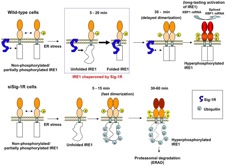
Figure 6 Schematic model depicting the role of the Sig-1R chaperone in the activation of IRE1. The Sig-1R molecular chaperone enhances its association with IRE1 to correct or stabilize the conformation of IRE1 when cells are facing ER stress (i.e., as indicated in the blue-lined rectangle). This transient association of the Sig-1R with IRE1 interferes with the dimerization of IRE1, leading to a delay in the transautophosphorylation of IRE1 (30 min). This delayed dimerization/phosphorylation, however, ensures a long-lasting active form of IRE1 (the cytoplasmic domain filled in red) which splices the XBP1 mRNA. In lower panels, when Sig-1R knockdown cells encounter ER stress, IRE1, although being misfolded, can still quickly dimerize and transautophosphorylate (5–15 min lower panels). The conformationally awry pIRE1, which may still possess endonuclease activity albeit being less compared to controls, is however readily ubiquitinated and degraded by proteasomes.
Figure 6 Schematic model depicting the role of the Sig-1R chaperone in the activation of IRE1. The Sig-1R molecular chaperone enhances its association with IRE1 to correct or stabilize the conformation of IRE1 when cells are facing ER stress (i.e., as indicated in the blue-lined rectangle). This transient association of the Sig-1R with IRE1 interferes with the dimerization of IRE1, leading to a delay in the transautophosphorylation of IRE1 (30 min). This delayed dimerization/phosphorylation, however, ensures a long-lasting active form of IRE1 (the cytoplasmic domain filled in red) which splices the XBP1 mRNA. In lower panels, when Sig-1R knockdown cells encounter ER stress, IRE1, although being misfolded, can still quickly dimerize and transautophosphorylate (5–15 min lower panels). The conformationally awry pIRE1, which may still possess endonuclease activity albeit being less compared to controls, is however readily ubiquitinated and degraded by proteasomes.CC0 · PubMed Central

Verification

Claims the paper makes

The claims and statistics of this paper haven’t been checked yet.

Run the check — free, no account needed. It reads the open-access text and checks the sources it cites.

A few free checks a day · papers already verified open instantly · results are public

Claims other papers make about this one

Each is checked against this paper’s own text: does it say what the citing paper says it does?

Self-check

Percentages, sums and reported differences re-computed from the paper’s own numbers.

The self-check needs the paper’s full text.

Citations

Cites

1 retracted

Show all 51 references

Cited by

Sorted by citations

See all 320

Metadata

Authors
Mori, T., Hayashi, T., Hayashi, E., & Su, T.-P.
Affiliations
United States Department of Health and Human Services (US)
Corresponding author
Su, Tsung-Ping
Venue
PLoS ONE
Published
2013-10-18
Volume
8
Issue
10
Pages
e76941
ISSN
1932-6203 (ISSN-L)
OpenAlex
W1994289493
Cited by
317
References
51
Citations by year
2026: 22 · 2025: 24 · 2024: 20 · 2023: 29 · 2022: 26 · 2021: 38 · 2020: 33 · 2019: 34 · 2018: 18 · 2017: 29 · 2016: 16 · 2015: 10 · 2014: 17
FWCI
8.51
Type
journal article
Language
en
Open access
Locations
  • PLoS ONE (publishedVersion, public-domain)
  • Europe PMC (PubMed Central) (publishedVersion, public-domain)
  • PubMed Central (publishedVersion, public-domain)
  • PubMed (publishedVersion)
  • DOAJ (DOAJ: Directory of Open Access Journals) (submittedVersion, cc-by-sa)
  • Figshare (submittedVersion, cc-by)
Funders
  • U.S. Department of Health and Human Services
  • National Institutes of Health
  • National Institute on Drug Abuse
Topics
  • Life Sciences › Biochemistry, Genetics and Molecular Biology › Cell Biology › Endoplasmic Reticulum Stress and Disease
  • Life Sciences › Pharmacology, Toxicology and Pharmaceutics › Pharmacology › Pharmacological Receptor Mechanisms and Effects
  • Life Sciences › Neuroscience › Sensory Systems › Ion Channels and Receptors
Keywords
sigma-1 receptor, mitochondria-associated membranes, endoplasmic reticulum, IRE1, endoplasmic reticulum stress, mitochondrial reactive oxygen species, molecular chaperones, protein stabilization, endonuclease activation, cell survival
MeSH terms
Sigma-1 Receptor, Animals, Cell Nucleus, Cell Survival, Cricetulus, Endoplasmic Reticulum, Endoribonucleases, Cricetinae, HeLa Cells, Humans, Immunohistochemistry, Intracellular Membranes, Mitochondria, Mutation, Protein Binding, Blotting, Western, Signal Transduction, CHO Cells, Protein Serine-Threonine Kinases, Reactive Oxygen Species, Receptors, sigma, Microscopy, Confocal, Molecular Chaperones, RNA Interference, Cell Line, Tumor, Mice

About these checks

Registry, retraction, Cites and statistics results are computed, not judged, with one exception: a printed reference that no index lists is read once by a model before it counts as a citation error. Claims are extracted by a model and kept only when their quote appears verbatim in the text; each cited source is then checked, with a model’s help, for whether it says what the claim relies on. Those verdicts can be wrong — read the claim’s quote and the cited source before relying on one.

Statistics and arithmetic are recomputed, not judged.

Confidence bands are shown only for fields calibrated on our annotated test set. None is calibrated yet, so no band is shown.

What counts as a claim ›How we measure confidence ›How we check this ›

Are you an author, or do you see a problem with this page? Request a review