

The DOI matches its Crossref record; 5 of the 5 references it cites were found in a registry or an index.
Impact of Helminth Infections and Nutritional Constraints on the Small Intestine Microbiota
Overview
The claims and statistics of this paper haven’t been checked yet.
Run the check — free, no account needed. It reads the open-access text and checks the sources it cites.
Integrity signals
Registry
Cites
Statistics
Figures
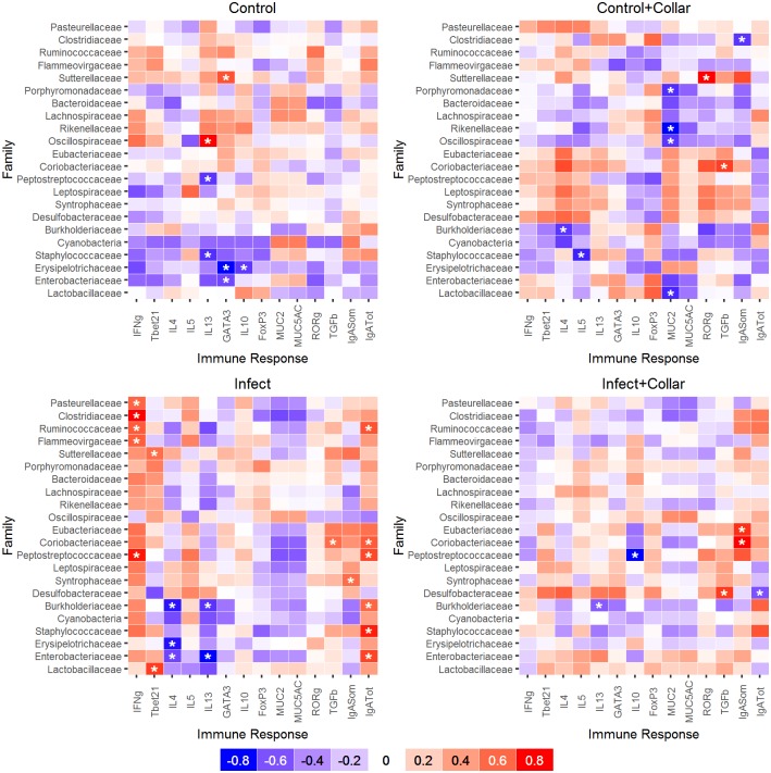
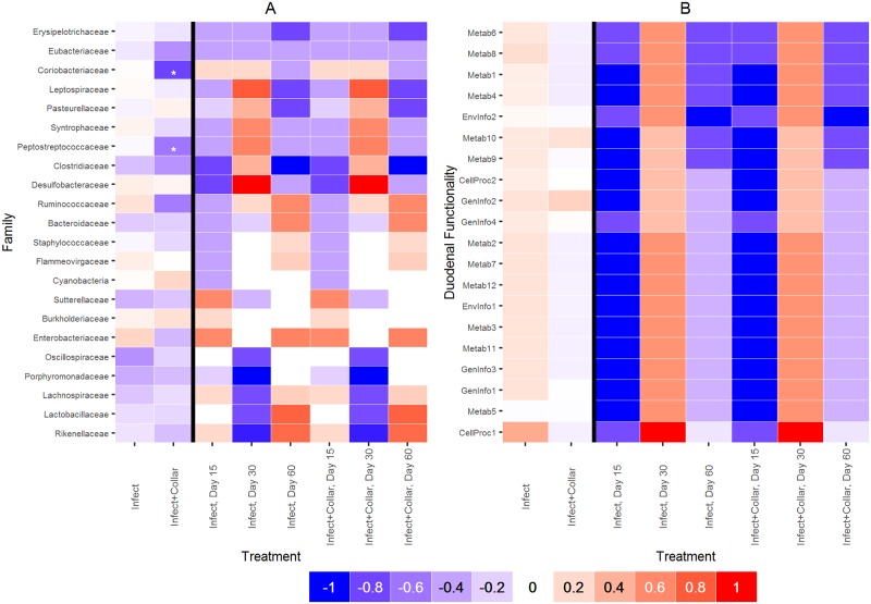
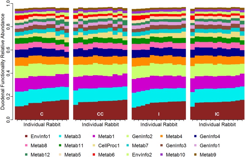
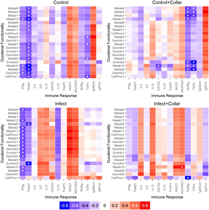
Verification
Claims the paper makes
The claims and statistics of this paper haven’t been checked yet.
Run the check — free, no account needed. It reads the open-access text and checks the sources it cites.
Claims other papers make about this one
Each is checked against this paper’s own text: does it say what the citing paper says it does?
In the citing paper
“These bacteria produce short-chain fatty acids (SCFAs), which, in turn, can stimulate Treg populations.”
From Parasites and allergies: a complex bidirectional relationship from evolutionary origins to modern therapeutics (2026) · cites this paper as Cattadori et al. 2016
In the citing paper
“For instance, during chronic nematode infections, studies have shown increased colonization of the small intestine by Lactobacillus species and increased growth of Clostridium species in the large intestine.”
From Parasites and allergies: a complex bidirectional relationship from evolutionary origins to modern therapeutics (2026) · cites this paper as Cattadori et al. 2016
In the citing paper
“Changes in the microbiome induced by parasites may also influence the risk of food allergies and other allergic conditions.”
From Parasites and allergies: a complex bidirectional relationship from evolutionary origins to modern therapeutics (2026) · cites this paper as Cattadori et al. 2016
In the citing paper
“Studies have demonstrated that helminth infections, intestinal microbiota, and nutrition are interrelated.107”
From Global issues in allergy and immunology: Parasitic infections and allergy (2017) · cites this paper as [107]
Self-check
Percentages, sums and reported differences re-computed from the paper’s own numbers.
The self-check needs the paper’s full text.
Citations
Cites
Human nutrition, the gut microbiome and the immune system
Kau, A. L., Ahern, P. P., Griffin, N. W., Goodman, A. L., & Gordon, J. I. (2011) · Nature · doi:10.1038/nature10213 · Cited by 2,588
Role of the gut microbiota in immunity and inflammatory disease
Kamada, N., Seo, S.-U., Chen, G. Y., & Núñez, G. (2013) · Nature reviews. Immunology · doi:10.1038/nri3430 · Cited by 2,256
The epidemiology of global micronutrient deficiencies
Bailey, R. L., West Jr., K. P., & Black, R. E. (2015) · Annals of Nutrition and Metabolism · doi:10.1159/000371618 · Cited by 2,035
An ecological and evolutionary perspective on human–microbe mutualism and disease
Dethlefsen, L., McFall-Ngai, M., & Relman, D. A. (2007) · Nature · doi:10.1038/nature06245 · Cited by 1,660
Oral microbiome profiles: 16S rRNA pyrosequencing and microarray assay comparison
Ahn, J., Yang, L., Paster, B. J., Ganly, I., Morris, L., Pei, Z., & Hayes, R. B. (2011) · PLoS ONE · doi:10.1371/journal.pone.0022788 · Cited by 183
Research works printed but not linked: 64
- EK Costello, 2012, The application of ecological theory toward an understanding of the human microbiome, Science, 336, 1255, 10.1126/science.1224203 · DOI · Paper page
- KP Lemon, 2012, Microbiota-targeted therapies: an ecological perspective, Sci Transl Med, 4, 137rv5, 10.1126/scitranslmed.3004183 · DOI
- CA Lozupone, 2012, Diversity, stability and resilience of the human gut microbiota, Nature, 489, 220, 10.1038/nature11550 · DOI · Paper page
- I Sekirov, 2010, Gut microbiota in health and disease, Physiol Rev, 90, 859, 10.1152/physrev.00045.2009 · DOI · Paper page
- ST Walk, 2010, Alteration of the murine gut microbiota during infection with the parasitic helminth Heligmosomoides polygyrus, Inflamm Bowel Dis, 16, 1841, 10.1002/ibd.21299 · DOI
- S Wu, 2012, Worm burden-dependent disruption of the porcine colon microbiota by Trichuris suis infection, PLoS One, 7, e35470, 10.1371/journal.pone.0035470 · DOI
- F Berrilli, 2012, Interactions between parasites and microbial communities in the human gut, Front Cell Infect Microbiol, 2, 141, 10.3389/fcimb.2012.00141 · DOI
- RW Li, 2012, Alterations in the porcine colon microbiota induced by the gastrointestinal nematode Trichuris suis, Infect Immun, 80, 2150, 10.1128/IAI.00141-12 · DOI
- SC Lee, 2014, Helminth colonization is associated with increased diversity of the gut microbiota, PLoS Negl Trop Dis, 8, e2880, 10.1371/journal.pntd.0002880 · DOI
- V Tremaroli, 2012, Functional interactions between the gut microbiota and host metabolism, Nature, 489, 242, 10.1038/nature11552 · DOI · Paper page
- PJ Turnbaugh, 2009, The core gut microbiome, energy balance and obesity, J Physiol, 587, 4153, 10.1113/jphysiol.2009.174136 · DOI · Paper page
- SL Schnorr, 2014, Gut microbiome of the Hadza hunter-gatherers, Nat Commun, 5, 10.1038/ncomms4654 · DOI · Paper page
- C De Filippo, 2010, Impact of diet in shaping gut microbiota revealed by a comparative study in children from Europe and rural Africa, Proc Natl Acad Sci U S A, 107, 14691, 10.1073/pnas.1005963107 · DOI · Paper page
- RL Coop, 2001, Influence of host nutrition on the development and consequences of nematode parasitism in ruminants, Trends Parasitol, 17, 325, 10.1016/S1471-4922(01)01900-6 · DOI
- P Katona, 2008, The interaction between nutrition and infection, Clin Infect Dis, 46, 1582, 10.1086/587658 · DOI
- H Hirakawa, 2001, Coprophagy in leporids and other mammalian herbivores, Mammal Review, 31, 61, 10.1046/j.1365-2907.2001.00079.x · DOI
- LJ Brandt, 2013, American Journal of Gastroenterology Lecture: Intestinal microbiota and the role of fecal microbiota transplant (FMT) in treatment of C. difficile infection, Am J Gastroenterol, 108, 177, 10.1038/ajg.2012.450 · DOI
- Y Wei, 2015, Fecal microbiota transplantation restores dysbiosis in patients with methicillin resistant Staphylococcus aureus enterocolitis, BMC Infect Dis, 15, 265, 10.1186/s12879-015-0973-1 · DOI
- EN Bergman, 1990, Energy contributions of volatile fatty acids from the gastrointestinal tract in various species, Physiol Rev, 70, 567, 10.1152/physrev.1990.70.2.567 · DOI · Paper page
- R Kulwich, 1953, The effect of coprophagy on the excretion of B vitamins by the rabbit, J Nutr, 49, 639, 10.1093/jn/49.4.639 · DOI
- T Yoshida, 1968, Efficiency of digestion in germ-free and conventional rabbits, Br J Nutr, 22, 723, 10.1079/BJN19680082 · DOI
- M Griffiths, 1963, The role of the soft pellets in the production of lactic acid in the rabbit stomach, J Nutr, 80, 171, 10.1093/jn/80.2.171 · DOI
- MJ Broadhurst, 2012, Therapeutic helminth infection of macaques with idiopathic chronic diarrhea alters the inflammatory signature and mucosal microbiota of the colon, PLoS Pathog, 8, e1003000, 10.1371/journal.ppat.1003000 · DOI
- JM Leung, 2013, A role for IL-22 in the relationship between intestinal helminths, gut microbiota and mucosal immunity, Int J Parasitol, 43, 253, 10.1016/j.ijpara.2012.10.015 · DOI
- HJ McSorley, 2012, Helminth infections and host immune regulation, Clin Microbiol Rev, 25, 585, 10.1128/CMR.05040-11 · DOI · Paper page
- PK Mishra, 2014, Systemic impact of intestinal helminth infections, Mucosal Immunol, 7, 753, 10.1038/mi.2014.23 · DOI
- B Mavrommatis, 2013, Counterpoise between the microbiome, host immune activation and pathology, Curr Opin Immunol, 25, 456, 10.1016/j.coi.2013.05.009 · DOI
- T Yanagibashi, 2009, Bacteroides induce higher IgA production than Lactobacillus by increasing activation-induced cytidine deaminase expression in B cells in murine Peyer's patches, Biosci Biotechnol Biochem, 73, 372, 10.1271/bbb.80612 · DOI
- A Iwasaki, 1999, Freshly isolated Peyer's patch, but not spleen, dendritic cells produce interleukin 10 and induce the differentiation of T helper type 2 cells, J Exp Med, 190, 229, 10.1084/jem.190.2.229 · DOI
- BL Kelsall, 2005, Involvement of intestinal dendritic cells in oral tolerance, immunity to pathogens, and inflammatory bowel disease, Immunol Rev, 206, 132, 10.1111/j.0105-2896.2005.00292.x · DOI
- AJ Macpherson, 2004, Induction of protective IgA by intestinal dendritic cells carrying commensal bacteria, Science, 303, 1662, 10.1126/science.1091334 · DOI · Paper page
- HR Christensen, 2002, Lactobacilli differentially modulate expression of cytokines and maturation surface markers in murine dendritic cells, J Immunol, 168, 171, 10.4049/jimmunol.168.1.171 · DOI
- F Finkelman, 1997, Cytokine regulation of host defense against parasitic gastrointestinal nematodes: lessons from studies with rodent models, Annu Rev Immunol, 15, 505, 10.1146/annurev.immunol.15.1.505 · DOI
- LK Scalfone, 2013, Participation of MyD88 and interleukin-33 as innate drivers of Th2 immunity to Trichinella spiralis, Infect Immun, 81, 1354, 10.1128/IAI.01307-12 · DOI
- F Audebert, 2003, Intestinal migrations of Trichostrongylus retortaeformis (Trichostrongylina, Trichostrongylidae) in the rabbit, Vet Parasitol, 112, 131, 10.1016/S0304-4017(02)00386-2 · DOI
- AT van Kuren, 2013, Variability in the intensity of nematode larvae from gastrointestinal tissues of a natural herbivore, Parasitology, 140, 632, 10.1017/S0031182012001898 · DOI
- IM Cattadori, 2005, Peak shift and epidemiology in a seasonal host-nematode system, Proc Biol Sci, 272, 1163, 10.1098/rspb.2004.3050 · DOI
- SJ Cornell, 2008, Seasonality, cohort-dependence and the development of immunity in a natural host-nematode system, Proc Biol Sci, 275, 511, 10.1098/rspb.2007.1415 · DOI
- L Murphy, 2011, Explaining patterns of infection in free-living populations using laboratory immune experiments, Parasite Immunol, 33, 287, 10.1111/j.1365-3024.2011.01281.x · DOI
- F Audebert, 2000, Morphogenesis and distribution of Trichostrongylus retortaeformis in the intestine of the rabbit, J Helminthol, 74, 95, 10.1017/S0022149X00000135 · DOI
- A Klindworth, 2013, Evaluation of general 16S ribosomal RNA gene PCR primers for classical and next-generation sequencing-based diversity studies, Nucleic Acids Res, 41, e1, 10.1093/nar/gks808 · DOI · Paper page
- CW Nossa, 2010, Design of 16S rRNA gene primers for 454 pyrosequencing of the human foregut microbiome, World J Gastroenterol, 16, 4135, 10.3748/wjg.v16.i33.4135 · DOI
- J Thakar, 2012, Network model of immune responses reveals key effectors to single and co-infection dynamics by a respiratory bacterium and a gastrointestinal helminth, PLoS Comput Biol, 8, e1002345, 10.1371/journal.pcbi.1002345 · DOI
- KJ Livak, 2001, Analysis of relative gene expression data using real-time quantitative PCR and the 2(-Delta Delta C(T)) Method, Methods, 25, 402, 10.1006/meth.2001.1262 · DOI · Paper page
- PD Schloss, 2009, Introducing mothur: open-source, platform-independent, community-supported software for describing and comparing microbial communities, Appl Environ Microbiol, 75, 7537, 10.1128/AEM.01541-09 · DOI · Paper page
- SF Altschul, 1990, Basic local alignment search tool, J Mol Biol, 215, 403, 10.1016/S0022-2836(05)80360-2 · DOI · Paper page
- DH Huson, 2007, MEGAN analysis of metagenomic data, Genome Res, 17, 377, 10.1101/gr.5969107 · DOI
- JG Caporaso, 2010, QIIME allows analysis of high-throughput community sequencing data, Nat Methods, 7, 335, 10.1038/nmeth.f.303 · DOI · Paper page
- RC Edgar, 2010, Search and clustering orders of magnitude faster than BLAST, Bioinformatics, 26, 2460, 10.1093/bioinformatics/btq461 · DOI
- MG Langille, 2013, Predictive functional profiling of microbial communities using 16S rRNA marker gene sequences, Nat Biotechnol, 31, 814, 10.1038/nbt.2676 · DOI · Paper page
- M Kanehisa, 2012, KEGG for integration and interpretation of large-scale molecular data sets, Nucleic Acids Res, 40, D109, 10.1093/nar/gkr988 · DOI · Paper page
- PJ McMurdie, 2014, Waste not, want not: why rarefying microbiome data is inadmissible, PLoS Comput Biol, 10, e1003531, 10.1371/journal.pcbi.1003531 · DOI
- K Hovind-Hougen, 1979, Leptospiraceae, a New Family to Include Leptospira Noguchi 1917 and Leptonema gen. nov, International Journal of Systematic and Evolutionary Microbiology, 29, 245
- Berg JM, Tymockzo JL, Stryer L,. Biochemistry 7th Edition. 2011.
- ITW Harley, 2012, Obesity and the gut microbiome: Striving for causality, Molecular Metabolism, 1, 21, 10.1016/j.molmet.2012.07.002 · DOI
- RE Ley, 2006, Microbial ecology: human gut microbes associated with obesity, Nature, 444, 1022, 10.1038/4441022a · DOI · Paper page
- EF Murphy, 2010, Composition and energy harvesting capacity of the gut microbiota: relationship to diet, obesity and time in mouse models, Gut, 59, 1635, 10.1136/gut.2010.215665 · DOI
- M Vernay, 1986, Influence of caecotrophy (coprophagia) on the production, absorption and utilization of organic acids in the rabbit, Reprod Nutr Dev, 26, 1137, 10.1051/rnd:19860705 · DOI
- B Zeng, 2015, The bacterial communities associated with fecal types and body weight of rex rabbits, Sci Rep, 5, 9342, 10.1038/srep09342 · DOI
- J Kreisinger, 2015, Interactions between multiple helminths and the gut microbiota in wild rodents, Philos Trans R Soc Lond B Biol Sci, 370, 10.1098/rstb.2014.0295 · DOI
- WS Garrett, 2007, Communicable ulcerative colitis induced by T-bet deficiency in the innate immune system, Cell, 131, 33, 10.1016/j.cell.2007.08.017 · DOI
- Y Belkaid, 2014, Role of the microbiota in immunity and inflammation, Cell, 157, 121, 10.1016/j.cell.2014.03.011 · DOI · Paper page
- J Petersson, 2011, Importance and regulation of the colonic mucus barrier in a mouse model of colitis, Am J Physiol Gastrointest Liver Physiol, 300, G327, 10.1152/ajpgi.00422.2010 · DOI
- IM Cattadori, 2014, Infections do not predict shedding in co-infections with two helminths from a natural system, Ecology, 95, 1684, 10.1890/13-1538.1 · DOI
Cited by
Sorted by citations
Global issues in allergy and immunology: Parasitic infections and allergy
Cruz, A. A., Cooper, P. J., Figueiredo, C. A., Alcantara-Neves, N. M., Rodrigues, L. C., & Barreto, M. L. (2017) · Journal of Allergy and Clinical Immunology · doi:10.1016/j.jaci.2017.09.005 · Cited by 82
Corrêa, P. S., Mendes, L. W., Lemos, L. N., Crouzoulon, P., Niderkorn, V., Hoste, H., Costa-Júnior, L. M., Tsai, S. M., Faciola, A. P., Abdalla, A. L., et al. (2020) · FEMS Microbiology Ecology · doi:10.1093/femsec/fiaa024 · Cited by 39
Packi, K., Świerczewski, D., Klimczak, S., Ochab, D., Gałęziewska, J., Gromek, P., Kruczkowska, W., Płuciennik, E., Kałuzińska-Kołat, Ż., & Kołat, D. (2026) · Antonie van Leeuwenhoek · doi:10.1007/s10482-026-02425-4
Metadata
- Authors
- Cattadori, I. M., Sebastian, A., Hao, H., Katani, R., Albert, I., Eilertson, K. E., Kapur, V., Pathak, A., & Mitchell, S.
- Venue
- PLOS ONE
- Publisher
- Public Library of Science (PLoS)
- Published
- 2016-07-20
- Volume
- 11
- Issue
- 7
- Pages
- e0159770
- ISSN
- 1932-6203
- PMC
- PMC4954658
- Cited by
- 52
- References
- 5
- Language
- en
- Funders
- The Pennsylvania Department of Health Tobacco Cure Funds and The Pennsylvania State University Huck Institutes of the Life Sciences SEED grant
About these checks
Registry, retraction, Cites and statistics results are computed, not judged, with one exception: a printed reference that no index lists is read once by a model before it counts as a citation error. Claims are extracted by a model and kept only when their quote appears verbatim in the text; each cited source is then checked, with a model’s help, for whether it says what the claim relies on. Those verdicts can be wrong — read the claim’s quote and the cited source before relying on one.
Statistics and arithmetic are recomputed, not judged.
Confidence bands are shown only for fields calibrated on our annotated test set. None is calibrated yet, so no band is shown.
What counts as a claim ›How we measure confidence ›How we check this ›
Are you an author, or do you see a problem with this page? Request a review
