

The DOI matches its Crossref record; 54 of the 54 references it cites were found in a registry or an index.
Exosomes from conditioned media of bone marrow-derived mesenchymal stem cells promote bone regeneration by enhancing angiogenesis
Open accessAbstract
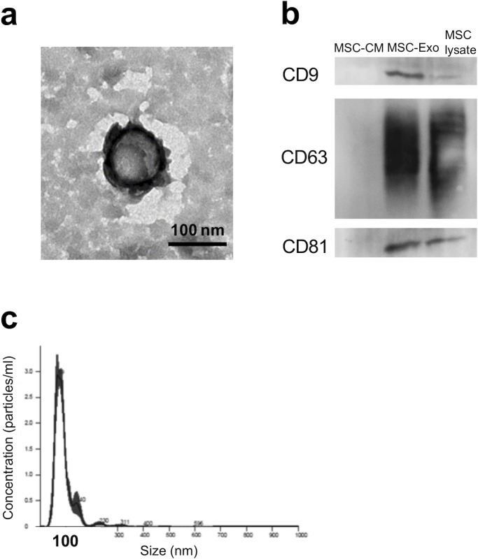
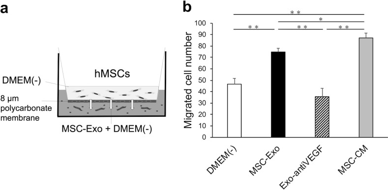
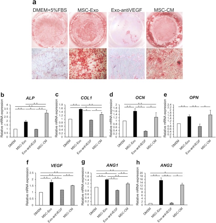
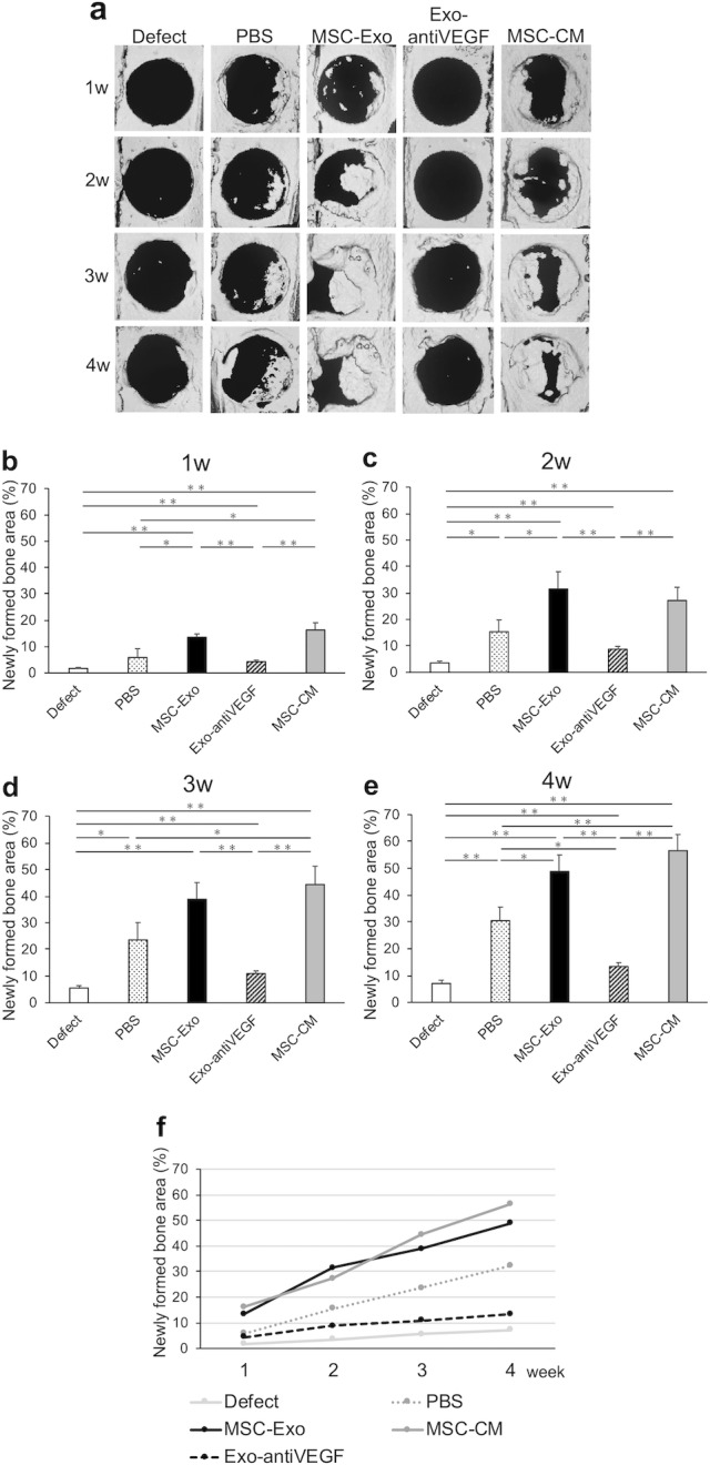
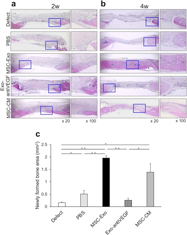
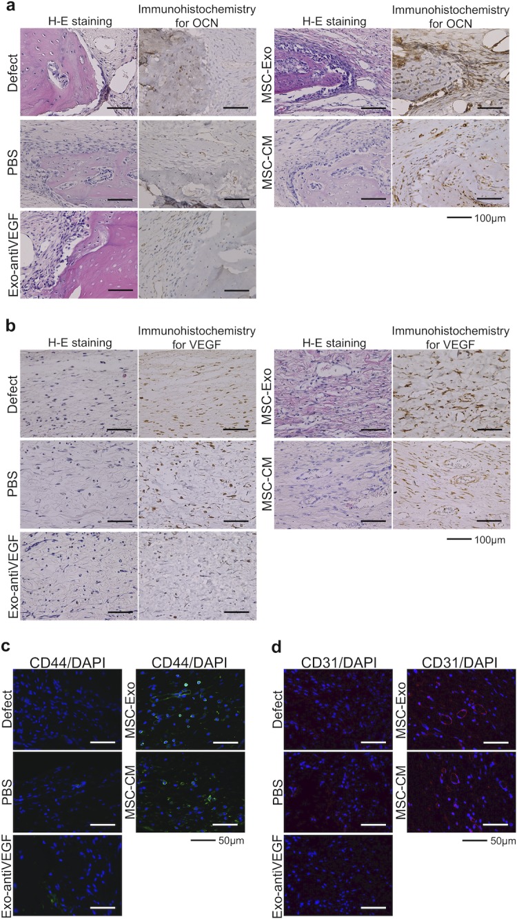
Growth factors in serum-free conditioned media from human bone marrow-derived mesenchymal stem cells (MSC-CM) are known to be effective in bone regeneration. However, the secretomes in MSC-CM that act as active ingredients for bone regeneration, as well as their mechanisms, remains unclear. Exosomes, components of MSC-CM, provide the recipient cells with genetic information and enhance the recipient cellular paracrine stimulation, which contributes to tissue regeneration. We hypothesized that MSC-CM-derived exosomes (MSC-Exo) promoted bone regeneration, and that angiogenesis was a key step. Here, we prepared an MSC-Exo group, MSC-CM group, and Exo-antiVEGF group (MSC-Exo with angiogenesis inhibitor), and examined the osteogenic and angiogenic potential in MSCs. Furthermore, we used a rat model of calvaria bone defect and implanted each sample to evaluate bone formation weekly, until week 4 after treatment. Results showed that MSC-Exo enhanced cellular migration and osteogenic and angiogenic gene expression in MSCs compared to that in other groups. In vivo, early bone formation by MSC-Exo was also confirmed. Two weeks after implantation, the newly formed bone area was 31.5 ± 6.5% in the MSC-Exo group while those in the control and Exo-antiVEGF groups were 15.4 ± 4.4% and 8.7 ± 1.1%, respectively. Four weeks after implantation, differences in the area between the MSC-Exo group and the Exo-antiVEGF or control groups were further broadened. Histologically, notable accumulation of osteoblast-like cells and vascular endothelial cells was observed in the MSC-Exo group; however, fewer cells were found in the Exo-antiVEGF and control groups. In conclusion, MSC-Exo promoted bone regeneration during early stages, as well as enhanced angiogenesis. Considering the tissue regeneration with transplanted cells and their secretomes, this study suggests that exosomes might play an important role, especially in angiogenesis.
Overview
The claims and statistics of this paper haven’t been checked yet.
Run the check — free, no account needed. It reads the open-access text and checks the sources it cites.
Integrity signals
Registry
Cites
Statistics
Figures
Verification
Claims the paper makes
The claims and statistics of this paper haven’t been checked yet.
Run the check — free, no account needed. It reads the open-access text and checks the sources it cites.
Claims other papers make about this one
Each is checked against this paper’s own text: does it say what the citing paper says it does?
The cited source says what the claim attributes to it.
In the citing paper
“osteogenesis is closely related with vascularization due to cell-to-cell communication between vascular endothelial cells and osteoblasts [99].”
From Mesenchymal stem cell secretome for regenerative medicine: Where do we stand? (2025) · cites this paper as [99]
Self-check
Percentages, sums and reported differences re-computed from the paper’s own numbers.
The self-check needs the paper’s full text.
Citations
Cites
Valadi, H., Ekström, K., Bossios, A., Sjöstrand, M., Lee, J. J., & Lötvall, J. O. (2007) · Nature Cell Biology · doi:10.1038/ncb1596 · Cited by 12,834
Extracellular vesicles: Exosomes, microvesicles, and friends
Raposo, G., & Stoorvogel, W. (2013) · Journal of Cell Biology · doi:10.1083/jcb.201211138 · Cited by 8,099
A perivascular origin for mesenchymal stem cells in multiple human organs
Crisan, M., Yap, S., Casteilla, L., Chen, C.-W., Corselli, M., Park, T. S., Andriolo, G., Sun, B., Zheng, B., Zhang, L., et al. (2008) · Cell Stem Cell · doi:10.1016/j.stem.2008.07.003 · Cited by 3,991 · cited 1×
“Stem cells are present in various organs and tissues, and they play an important role in maintaining homeostasis in the healthy body [35].”
BackgroundVascular endothelial growth factor (VEGF) and its receptors
Neufeld, G., Cohen, T., Gengrinovitch, S., & Gluzman‐Poltorak, Z. (1999) · The FASEB Journal · doi:10.1096/fasebj.13.1.9 · Cited by 3,511
Basic and Therapeutic Aspects of Angiogenesis
Potente, M., Gerhardt, H., & Carmeliet, P. (2011) · Cell · doi:10.1016/j.cell.2011.08.039 · Cited by 2,771
Exosome secreted by MSC reduces myocardial ischemia/reperfusion injury
Lai, R. C., Arslan, F., Lee, M. M., Sze, N. S. K., Choo, A., Chen, T. S., Salto-Tellez, M., Timmers, L., Lee, C. N., El Oakley, R. M., et al. (2010) · Stem Cell Research · doi:10.1016/j.scr.2009.12.003 · Cited by 2,215
Vascularization in tissue engineering
Rouwkema, J., Rivron, N. C., & van Blitterswijk, C. (2008) · Trends in biotechnology · doi:10.1016/j.tibtech.2008.04.009 · Cited by 1,218
Mesenchymal stem cells: a new trend for cell therapy
Wei, X., Yang, X. F., Han, Z., Qu, F.-F., Shao, L., & Shi, Y. (2013) · Acta Pharmacologica Sinica · doi:10.1038/aps.2013.50 · Cited by 964
Exosomes Derived from Mesenchymal Stem Cells
Yu, B., Zhang, X., & Li, X. (2014) · International Journal of Molecular Sciences · doi:10.3390/ijms15034142 · Cited by 788
Zhang, Y., Chopp, M., Meng, Y., Katakowski, M., Xin, H., Mahmood, A., & Xiong, Y. (2015) · Journal of neurosurgery · doi:10.3171/2014.11.jns14770 · Cited by 737
Cited by
Sorted by citations
Kou, M., Huang, L., Yang, J., Chiang, Z., Chen, S., Liu, J., Guo, L., Zhang, X., Zhou, X., Xu, X., et al. (2022) · Cell Death & Disease · doi:10.1038/s41419-022-05034-x · Cited by 640
Mesenchymal stem cells: Amazing remedies for bone and cartilage defects
Kangari, P., Talaei-Khozani, T., Razeghian-Jahromi, I., & Razmkhah, M. (2020) · Stem Cell Research & Therapy · doi:10.1186/s13287-020-02001-1 · Cited by 286
Osteoblast Differentiation and Signaling: Established Concepts and Emerging Topics
Ponzetti, M., & Rucci, N. (2021) · International Journal of Molecular Sciences · doi:10.3390/ijms22136651 · Cited by 310
Wu, D., Chang, X., Tian, J., Kang, L., Wu, Y., Liu, J., Wu, X., Huang, Y., Gao, B., Wang, H., et al. (2021) · Journal of Nanobiotechnology · doi:10.1186/s12951-021-00958-6 · Cited by 266
Mesenchymal stem cell secretome for regenerative medicine: Where do we stand?
Trigo, C. M., Rodrigues, J. S., Camões, S. P., Solá, S., & Miranda, J. P. (2025) · Journal of Advanced Research · doi:10.1016/j.jare.2024.05.004 · Cited by 252
“In fact, osteogenesis is closely related with vascularization due to cell-to-cell communication between vascular endothelial cells and osteoblasts [99].”
Maqsood, M., Kang, M., Wu, X., Chen, J., Teng, L., & Qiu, L. (2020) · Life Sciences · doi:10.1016/j.lfs.2020.118002 · Cited by 207
Mesenchymal stem cells secretome: The cornerstone of cell-free regenerative medicine
González-González, A., García‐Sánchez, D., Dotta, M., Rodríguez-Rey, J. C., & Pérez-Campo, F. M. (2020) · World Journal of Stem Cells · doi:10.4252/wjsc.v12.i12.1529 · Cited by 211
Hoang, D. H., Nguyen, T. D., Nguyen, H.-P., Nguyen, X., Do, P. T. X., Dang, V. D., Dam, P. T. M., Bui, H. T. H., Trinh, M. Q., Vu, D. M., et al. (2020) · Frontiers in Molecular Biosciences · doi:10.3389/fmolb.2020.00119 · Cited by 177
Tan, S. H. S., Wong, J. R. Y., Sim, S. J. Y., Tjio, C. K. E., Wong, K. L., Chew, J. R. J., Hui, J. H. P., & Toh, W. S. (2020) · Materials Today Bio · doi:10.1016/j.mtbio.2020.100067 · Cited by 152
Application of stem cells in regeneration medicine
Jin, Y. H., Li, S., Yu, Q., Chen, T., & Liu, D. (2023) · MedComm · doi:10.1002/mco2.291 · Cited by 149
Malekpour, K., Hazrati, A., Zahar, M., Markov, A. A., Zekiy, A. O., Navashenaq, J. G., Roshangar, L., & Ahmadi, M. (2021) · Stem Cell Reviews and Reports · doi:10.1007/s12015-021-10185-z · Cited by 120
Oral Bone Tissue Regeneration: Mesenchymal Stem Cells, Secretome, and Biomaterials
Gugliandolo, A., Fonticoli, L., Trubiani, O., Rajan, T. S., Marconi, G. D., Bramanti, P., Mazzon, E., Pizzicannella, J., & Diomede, F. (2021) · International Journal of Molecular Sciences · doi:10.3390/ijms22105236 · Cited by 112
Matrix Vesicles: Role in Bone Mineralization and Potential Use as Therapeutics
Ansari, S., de Wildt, B. W. M., Vis, M. A. M., de Korte, C. E., Ito, K., Hofmann, S., & Yuana, Y. (2021) · Pharmaceuticals · doi:10.3390/ph14040289 · Cited by 109
Extracellular vesicles in nanomedicine and regenerative medicine: A review over the last decade
Moghassemi, S., Dadashzadeh, A., Sousa, M. J., Vlieghe, H., Yang, J., León-Félix, C. M., & Amorim, C. A. (2024) · Bioactive Materials · doi:10.1016/j.bioactmat.2024.02.021 · Cited by 113
MSCs vs. iPSCs: Potential in therapeutic applications
Thanaskody, K., Jusop, A. S., Tye, G. J., Zaman, W. S. W. K., Dass, S. A., & Nordin, F. (2022) · Frontiers in Cell and Developmental Biology · doi:10.3389/fcell.2022.1005926 · Cited by 106
Understanding exosomes: Part 1—Characterization, quantification and isolation techniques
Miron, R. J., & Zhang, Y. (2023) · Periodontology 2000 · doi:10.1111/prd.12520 · Cited by 102
Understanding exosomes: Part 2—Emerging leaders in regenerative medicine
Miron, R. J., Estrin, N. E., Sculean, A., & Zhang, Y. (2024) · Periodontology 2000 · doi:10.1111/prd.12561 · Cited by 89
Biomaterial-based extracellular vesicle delivery for therapeutic applications
Priya, M. V., & Holmes, C. A. (2021) · Acta Biomaterialia · doi:10.1016/j.actbio.2021.01.010 · Cited by 80
Sun, J., Sun, X., Chen, J., Liao, X., He, Y., Wang, J., Chen, R., Hu, S., & Qiu, C. (2021) · Stem Cell Research & Therapy · doi:10.1186/s13287-020-02068-w · Cited by 78
Ying, C., Wang, R., Wang, Z., Tao, J., Yin, W., Zhang, J., Yi, C., Qi, X., & Han, D. (2020) · Frontiers in Bioengineering and Biotechnology · doi:10.3389/fbioe.2020.565561 · Cited by 75
Mesenchymal Stem Cell-Derived Extracellular Vesicles for Bone Defect Repair
Wang, D., Cao, H., Hua, W., Gao, L., Yuan, Y. J., Zhou, X., & Zeng, Z. (2022) · Membranes · doi:10.3390/membranes12070716 · Cited by 74
Clinical Potential of Mesenchymal Stem Cell-Derived Exosomes in Bone Regeneration
Torrecillas-Baena, B., Pulido-Escribano, V., Dorado, G., Gálvez-Moreno, M. Á., Camacho-Cardeñosa, M., & Casado‐Díaz, A. (2023) · Journal of Clinical Medicine · doi:10.3390/jcm12134385 · Cited by 75
Engineered extracellular vesicles for bone therapy
Jiang, Y., Li, J., Xu, X. J., Yin, Z., Xu, K., & Su, J. (2022) · Nano Today · doi:10.1016/j.nantod.2022.101487 · Cited by 68
Williams, T., Salmanian, G., Burns, M., Maldonado, V. V., Smith, E., Porter, R. M., Song, Y. H., & Samsonraj, R. M. (2022) · Biochimie · doi:10.1016/j.biochi.2022.11.011 · Cited by 71
Exosomes and Their Role in Cancer Progression
Liu, Y., Shi, K., Chen, Y., Wu, X.-R., Chen, Z., Cao, K., Tao, Y.-G., Chen, X., Liao, J., & Zhou, J. (2021) · Frontiers in Oncology · doi:10.3389/fonc.2021.639159 · Cited by 37
Gao, W., He, R., Ren, J., Zhang, W., Wang, K., Zhu, L., & Liang, T. (2021) · FEBS Open Bio · doi:10.1002/2211-5463.13142 · Cited by 66
Current Status of Angiogenic Cell Therapy and Related Strategies Applied in Critical Limb Ischemia
Beltrán-Camacho, L., Rojas-Torres, M., & Durán-Ruiz, M. C. (2021) · International Journal of Molecular Sciences · doi:10.3390/ijms22052335 · Cited by 66
Imanishi, Y., Hata, M., Matsukawa, R., Aoyagi, A., Omi, M., Mizutani, M., Naruse, K., Ozawa, S., Honda, M. J., Matsubara, T., et al. (2021) · Inflammation and Regeneration · doi:10.1186/s41232-021-00163-w · Cited by 67
Huang, Y., Lu, D., Ma, W., Liu, J., Ning, Q., Tang, F., & Long, L. (2022) · Molecular Immunology · doi:10.1016/j.molimm.2022.01.002 · Cited by 61
Liu, Y., & Holmes, C. A. (2021) · Frontiers in Cell and Developmental Biology · doi:10.3389/fcell.2021.648098 · Cited by 60
Chen, W., Xie, G., Lu, Y., Wang, J., Feng, B., Wang, Q., Xu, K., & Bao, J. (2021) · Biomaterials · doi:10.1016/j.biomaterials.2021.121260 · Cited by 56
Effects of microenvironment and biological behavior on the paracrine function of stem cells
Xue, Z., Liao, Y., & Li, Y. (2023) · Genes & Diseases · doi:10.1016/j.gendis.2023.03.013 · Cited by 61
Padinharayil, H., Varghese, J., Wilson, C. M., & George, A. (2024) · Life Sciences · doi:10.1016/j.lfs.2024.122542 · Cited by 58
Gholami, L., Nooshabadi, V. T., Shahabi, S., Jazaeri, M. S., Tarzemany, R., Afsartala, Z., & Khorsandi, K. (2021) · Cell & Bioscience · doi:10.1186/s13578-020-00527-8 · Cited by 57
Liu, S., Xu, X., Liang, S., Chen, Z., Zhang, Y., Qian, A., & Hu, L. (2020) · Frontiers in Cell and Developmental Biology · doi:10.3389/fcell.2020.00619 · Cited by 51
Huang, B., Su, Y., Shen, E., Song, M., Liu, D., & Qi, H. (2021) · Life Sciences · doi:10.1016/j.lfs.2021.119208 · Cited by 51
Yuan, N., Ge, Z., Ji, W., & Li, J. (2021) · BioMed Research International · doi:10.1155/2021/6655225 · Cited by 50
Exosomes for angiogenesis induction in ischemic disorders
Moeinabadi‐Bidgoli, K., Rezaee, M., Hossein‐Khannazer, N., Babajani, A., Aghdaei, H. A., Arki, M. K., Afaghi, S., Niknejad, H., & Vosough, M. (2023) · Journal of Cellular and Molecular Medicine · doi:10.1111/jcmm.17689 · Cited by 51
Osteoblasts-derived exosomes regulate osteoclast differentiation through miR-503-3p/Hpse axis
Wang, Q., Shen, X., Chen, Y., Chen, J., & Li, Y. (2021) · Acta Histochemica · doi:10.1016/j.acthis.2021.151790 · Cited by 53
Cheng, P., Cao, T., Zhao, X., Lu, W., Miao, S., Ning, F., Wang, D., Gao, Y., Wang, L., Pei, G., et al. (2021) · Bioactive Materials · doi:10.1016/j.bioactmat.2021.10.021 · Cited by 47
Sun, X., Mao, Y., Liu, B., Gu, K., Liu, H., Du, W., Li, R., & Zhang, J. (2023) · Journal of Personalized Medicine · doi:10.3390/jpm13020180 · Cited by 50
Brunello, G., Zanotti, F., Trentini, M., Zanolla, I., Pishavar, E., Favero, V., Favero, R., Favero, L., Bressan, E. A., Bonora, M., et al. (2022) · Pharmaceutics · doi:10.3390/pharmaceutics14050908 · Cited by 46
Zhu, Q., Tang, Y., Zhou, T., Yang, L., Zhang, G., Meng, Y., Zhang, H., Gao, J., Wang, C., Su, Y., et al. (2023) · Pharmacological Research · doi:10.1016/j.phrs.2023.106798 · Cited by 44
The importance of cellular and exosomal miRNAs in mesenchymal stem cell osteoblastic differentiation
Shirazi, S., Huang, C.-C., Kang, M., Lu, Y., Ravindran, S., & Cooper, L. F. (2021) · Scientific Reports · doi:10.1038/s41598-021-85306-2 · Cited by 43
Exosomes: a tool for bone tissue engineering
Huber, J., Griffin, M. F., Longaker, M. T., & Quarto, N. (2022) · Tissue Engineering Part B Reviews · doi:10.1089/ten.teb.2020.0246 · Cited by 41
Ana, I. D., Barlian, A., Hidajah, A. C., Wijaya, C. H., Notobroto, H. B., & Wungu, T. D. K. (2021) · Future Science OA · doi:10.2144/fsoa-2021-0050 · Cited by 43
Smolinská, V., Boháč, M., & Danišovič, Ľ. (2023) · Physiological Research · doi:10.33549/physiolres.935186 · Cited by 44
Understanding exosomes: Part 3—therapeutic + diagnostic potential in dentistry
Miron, R. J., Estrin, N. E., Sculean, A., & Zhang, Y. (2024) · Periodontology 2000 · doi:10.1111/prd.12557 · Cited by 43
Osteogenically-induced exosomes stimulate osteogenesis of human adipose-derived stem cells
Zhu, M., Liu, Y., Qin, H., Tong, S., Sun, Q., Wang, T., Zhang, H., Cui, M., & Guo, S. (2020) · Cell and Tissue Banking · doi:10.1007/s10561-020-09867-8 · Cited by 40
Recellularization of Native Tissue Derived Acellular Scaffolds with Mesenchymal Stem Cells
Ahmed, E., Saleh, T., & Xu, M. (2021) · Cells · doi:10.3390/cells10071787 · Cited by 44
Cutting Edge Endogenous Promoting and Exogenous Driven Strategies for Bone Regeneration
Macías, I., Alcorta-Sevillano, N., Infante, A., & Rodrı́guez, C. I. (2021) · International Journal of Molecular Sciences · doi:10.3390/ijms22147724 · Cited by 40
Generation of bioactive MSC-EVs for bone tissue regeneration by tauroursodeoxycholic acid treatment
Cha, K., Cho, W., Park, S., Ahn, J., Park, H., Baek, I., Lee, M., Lee, S., Arai, Y., & Lee, S. (2023) · Journal of Controlled Release · doi:10.1016/j.jconrel.2022.12.053 · Cited by 38
Niedermair, T., Lukas, C., Li, S., Stöckl, S., Craiovan, B. S., Brochhausen, C., Federlin, M., Herrmann, M., & Grässel, S. (2020) · Frontiers in Bioengineering and Biotechnology · doi:10.3389/fbioe.2020.615520 · Cited by 38
Re, F., Gabusi, E., Manferdini, C., Russo, D., & Lisignoli, G. (2021) · Biology · doi:10.3390/biology10070579 · Cited by 37
Wang, X., Ao, J., Lu, H., Zhao, Q., Ma, Y., Zhang, J., Ren, H., & Zhang, Y. (2020) · International Journal of Nanomedicine · doi:10.2147/ijn.s248741 · Cited by 37
Zhao, Q., Zhang, Y., Xiao, L., Lu, H., Ma, Y., Liu, Q., & Wang, X. (2021) · Regenerative Biomaterials · doi:10.1093/rb/rbab010 · Cited by 37
Lu, H., Zhang, Y., Xiong, S., Zhou, Y., Xiao, L., Ma, Y., Xiao, Y., & Wang, X. (2021) · Frontiers in Chemistry · doi:10.3389/fchem.2021.699802 · Cited by 36
Clinical Development of Regenerative Medicine Targeted for Intervertebral Disc Disease
Sakai, D., Schol, J., & Watanabe, M. (2022) · Medicina · doi:10.3390/medicina58020267 · Cited by 36
Angiogenesis in Regenerative Dentistry: Are We Far Enough for Therapy?
Baru, O., Nutu, A. M., Braicu, C., Cismaru, C. A., Berindan‐Neagoe, I., Buduru, S. D., & Badea, M. E. (2021) · International Journal of Molecular Sciences · doi:10.3390/ijms22020929 · Cited by 35
Cell-based therapies for rheumatoid arthritis: opportunities and challenges
Li, Y.-J., & Chen, Z. (2022) · Therapeutic Advances in Musculoskeletal Disease · doi:10.1177/1759720x221100294 · Cited by 34
Chopra, V., Thomas, J., Kaushik, S., Rajput, S., Guha, R., Mondal, B., Naskar, S., Mandal, D., Chauhan, G., Chattopadhyay, N., et al. (2023) · Small · doi:10.1002/smll.202204637 · Cited by 35
Therapeutic Potential of Extracellular Vesicles in Aging and Age-Related Diseases
Sanz‐Ros, J., Mas‐Bargues, C., Romero-García, N., Huete-Acevedo, J., Dromant, M., & Borrás, C. (2022) · International Journal of Molecular Sciences · doi:10.3390/ijms232314632 · Cited by 35
Engineering extracellular vesicles for ROS scavenging and tissue regeneration
Dayem, A. A., Yan, E., Do, M., Kim, Y., Lee, Y. K., Cho, S., & Kim, D. (2024) · Nano Convergence · doi:10.1186/s40580-024-00430-9 · Cited by 34
Xu, C., Li, Z., Kang, M., Chen, Y., Sheng, R., Aghaloo, T. L., & Lee, M. (2025) · Biomaterials · doi:10.1016/j.biomaterials.2025.123088 · Cited by 33
Mesenchymal Stem Cells and Their Exocytotic Vesicles
Cai, H., & Guo, H. (2023) · International Journal of Molecular Sciences · doi:10.3390/ijms24032085 · Cited by 32
Extracellular vesicles: From bone development to regenerative orthopedics
Davies, O. G. (2023) · Molecular Therapy · doi:10.1016/j.ymthe.2023.02.021 · Cited by 34
Emerging technologies in regenerative medicine: The future of wound care and therapy
Sharma, Y., Ghatak, S., Sen, C. K., & Mohanty, S. (2024) · Journal of Molecular Medicine · doi:10.1007/s00109-024-02493-x · Cited by 32
Extracellular Vesicles for the Treatment of Radiation Injuries
Nanduri, L. S. Y., Duddempudi, P. K., Yang, W.-L., Tamarat, R., & Guha, C. (2021) · Frontiers in Pharmacology · doi:10.3389/fphar.2021.662437 · Cited by 30
Influence of Periodontal Ligament Stem Cell-Derived Conditioned Medium on Osteoblasts
Novello, S., Tricot‐Doleux, S., Novella, A., Pellen‐Mussi, P., & Jeanne, S. (2022) · Pharmaceutics · doi:10.3390/pharmaceutics14040729 · Cited by 29
Moeinabadi‐Bidgoli, K., Rezaee, M., Rismanchi, H., Mohammadi, M. M., & Babajani, A. (2022) · Frontiers in Cell and Developmental Biology · doi:10.3389/fcell.2022.900418 · Cited by 33
Teo, K. Y. W., Tan, R., Wong, K. L., Hey, H. W. D., Hui, J. H. P., & Toh, W. S. (2023) · Cytotherapy · doi:10.1016/j.jcyt.2023.04.011 · Cited by 27
Application of mesenchymal stem cell-derived exosomes in kidney diseases
Deng, G., Yu, C. J., Zhu, S., Zhu, P., & Nasser, M. I. (2021) · Cellular Immunology · doi:10.1016/j.cellimm.2021.104358 · Cited by 26
Wu, S., Kuo, P., Rau, C., Huang, L., Lin, C.-W., Wu, Y.-C., Wu, C., Tsai, C.-W., Hsieh, T.-M., Liu, H.-T., et al. (2021) · International Journal of Molecular Sciences · doi:10.3390/ijms22168877 · Cited by 26
Xu, Y., Huang, R., Shi, W., Zhou, R., Xie, X., Wang, M., Wang, Y., Gu, P., Ni, N., & Bi, X. (2025) · Advanced Healthcare Materials · doi:10.1002/adhm.202500060 · Cited by 25
Immunomodulatory and Regenerative Functions of MSC-Derived Exosomes in Bone Repair
Arun, M., Rajasingh, S., Parani, M., & Rajasingh, J. (2025) · Bioengineering · doi:10.3390/bioengineering12080844 · Cited by 29
Mesenchymal stromal cell-derived extracellular vesicles for bone regeneration therapy
Murali, V. P., & Holmes, C. A. (2021) · Bone Reports · doi:10.1016/j.bonr.2021.101093 · Cited by 25
Kirkham, A. M., Bailey, A. J.-M., Tieu, A., Maganti, H. B., Montroy, J. R., Shorr, R., Campbell, T. M., Fergusson, D. A., Lalu, M. M., Elmoazzen, H., et al. (2021) · Stem Cell Reviews and Reports · doi:10.1007/s12015-021-10208-9 · Cited by 24
Liu, F., Sun, T., An, Y., Ming, L., Li, Y., Zhou, Z., & Shang, F. (2023) · Frontiers in Bioengineering and Biotechnology · doi:10.3389/fbioe.2023.1050916 · Cited by 34
Ren, J., Yu, R., Xue, J., Tang, Y., Su, S., Liao, C., Guo, Q., Guo, W., & Zheng, J. (2022) · International Journal of Nanomedicine · doi:10.2147/ijn.s377598 · Cited by 22
Exosomes: A New Hope for Angiogenesis-Mediated Bone Regeneration
Lange, M., Babczyk, P., & Tobiasch, E. (2024) · International Journal of Molecular Sciences · doi:10.3390/ijms25105204 · Cited by 22
Li, J., Ge, Z., Ji, W., Yuan, N., & Wang, K. (2020) · BioMed Research International · doi:10.1155/2020/4176926 · Cited by 22
Interests of Exosomes in Bone and Periodontal Regeneration: A Systematic Review
Gegout, P., Stutz, C., Olson, J., Batool, F., Petit, C., Tenenbaum, H., Benkirane-Jessel, N., & Huck, O. (2020) · Advances in experimental medicine and biology · doi:10.1007/5584_2020_593 · Cited by 23
Advances in the Study of Extracellular Vesicles for Bone Regeneration
Yao, J., Liu, Y., Du, J., Xu, J., Luo, Z., Liu, Y. F., & Guo, L. (2024) · International Journal of Molecular Sciences · doi:10.3390/ijms25063480 · Cited by 24
Farashah, M. S. G., Mohammadi, A., Javadi, M., Rad, J. S., Shakouri, S. K., Meshgi, S., & Roshangar, L. (2023) · Cell and Tissue Banking · doi:10.1007/s10561-022-10066-w · Cited by 24
Emerging roles of extracellular vesicles in oral and maxillofacial areas
Wang, Q., Sun, J., Jiang, H., & Yu, M. (2025) · International Journal of Oral Science · doi:10.1038/s41368-024-00341-9 · Cited by 22
Sinking Our Teeth in Getting Dental Stem Cells to Clinics for Bone Regeneration
Shoushrah, S. H., Transfeld, J. L., Tonk, C. H., Büchner, D., Witzleben, S. T., Sieber, M. A., Schulze, M., & Tobiasch, E. (2021) · International Journal of Molecular Sciences · doi:10.3390/ijms22126387 · Cited by 21
Wu, H., Chen, G., Zhang, G., Lv, Q., Gu, D., & Dai, M. (2023) · Biomedical Materials · doi:10.1088/1748-605x/acb412 · Cited by 20
Delivery of Growth Factors to Enhance Bone Repair
Ball, J. R., Shelby, T., Hernandez, F., Mayfield, C. K., & Lieberman, J. R. (2023) · Bioengineering · doi:10.3390/bioengineering10111252 · Cited by 21
Chen, Z., Xue, S., Zhang, S., Cheng, K., & Ye, Q. (2021) · Journal of Cellular Physiology · doi:10.1002/jcp.30274 · Cited by 20
Sartori, M., Graziani, G., Sassoni, E., Pagani, S., Boi, M., Maltarello, M. C., Baldini, N., & Fini, M. (2021) · Materials Science and Engineering C · doi:10.1016/j.msec.2021.112031 · Cited by 20
Exosomes as Neurological Nanosized Machines
Pishavar, E., Trentini, M., Zanotti, F., Camponogara, F., Tiengo, E., Zanolla, I., Bonora, M., & Zavan, B. (2022) · ACS Nanoscience Au · doi:10.1021/acsnanoscienceau.1c00062 · Cited by 19
Rudiansyah, M., El‐Sehrawy, A. A. M. A., Ahmad, I. S., Terefe, E. M., Abdelbasset, W. K., Bokov, D. O., Salazar, A., Rizaev, J. A., Muthanna, F. M. S., & Shalaby, M. N. (2022) · Life Sciences · doi:10.1016/j.lfs.2022.120717 · Cited by 19
Schol, J., Ambrosio, L., Tamagawa, S., Joyce, K., Ruiz‐Fernández, C., Nomura, A., & Sakai, D. (2024) · Scientific Reports · doi:10.1038/s41598-024-62792-8 · Cited by 22
Therapeutic properties of stem cell-derived exosomes in ischemic heart disease
Raissi-Dehkordi, N., Raissi-Dehkordi, N., & Farjoo, M. H. (2022) · European Journal of Pharmacology · doi:10.1016/j.ejphar.2022.174839 · Cited by 19
Zhou, Z., Guo, C., Sun, X., Ren, Z., & Tao, J. (2023) · Stem Cells International · doi:10.1155/2023/4980871 · Cited by 18
Xing, X., Han, S., Ni, Y., Cheng, G., Cheng, Y., Ni, X., Deng, Y., Li, Z., & Li, Z. (2021) · International Journal of Pharmaceutics · doi:10.1016/j.ijpharm.2021.121136 · Cited by 18
A Narrative Review of Cell-Based Approaches for Cranial Bone Regeneration
Falguera‐Uceda, M. I., Sánchez-Casanova, S., Escudero-Duch, C., & Vilaboa, N. E. (2022) · Pharmaceutics · doi:10.3390/pharmaceutics14010132 · Cited by 16
Niu, H. M., Gao, N., Dang, Y., Guan, Y., & Guan, J. (2022) · Acta Biomaterialia · doi:10.1016/j.actbio.2022.03.021 · Cited by 17
Cataldi, A., Amoroso, R., di Giacomo, V., Zara, S., Maccallini, C., & Gallorini, M. (2022) · International Journal of Molecular Sciences · doi:10.3390/ijms232314560 · Cited by 18
Therapeutic potential of mesenchymal stem cell-derived exosomes in skeletal diseases
Yang, X., Zhang, S., Lu, J., Chen, X., Zheng, T., He, R., Ye, C., & Xu, J. (2024) · Frontiers in Molecular Biosciences · doi:10.3389/fmolb.2024.1268019 · Cited by 19
Metadata
- Authors
- Takeuchi, R., Katagiri, W., Endo, S., & Kobayashi, T.
- Affiliations
- Niigata University (JP)
- Corresponding author
- Katagiri, Wataru
- Venue
- PLOS ONE
- Publisher
- Public Library of Science (PLoS)
- Published
- 2019-11-21
- Volume
- 14
- Issue
- 11
- Pages
- e0225472
- ISSN
- 1932-6203 (ISSN-L)
- PMC
- PMC6872157
- OpenAlex
- W2989744940
- Cited by
- 210
- References
- 54
- Citations by year
- 2026: 15 · 2025: 26 · 2024: 33 · 2023: 32 · 2022: 37 · 2021: 51 · 2020: 16
- FWCI
- 8.60
- Type
- journal article
- Language
- en
- Open access
- gold
- doi.org
- Licence
- Locations
- PLoS ONE (publishedVersion, cc-by)
- Europe PMC (PubMed Central) (publishedVersion, cc-by)
- PubMed Central (publishedVersion, cc-by)
- PubMed (publishedVersion)
- DOAJ (DOAJ: Directory of Open Access Journals) (submittedVersion, cc-by-sa)
- Figshare (submittedVersion, cc-by)
- Institutional Repositories DataBase (IRDB) (submittedVersion)
- Funders
- Japan Society for the Promotion of Science (18K09785)
- Topics
- Life Sciences › Biochemistry, Genetics and Molecular Biology › Molecular Biology › Extracellular vesicles in disease
- Life Sciences › Biochemistry, Genetics and Molecular Biology › Cell Biology › Mesenchymal stem cell research
- Physical Sciences › Engineering › Biomedical Engineering › Bone Tissue Engineering Materials
- Keywords
- exosomes, bone marrow-derived mesenchymal stem cells, conditioned medium, bone regeneration, angiogenesis, VEGF inhibitors, osteogenic differentiation, paracrine signaling, cell transplantation, secretome
- MeSH terms
- Animals, Bone Regeneration, Cell Movement, Cells, Cultured, Disease Models, Animal, Humans, Skull, Culture Media, Conditioned, Neovascularization, Physiologic, Cell Culture Techniques, Paracrine Communication, Angiogenesis Inhibitors, Mesenchymal Stem Cell Transplantation, Cell Proliferation, Rats, Exosomes, Mesenchymal Stem Cells
About these checks
Registry, retraction, Cites and statistics results are computed, not judged, with one exception: a printed reference that no index lists is read once by a model before it counts as a citation error. Claims are extracted by a model and kept only when their quote appears verbatim in the text; each cited source is then checked, with a model’s help, for whether it says what the claim relies on. Those verdicts can be wrong — read the claim’s quote and the cited source before relying on one.
Statistics and arithmetic are recomputed, not judged.
Confidence bands are shown only for fields calibrated on our annotated test set. None is calibrated yet, so no band is shown.
What counts as a claim ›How we measure confidence ›How we check this ›
Are you an author, or do you see a problem with this page? Request a review
