

The DOI matches its Crossref record; 46 of the 46 references it cites were found in a registry or an index.
Aspergillus fumigatus Triggers Inflammatory Responses by Stage-Specific β-Glucan Display
Open accessAbstract
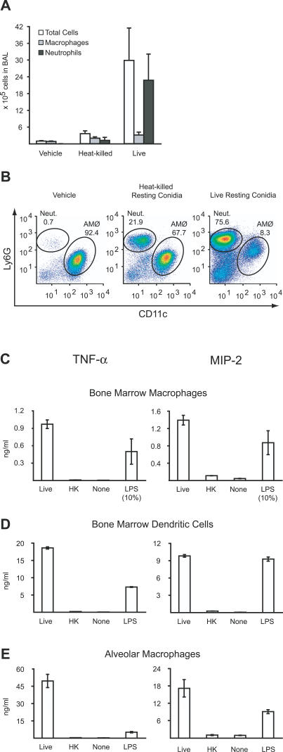
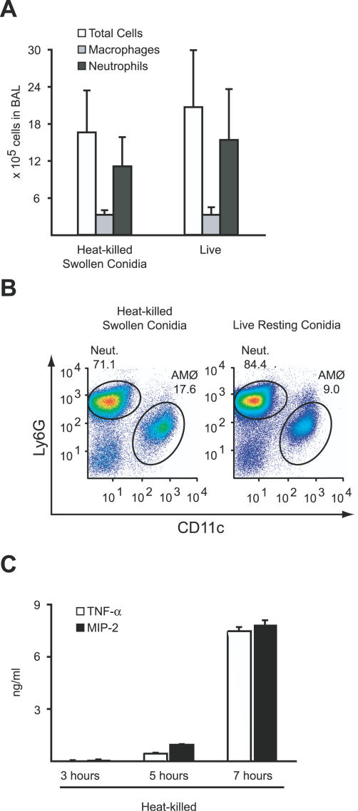
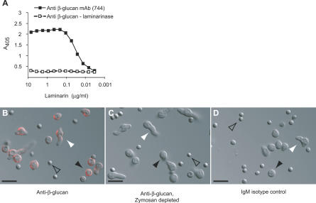
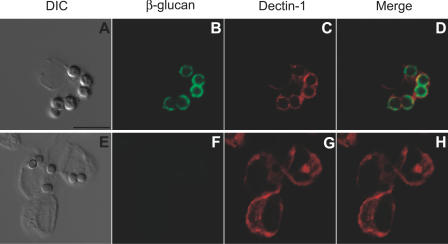
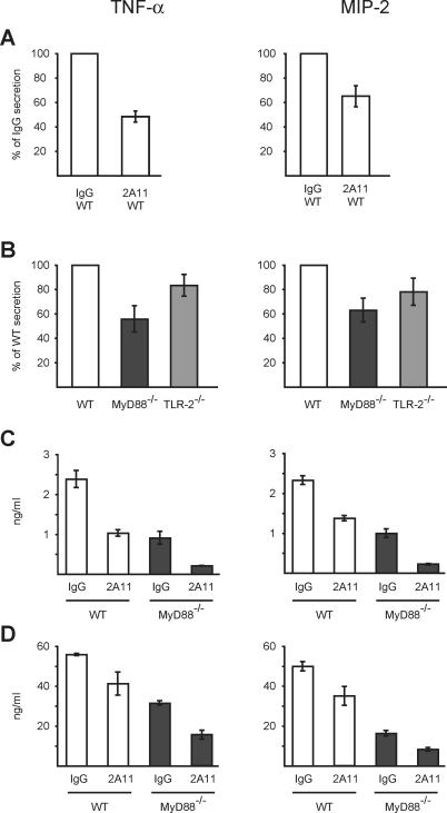
Inhalation of fungal spores (conidia) occurs commonly and, in specific circumstances, can result in invasive disease. We investigated the murine inflammatory response to conidia of Aspergillus fumigatus, the most common invasive mold in immunocompromised hosts. In contrast to dormant spores, germinating conidia induce neutrophil recruitment to the airways and TNF-alpha/MIP-2 secretion by alveolar macrophages. Fungal beta-glucans act as a trigger for the induction of these inflammatory responses through their time-dependent exposure on the surface of germinating conidia. Dectin-1, an innate immune receptor that recognizes fungal beta-glucans, is recruited in vivo to alveolar macrophage phagosomes that have internalized conidia with exposed beta-glucans. Antibody-mediated blockade of Dectin-1 partially inhibits TNF-alpha/MIP-2 induction by metabolically active conidia. TLR-2- and MyD88-mediated signals provide an additive contribution to macrophage activation by germinating conidia. Selective responsiveness to germinating conidia provides the innate immune system with a mechanism to restrict inflammatory responses to metabolically active, potentially invasive fungal spores.
Overview
The claims and statistics of this paper haven’t been checked yet.
Run the check — free, no account needed. It reads the open-access text and checks the sources it cites.
Integrity signals
Registry
Cites
Statistics
Figures
Verification
Claims the paper makes
The claims and statistics of this paper haven’t been checked yet.
Run the check — free, no account needed. It reads the open-access text and checks the sources it cites.
Claims other papers make about this one
Each is checked against this paper’s own text: does it say what the citing paper says it does?
In the citing paper
“Significant advances have been made in elucidating the main innate immune players and effector pathways for efficient control of Aspergillus infection in mouse models with the use of transgenic mice (189–202).”
From Aspergillus fumigatus and Aspergillosis in 2019 (2019) · cites this paper as [194]
In the citing paper
“The discovery of selective activation of Dectin-1/Syk signaling during germination of Aspergillus conidia due to stage-specific surface exposure of β-glucan (194, 246, 247) and the ability of PRR to regulate phagosome maturation (248) provided the conceptual framework of pathogen-associated molecular patterns (PAMPs) masking as a mechanism employed by fungi to escape macrophage killing.”
From Aspergillus fumigatus and Aspergillosis in 2019 (2019) · cites this paper as [194]
Self-check
Percentages, sums and reported differences re-computed from the paper’s own numbers.
The self-check needs the paper’s full text.
Citations
Cites
Janeway, C. A., & Medzhitov, R. (2002) · Annual Review of Immunology · doi:10.1146/annurev.immunol.20.083001.084359 · Cited by 8,394 · cited 1×
“The innate immune system confronts a wide spectrum of microbes, extending from the innocuous to the highly pathogenic [”
BackgroundAkira, S., & Takeda, K. (2004) · Nature reviews. Immunology · doi:10.1038/nri1391 · Cited by 8,312
Aspergillus fumigatus and Aspergillosis
Latgé, J.-P. (1999) · Clinical Microbiology Reviews · doi:10.1128/cmr.12.2.310 · Cited by 2,307 · cited 1×
“Aspergillus fumigatus is a mold that forms conidia with a diameter of 2–3 lm [4].”
BackgroundRomani, L. (2011) · Nature Reviews Immunology · doi:10.1038/nri2939 · Cited by 1,793
Collaborative Induction of Inflammatory Responses by Dectin-1 and Toll-like Receptor 2
Gantner, B. N., Simmons, R. M., Canavera, S. J., Akira, S., & Underhill, D. M. (2003) · The Journal of Experimental Medicine · doi:10.1084/jem.20021787 · Cited by 1,682
Brown, G. D., & Gordon, S. (2001) · Nature · doi:10.1038/35092620 · Cited by 1,579
MACROPHAGE RECEPTORS AND IMMUNE RECOGNITION
Taylor, P. R., Martínez‐Pomares, L., Stacey, M. A., Lin, H.-H., Brown, G. D., & Gordon, S. (2005) · Annual Review of Immunology · doi:10.1146/annurev.immunol.23.021704.115816 · Cited by 1,366
Beutler, B. (2003) · Molecular Immunology · doi:10.1016/j.molimm.2003.10.005 · Cited by 1,314
Dectin-1 Mediates the Biological Effects of β-Glucans
Brown, G. D., Herre, J., Williams, D. L., Willment, J. A., Marshall, A. S. J., & Gordon, S. (2003) · The Journal of Experimental Medicine · doi:10.1084/jem.20021890 · Cited by 1,209
Dectin-1 Is A Major β-Glucan Receptor On Macrophages
Brown, G. D., Taylor, P. R., Reid, D. M., Willment, J. A., Williams, D. L., Martínez‐Pomares, L., Wong, S. Y. C., & Gordon, S. (2002) · The Journal of Experimental Medicine · doi:10.1084/jem.20020470 · Cited by 1,015
Cited by
Sorted by citations
Aspergillus fumigatus and Aspergillosis in 2019
Latgé, J.-P., & Chamilos, G. (2019) · Clinical Microbiology Reviews · doi:10.1128/cmr.00140-18 · Cited by 1,400
Dectin-1 is required for β-glucan recognition and control of fungal infection
Taylor, P. R., Tsoni, S. V., Willment, J. A., Dennehy, K. M., Rosas, M., Findon, H., Haynes, K., Steele, C. H., Botto, M., Gordon, S., et al. (2006) · Nature Immunology · doi:10.1038/ni1408 · Cited by 1,185
Pathogenesis of Aspergillus fumigatus in Invasive Aspergillosis
Dagenais, T. R. T., & Keller, N. P. (2009) · Clinical Microbiology Reviews · doi:10.1128/cmr.00055-08 · Cited by 993
Surface hydrophobin prevents immune recognition of airborne fungal spores
Aimanianda, V., Bayry, J., Bozza, S., Kniemeyer, O., Perruccio, K., Elluru, S. R., Clavaud, C., Paris, S., Brakhage, A. A., Kaveri, S. V., et al. (2009) · Nature · doi:10.1038/nature08264 · Cited by 792
Dyken, S. J. V., & Locksley, R. M. (2013) · Annual Review of Immunology · doi:10.1146/annurev-immunol-032712-095906 · Cited by 743
The mycobiota: interactions between commensal fungi and the host immune system
Underhill, D. M., & Iliev, I. D. (2014) · Nature reviews. Immunology · doi:10.1038/nri3684 · Cited by 721
β‐glucan recognition by the innate immune system
Goodridge, H. S., Wolf, A. J., & Underhill, D. M. (2009) · Immunological Reviews · doi:10.1111/j.1600-065x.2009.00793.x · Cited by 713
Segal, B. H. (2009) · New England Journal of Medicine · doi:10.1056/nejmra0808853 · Cited by 664
Gringhuis, S. I., Kaptein, T. M., Wevers, B. A., Theelen, B., van der Vlist, M., Boekhout, T., & Geijtenbeek, T. B. H. (2012) · Nature Immunology · doi:10.1038/ni.2222 · Cited by 628
“fumigatus ligands for dectin-1 become exposed in a stage-specific manner (after germination”
BackgroundLuther, K., Torosantucci, A., Brakhage, A. A., Heesemann, J., & Ebel, F. (2006) · Cellular Microbiology · doi:10.1111/j.1462-5822.2006.00796.x · Cited by 582
“Very recently, a similar collaboration of dectin-1 and TLR2 was found in the innate immune response to the pathogenic fungi Coccidioides posadasii and A. fumigatus (Hohl et al., 2005; Steele et al., 2005; Viriyakosol et al., 2005; Gersuk et al., 2006).”
BackgroundInfluentialMartínez‐Pomares, L. (2012) · Journal of Leukocyte Biology · doi:10.1189/jlb.0512231 · Cited by 563
Medicinal importance of fungal β-(1→3), (1→6)-glucans
Chen, J., & Seviour, R. J. (2007) · Mycological Research · doi:10.1016/j.mycres.2007.02.011 · Cited by 539
Myeloid C-type lectins in innate immunity
Robinson, M. J., Sancho, D., Slack, E. M. C., LeibundGut‐Landmann, S., & Sousa, C. R. E. (2006) · Nature Immunology · doi:10.1038/ni1417 · Cited by 525
“Dectin-1 has been linked to TLR2-independent fungal responses in antibody-blocking studie”
BackgroundAspergillus fumigatus morphology and dynamic host interactions
van de Veerdonk, F. L., Gresnigt, M. S., Romani, L., Netea, M. G., & Latgé, J.-P. (2017) · Nature Reviews Microbiology · doi:10.1038/nrmicro.2017.90 · Cited by 490
Fungi and allergic lower respiratory tract diseases
Knutsen, A. P., Bush, R. K., Demain, J. G., Denning, D. W., Dixit, A., Fairs, A., Greenberger, P. A., Kariuki, B., Kita, H., Kurup, V. P., et al. (2012) · Journal of Allergy and Clinical Immunology · doi:10.1016/j.jaci.2011.12.970 · Cited by 486
Histoplasma capsulatum α-(1,3)-glucan blocks innate immune recognition by the β-glucan receptor
Rappleye, C. A., Eissenberg, L. G., & Goldman, W. E. (2007) · Proceedings of the National Academy of Sciences · doi:10.1073/pnas.0609848104 · Cited by 419
Requisite Role for the Dectin-1 β-Glucan Receptor in Pulmonary Defense against Aspergillus fumigatus
Werner, J. L., Metz, A. E., Horn, D., Schoeb, T. R., Hewitt, M. M., Schwiebert, L. M., Faro‐Trindade, I., Brown, G. D., & Steele, C. H. (2009) · The Journal of Immunology · doi:10.4049/jimmunol.0804250 · Cited by 417
“In our previous study, and confirmed by others (11, 12), Dectin-1 expressed by alveolar macrophages recognized unmasked -glucan moieties in A.”
ResultImmune responses to human fungal pathogens and therapeutic prospects
Lionakis, M. S., Drummond, R. A., & Hohl, T. M. (2023) · Nature Reviews Immunology · doi:10.1038/s41577-022-00826-w · Cited by 379
Self-citationGoodridge, H. S., Simmons, R. M., & Underhill, D. M. (2007) · The Journal of Immunology · doi:10.4049/jimmunol.178.5.3107 · Cited by 365
“Zymosan injection triggered severe chronic arthritis in genetically susceptible mice in a Dectin-1-dependent manner (34), and intratracheal administration of Aspergillus fumigatus conidia induced proinflammatory cytokine production, which was blocked by coadministration of either a soluble Dectin-1-Fc fusion protein o…”
BackgroundRandomized Controlled Trial of Oral Antifungal Treatment for Severe Asthma with Fungal Sensitization
Denning, D. W., O’Driscoll, B. R., Powell, G., Chew, F., Atherton, G. T., Vyas, A., Miles, J., Morris, J., & Niven, R. M. (2008) · American Journal of Respiratory and Critical Care Medicine · doi:10.1164/rccm.200805-737oc · Cited by 342
“Some direct immunologic effects of azoles have been reported in vitro, including suppression of type 2 cytokine production (IL-4 and IL-5) in T cells by itraconazole (26) and upregulation and/or production of numerous chemokines and inflammation-related cytokines, including IFN-g and tumor necrosis factor-a, by vorico…”
BackgroundInflammatory Monocytes Facilitate Adaptive CD4 T Cell Responses during Respiratory Fungal Infection
Hohl, T. M., Rivera, A., Lipuma, L., Gallegos, A., Shi, C., Mack, M., & Pamer, E. G. (2009) · Cell Host & Microbe · doi:10.1016/j.chom.2009.10.007 · Cited by 337
“TLR-mediated recognition of undefined fungal components and Dectin-1-mediated interactions with fungal b-glucans (Hohl et al., 2005) contribute to monocyte activation.”
Self-citationGersuk, G. M., Underhill, D. M., Zhu, L., & Marr, K. A. (2006) · The Journal of Immunology · doi:10.4049/jimmunol.176.6.3717 · Cited by 334
Hydrophobins—Unique Fungal Proteins
Bayry, J., Aimanianda, V., Guijarro, J. I., Sunde, M., & Latgé, J. (2012) · PLoS Pathogens · doi:10.1371/journal.ppat.1002700 · Cited by 323
Dynamic, Morphotype-Specific Candida albicans β-Glucan Exposure during Infection and Drug Treatment
Wheeler, R. T., Kombe, D., Agarwala, S. D., & Fink, G. R. (2008) · PLoS Pathogens · doi:10.1371/journal.ppat.1000227 · Cited by 309
Sadier, N. S., Padilla, E., Langsley, G., & Ojcius, D. M. (2010) · PLoS ONE · doi:10.1371/journal.pone.0010008 · Cited by 302
Latgé, J. (2010) · Cellular Microbiology · doi:10.1111/j.1462-5822.2010.01474.x · Cited by 303
“For example, Dectin-1 at the surface of the myeloid cells does not bind to conidia of A. fumigatus, whereas after conidial phagocytosis, the phagolysosome is strongly labelled with an anti-Dectin-1 antibody (Hohl et al., 2005).”
BackgroundBeyond pattern recognition: five immune checkpoints for scaling the microbial threat
Blander, J. M., & Sander, L. E. (2012) · Nature reviews. Immunology · doi:10.1038/nri3167 · Cited by 300
“Following germination, RodA is presumably degraded, exposing β-1,3-glucans (particularly at sites of hyphal extension), which signal a potentially threatening scenario and trigger the activation of PRRs such as dectin 1 (Ref”
Transcriptional programming of the dendritic cell network
Belz, G. T., & Nutt, S. L. (2012) · Nature reviews. Immunology · doi:10.1038/nri3149 · Cited by 282
“Under inflammatory conditions, circulating blood monocytes can be rapidly mobilized and can differentiate into cells that possess many prototypical features of DC”
BackgroundCurrent Perspectives on Echinocandin Class Drugs
Perlin, D. S. (2011) · Future Microbiology · doi:10.2217/fmb.11.19 · Cited by 274
“The innate immune system ignores the less threatening dormant conidia, but responds vigorously to germinating condia and hyphae [155].”
BackgroundKarki, R., Man, S. M., Malireddi, R. K. S., Gurung, P., Vogel, P., Lamkanfi, M., & Kanneganti, T.-D. (2015) · Cell Host & Microbe · doi:10.1016/j.chom.2015.01.006 · Cited by 272
“, 2008) and C-type lectin receptors (CLRs) dectin-1 (Hohl et al., 2005; Steele et al., 2005) and dectin-2 (Barrett et al.”
C-type lectin receptors in antifungal immunity
Willment, J. A., & Brown, G. D. (2007) · Trends in Microbiology · doi:10.1016/j.tim.2007.10.012 · Cited by 277
Immune defence to invasive fungal infections: A comprehensive review
Pathakumari, B., Liang, G., & Liu, W. (2020) · Biomedicine & Pharmacotherapy · doi:10.1016/j.biopha.2020.110550 · Cited by 278
Eduard, W. (2009) · Critical Reviews in Toxicology · doi:10.3109/10408440903307333 · Cited by 251
“…spores were applied� However, when spores were incubated until swollen before heat-killing, a similar inflammatory response was seen as induced by viable spores (Hohl et al�, 2005)� The applied dose (1 × 10⁷ spores/animal, approximately 4 × 10⁵ spores/g bw) was high compared to the doses used…”
BackgroundMethodInfluentialAspergillus fumigatus : Principles of Pathogenesis and Host Defense
Hohl, T. M., & Feldmesser, M. (2007) · Eukaryotic Cell · doi:10.1128/ec.00274-07 · Cited by 237
“observed for macrophages isolated from other sources (50, 78, 97).”
Self-citationInnate Immunity toAspergillusSpecies
Park, S. J., & Mehrad, B. (2009) · Clinical Microbiology Reviews · doi:10.1128/cmr.00014-09 · Cited by 242
“recognition by the host: the binding and ingestion of resting conidia, for example, induce very little inflammatory response (64, 74, 157), and optimal CD4 T-cell responses appear to occur only in response to live conidia (136).”
BackgroundWüthrich, M., Deepe, G. S., & Klein, B. S. (2012) · Annual Review of Immunology · doi:10.1146/annurev-immunol-020711-074958 · Cited by 234
“albicans) have developed mechanisms to mask their b-glucan exposure and limit detection by immune cells (Hohl et al. 2005; Steele et al. 2005; Wheeler et al. 2008).”
Advances in combating fungal diseases: vaccines on the threshold
Cutler, J. E., Deepe, G. S., & Klein, B. S. (2006) · Nature Reviews Microbiology · doi:10.1038/nrmicro1537 · Cited by 223
Aspergillus Cell Wall Melanin Blocks LC3-Associated Phagocytosis to Promote Pathogenicity
Akoumianaki, T., Kyrmizi, I., Valsecchi, I., Gresnigt, M. S., Samonis, G., Drakos, E., Boumpas, D., Muszkieta, L., Prevost, M.-C., Kontoyiannis, D. P., et al. (2016) · Cell Host & Microbe · doi:10.1016/j.chom.2015.12.002 · Cited by 218
“Surface exposure of these PAMPs upon biological (germination) or genetic removal of rodlet protein RodA (DrodA) (Aimanianda et al., 2009; Hohl et al., 2005; Steele et al., 2005) triggers potent immune cell activation.”
BackgroundMircescu, M. M., Lipuma, L., van Rooijen, N., Pamer, E. G., & Hohl, T. M. (2009) · The Journal of Infectious Diseases · doi:10.1086/600380 · Cited by 216
“A. fumigatus strain AF293 conidial suspensions were administered to anesthesized mice via the intratracheal route as described elsewhere [ 21 ].”
MethodSelf-citationNOX2-dependent regulation of inflammation
Singel, K. L., & Segal, B. H. (2016) · Clinical Science · doi:10.1042/cs20150660 · Cited by 224
Beyond autophagy: LC3-associated phagocytosis and endocytosis
Peña-Martinez, C., Rickman, A. D., & Heckmann, B. L. (2022) · Science Advances · doi:10.1126/sciadv.abn1702 · Cited by 227
Latgé, J.-P., Beauvais, A., & Chamilos, G. (2017) · Annual Review of Microbiology · doi:10.1146/annurev-micro-030117-020406 · Cited by 207
Tsoni, S. V., & Brown, G. D. (2008) · Annals of the New York Academy of Sciences · doi:10.1196/annals.1443.019 · Cited by 210
Collaboration between the innate immune receptors dectin‐1, TLRs, and Nods
Underhill, D. M. (2007) · Immunological Reviews · doi:10.1111/j.1600-065x.2007.00548.x · Cited by 195
Carrion, S. D. J., Leal, S. M., Ghannoum, M. A., Aimanianda, V., Latgé, J., & Pearlman, E. M. (2013) · The Journal of Immunology · doi:10.4049/jimmunol.1300748 · Cited by 192
“gatus (29, 30), less is known about the role of Dectin-2 during fungal infection.”
BackgroundZarember, K. A., Sugui, J. A., Chang, Y. C., Kwon-Chung, K. J., & Gallin, J. I. (2007) · The Journal of Immunology · doi:10.4049/jimmunol.178.10.6367 · Cited by 193
Lamaris, G. A., Lewis, R. E., Chamilos, G., May, G. S., Safdar, A., Walsh, T. J., RAAD, I. I., & Kontoyiannis, D. P. (2008) · The Journal of Infectious Diseases · doi:10.1086/589305 · Cited by 192
LeibundGut‐Landmann, S., Wüthrich, M., & Hohl, T. M. (2012) · Current Opinion in Immunology · doi:10.1016/j.coi.2012.04.007 · Cited by 190
Self-citationAspergillus Fumigatus : Cell Wall Polysaccharides, their Biosynthesis and Organization
Gastebois, A., Clavaud, C., Aimanianda, V., & Latgé, J. (2009) · Future Microbiology · doi:10.2217/fmb.09.29 · Cited by 185
Hyphal Growth in Human Fungal Pathogens and Its Role in Virulence
Brand, A. (2011) · International Journal of Microbiology · doi:10.1155/2012/517529 · Cited by 184
Aspergillus fumigatus: contours of an opportunistic human pathogen
McCormick, A., Loeffler, J., & Ebel, F. (2010) · Cellular Microbiology · doi:10.1111/j.1462-5822.2010.01517.x · Cited by 185
“Interestingly, it is not traceable on hyphae (Hohl et al., 2005), a fact that has been discussed as a fungal stealth strategy.”
BackgroundDistinct Roles for Dectin-1 and TLR4 in the Pathogenesis of Aspergillus fumigatus Keratitis
Leal, S. M., Cowden, S., Hsia, Y. C., Ghannoum, M. A., Momany, M., & Pearlman, E. M. (2010) · PLoS Pathogens · doi:10.1371/journal.ppat.1000976 · Cited by 180
Herbst, S., Shah, A., Mazón, M. J., Marzola, V., Jensen, B., Reed, A. K., Birrell, M. A., Saijo, S., Mostowy, S., Shaunak, S., et al. (2015) · EMBO Molecular Medicine · doi:10.15252/emmm.201404556 · Cited by 178
“…to AF appear more complexand involve both Dectin-1 and Dectin-2, as well as Toll-like receptors, complement and opsonins (Richardson et al, 1991; Madan et al, 1997; Hohl et al, 2005; Luther et al, 2007; Werner et al, 2009; Moalli et al, 2010; Hasenberg et al, 2011; Carrion Sdeet al, 2013).”
BackgroundSelective C-Rel Activation via Malt1 Controls Anti-Fungal TH-17 Immunity by Dectin-1 and Dectin-2
Gringhuis, S. I., Wevers, B. A., Kaptein, T. M., van Capel, T. M. M., Theelen, B., Boekhout, T., de Jong, E. C., & Geijtenbeek, T. B. H. (2011) · PLoS Pathogens · doi:10.1371/journal.ppat.1001259 · Cited by 179
Recognition of fungal pathogens by Toll-like receptors
Netea, M. G., de Graaf, C., van der Meer, J. W. M., & Kullberg, B. J. (2004) · European Journal of Clinical Microbiology & Infectious Diseases · doi:10.1007/s10096-004-1192-7 · Cited by 172
Immune responses to commensal and environmental microbes
Pamer, E. G. (2007) · Nature Immunology · doi:10.1038/ni1526 · Cited by 173
“fumigatus spores induce recruitment of inflammatory cells to the airways, and macrophages and dendritic cells distinguish between germinating and dormant spores in a TLR- and Dectin-1-dependent fashio”
Self-citationThywißen, A., Heinekamp, T., Dahse, H., Schmaler-Ripcke, J., Nietzsche, S., Zipfel, P. F., & Brakhage, A. A. (2011) · Frontiers in Microbiology · doi:10.3389/fmicb.2011.00096 · Cited by 169
“In recent years, the process by which conidia are recognized and ingested by professional phagocytes (Hohl et al., 2005; Luther et al., 2008; Bhatia et al., 2011) as well as non-professional phagocytes (Paris et al., 1997; Filler and Sheppard, 2006) has been studied extensively.”
The unexpected role of bioaerosols in the Oxidative Potential of PM
Samaké, A., Uzu, G., Martins, J., Calas, A., Vince, E., Parat, S., & Jaffrezo, J. ‐. (2017) · Scientific Reports · doi:10.1038/s41598-017-11178-0 · Cited by 158
Cushion, M. T., Linke, M. J., Ashbaugh, A., Sesterhenn, T., Collins, M. S., Lynch, K., Brubaker, R., & Walzer, P. D. (2010) · PLoS ONE · doi:10.1371/journal.pone.0008524 · Cited by 158
Rivera, A., Hohl, T. M., Collins, N., Leiner, I. M., Gallegos, A. M., Saijo, S., Coward, J., Iwakura, Y., & Pamer, E. G. (2011) · The Journal of Experimental Medicine · doi:10.1084/jem.20100906 · Cited by 157
“Germinating A. fumigatus spores activate TLRs and Dectin-1 (Hohl et al., 2005; Rivera et al., 2005; Steele et al., 2005; Gersuk et al., 2006) and induce robust recruitment of inflammatory cells to JEM VOL. 208, February 14, 2011 Article 371 TCR-tg cells that were adoptively transferred into WT-B6 mice differentiated i…”
InfluentialSelf-citationDynamics of cell wall components of Magnaporthe grisea during infectious structure development
Fujikawa, T., Kuga, Y., Yano, S., Yoshimi, A., Tachiki, T., Abe, K., & Nishimura, M. (2009) · Molecular Microbiology · doi:10.1111/j.1365-2958.2009.06786.x · Cited by 155
“…b-1,3-glucan-specific antibody (Biosupplies, Parkville, Australia) (0.1 mg ml-1 in PBS buffer) as the primary antibody and 20 ml Alexa Fluor 594 goat anti-mouse IgG (H + l) antibody (Invitrogen) (0.1 mg ml-1 in PBS buffer) as the secondary antibody (Hohl et al., 2005; Wheeler and Fink, 2006).”
MethodInfluentialFungal antioxidant pathways promote survival against neutrophils during infection
Leal, S. M., Vareechon, C., Cowden, S., Cobb, B. A., Latgé, J.-P., Momany, M., & Pearlman, E. (2012) · Journal of Clinical Investigation · doi:10.1172/jci63239 · Cited by 146
“However, we have shown that dectin-1–/– mice exhibit defects in fungal killing in vivo (34, 56).”
BackgroundInteractions of Aspergillus fumigatus Conidia with Airway Epithelial Cells: A Critical Review
Croft, C. A., Culibrk, L., Moore, M. M., & Tebbutt, S. J. (2016) · Frontiers in Microbiology · doi:10.3389/fmicb.2016.00472 · Cited by 142
“Furthermore, inhibitors of actin polymerization and an anti-Dectin-1 antibody decreased both the amount of conidia internalized and the monolayer decay (Bertuzzi et al., 2014).”
BackgroundInfluentialKyrmizi, I., Gresnigt, M. S., Akoumianaki, T., Samonis, G., Sidiropoulos, P., Boumpas, D., Netea, M. G., van de Veerdonk, F. L., Kontoyiannis, D. P., & Chamilos, G. (2013) · The Journal of Immunology · doi:10.4049/jimmunol.1300132 · Cited by 142
“fumigatus spores (28), we tested whether this could be the trigger for LC3 II recruitment in fungal phagosomes.”
BackgroundInfluentialAspergillus fumigatus Conidial Melanin Modulates Host Cytokine Response
Chai, L. Y. A., Netea, M. G., Sugui, J. A., Vonk, A. G., van de Sande, W. W. J., Warris, A., Kwon-Chung, K. J., & Kullberg, B. J. (2009) · Immunobiology · doi:10.1016/j.imbio.2009.10.002 · Cited by 144
“This has been corroborated recently by other groups, which had demonstrated a high concentration of accessible b-glucan on the conidial surface of a pksP deletant strain or on swollen conidia (Luther et al. 2007, Hohl et al. 2005).”
The Interaction of Human Pathogenic Fungi With C-Type Lectin Receptors
Goyal, S., Castrillón-Betancur, J. C., Klaile, E., & Slevogt, H. (2018) · Frontiers in Immunology · doi:10.3389/fimmu.2018.01261 · Cited by 148
“fumigatus conidia and germ tubes stimulated NF-κB activation, mediated the secretion of proinflammatory cytokines involved in the recruitment of neutrophils, and led to ROS production by human monocyte-derived macrophages, murine macrophages, and alveolar macrophages (AMs) (89, 92, 93).”
BackgroundRosas, M., Liddiard, K., Kimberg, M., Faro‐Trindade, I., McDonald, J. U., Williams, D. L., Brown, G. D., & Taylor, P. R. (2008) · The Journal of Immunology · doi:10.4049/jimmunol.181.5.3549 · Cited by 142
Regulation of innate immunity by NADPH oxidase
Segal, B. H., Grimm, M. J., Khan, A. N. H., Han, W., & Blackwell, T. S. (2012) · Free Radical Biology and Medicine · doi:10.1016/j.freeradbiomed.2012.04.022 · Cited by 140
Innate Immune Activation and CD4+ T Cell Priming during Respiratory Fungal Infection
Rivera, A., Ro, G. S., Epps, H. L. V., Simpson, T. R., Leiner, I. M., Sant’Angelo, D. B., & Pamer, E. G. (2006) · Immunity · doi:10.1016/j.immuni.2006.08.016 · Cited by 137
Self-citationHopke, A., Nicke, N., Hidu, E. E., Degani, G., Popolo, L., & Wheeler, R. T. (2016) · PLOS Pathogens · doi:10.1371/journal.ppat.1005644 · Cited by 131
Ben‐Ami, R., Lewis, R. E., & Kontoyiannis, D. P. (2010) · British Journal of Haematology · doi:10.1111/j.1365-2141.2010.08283.x · Cited by 132
“Recognition of BG by these innate cells triggers a robust inflammatory response that is independent of MyD88 signaling (Hohl et al, 2005).”
BackgroundChamilos, G., Ganguly, D., Lande, R., Gregorio, J. D., Meller, S., Goldman, W. E., Gilliet, M. F., & Kontoyiannis, D. P. (2010) · PLoS ONE · doi:10.1371/journal.pone.0012955 · Cited by 133
Fungal Chitin from Asthma-Associated Home Environments Induces Eosinophilic Lung Infiltration
Dyken, S. J. V., Garcia, D. A., Porter, P., Huang, X., Quinlan, P. J., Blanc, P. D., Corry, D. B., & Locksley, R. M. (2011) · The Journal of Immunology · doi:10.4049/jimmunol.1100972 · Cited by 134
“This innate response to insoluble hyphal fungal fragments, which, along with conidia, can become airborne and inhaled in the indoor environment (15), suggested a key role for exposed cell wall elements, consistent with prior studies demonstrating the requirement for conidial swelling or subsequent hyphal branching to…”
ResultImmune Interactions with Pathogenic and Commensal Fungi: A Two-Way Street
Underhill, D. M., & Pearlman, E. (2015) · Immunity · doi:10.1016/j.immuni.2015.10.023 · Cited by 130
“Dectin-1 is not activated by dormant Aspergillus conidia, which do not express b-glucan on the cell surface; however, after germination, b-glucan is exposed on the cell wall surface and can then be detected by Dectin-1 (Gersuk et al., 2006; Hohl et al., 2005; Steele et al., 2005).”
BackgroundThe innate immune response to Aspergillus fumigatus at the alveolar surface
Margalit, A., & Kavanagh, K. A. (2015) · FEMS Microbiology Reviews · doi:10.1093/femsre/fuv018 · Cited by 137
“Elsewhere, it was reported that, Dectin-1 signaling pathways synergize with MyD88 signaling pathways to provide an optimum inflammatory response against swollen, but not resting A. fumigatus conidia (Hohl et al. 2005).”
BackgroundInfluentialChapter 5 Emergence of the Th17 Pathway and Its Role in Host Defense
O’Quinn, D. B., Palmer, M. T., Lee, Y. K., & Weaver, C. T. (2008) · Advances in immunology · doi:10.1016/s0065-2776(08)00605-6 · Cited by 130
Rapaka, R. R., Ricks, D. M., Alcorn, J. F., Chen, K., Khader, S. A., Zheng, M., Plevy, S. E., Bengtén, E., & Kolls, J. K. (2010) · The Journal of Experimental Medicine · doi:10.1084/jem.20100034 · Cited by 127
“…of surface -1,3 glucan, were poorly reactive with natural IgM, whereas conidia matured to a swollen, prehyphal state, leading to cell wall remodeling, and the display of high quantities of surface -1,3 glucan (Hohl et al., 2005; Steele et al., 2005) bound significant quantities of IgM (Fig.”
BackgroundPathogenic Fungi Regulate Immunity by Inducing Neutrophilic Myeloid-Derived Suppressor Cells
Rieber, N. P., Singh, A., Öz, H. H., Carevic, M., Bouzani, M., Amich, J., Ost, M., Ye, Z., Ballbach, M., Schäfer, I., et al. (2015) · Cell Host & Microbe · doi:10.1016/j.chom.2015.02.007 · Cited by 130
“The potency of A. fumigatus to induce MDSCs was most pronounced for germ tubes and hyphae, morphotypes charac- teristic for invasive fungal infections (Figure 1A) (Aimanianda et al., 2009; Hohl et al., 2005; Moyes et al., 2010).”
BackgroundKarthikeyan, R. S. G., Leal, S. M., Prajna, N. V., Dharmalingam, K., Geiser, D. M., Pearlman, E. M., & Lalitha, P. (2011) · The Journal of Infectious Diseases · doi:10.1093/infdis/jir426 · Cited by 125
“Expression of b-Glucan and Characterization of the Cellular Infiltrate in Posttransplant Corneas Infected With Aspergillus or Fusarium Because Dectin-1 recognizes b-glucan on germinating conidia and hyphae [20], we next examined b-glucan expression in posttransplant infected corneas.”
BackgroundJhingran, A., Mar, K. B., Kumasaka, D. K., Knoblaugh, S. E., Ngo, L. Y., Segal, B. H., Iwakura, Y., Lowell, C. A., Hamerman, J. A., Lin, X., et al. (2012) · Cell Reports · doi:10.1016/j.celrep.2012.10.026 · Cited by 124
“and classified as described in Hohl et al. (2009). Tissue processing did not result in leukocyte uptake of exogenously added FLARE conidia.”
BackgroundMethodInfluentialSelf-citationRamirez-Ortiz, Z. G., Lee, C. K., Wang, J. P., Boon, L., Specht, C. A., & Levitz, S. M. (2011) · Cell Host & Microbe · doi:10.1016/j.chom.2011.04.007 · Cited by 121
“DGliZ, GliZ Comp, DLaeA, and LaeA Comp (Bok et al., 2005; Bok and Keller, 2004; Bouhired et al., 2007; Hohl et al., 2005) were generous gifts of Nancy Keller (University of Wisconsin, Madison, WI).”
BackgroundInterference of Aspergillus fumigatus with the immune response
Heinekamp, T., Schmidt, H., Lapp, K., Pähtz, V., Shopova, I., Köster-Eiserfunke, N., Krüger, T., Kniemeyer, O., & Brakhage, A. A. (2015) · Seminars in Immunopathology · doi:10.1007/s00281-014-0465-1 · Cited by 123
“Swelling and germination of conidia are accompanied by a subsequent loss of the protective melanin and hydrophobin layer and thereby lead to an increasing exposure of immunological surface components and enhanced phagocytosis [16, 18].”
BackgroundHohl, T. M., Feldmesser, M., Perlin, D. S., & Pamer, E. G. (2008) · The Journal of Infectious Diseases · doi:10.1086/589304 · Cited by 122
“fumigatus strains Af293 and ATCC 90906 were cultured as described elsewhere [20].”
BackgroundMethodResultInfluentialSelf-citationBhatia, S., Fei, M., Yarlagadda, M., Qi, Z., Akira, S., Saijo, S., Iwakura, Y., van Rooijen, N., Gibson, G. A., Croix, C. M. S., et al. (2011) · PLoS ONE · doi:10.1371/journal.pone.0015943 · Cited by 125
Adaptive Immunity to FungiPreprint
Verma, A. H., Wüthrich, M. D., Deepe, G. S., & Klein, B. S. (2014) · Cold Spring Harbor Perspectives in Medicine · doi:10.1101/cshperspect.a019612 · Cited by 123
“albicans) have developed mechanisms to mask their b-glucan exposure and limit detection by immune cells (Hohl et al. 2005; Steele et al. 2005; Wheeler et al. 2008).”
Chai, L. Y. A., de Boer, M. G. J., van der Velden, W. J. F. M., Plantinga, T. S., van Spriel, A. B., Jacobs, C. W. M., Halkes, C. J. M., Vonk, A. G., Blijlevens, N. M. A., van Dissel, J. T., et al. (2011) · The Journal of Infectious Diseases · doi:10.1093/infdis/jiq102 · Cited by 120
“Results from in vitro and murine models have shown that Dectin-1 is pivotal in host defense against Aspergillus infection [8–10].”
BackgroundOlynych, T., Jakeman, D. L., & Marshall, J. S. (2006) · Journal of Allergy and Clinical Immunology · doi:10.1016/j.jaci.2006.06.008 · Cited by 116
Fungal pathogens—a sweet and sour treat for toll-like receptors
Bourgeois, C., & Kuchler, K. (2012) · Frontiers in Cellular and Infection Microbiology · doi:10.3389/fcimb.2012.00142 · Cited by 117
“Cell wall remodeling upon environmental stress or during hyphae formation may change PAMP composition and alter accessibility for TLRs as observed for many species (Hohl et al., 2005; Collette and Lorenz, 2011).”
BackgroundInvasive aspergillosis in glucocorticoid-treated patients
Lewis, R. E., & Kontoyiannis, D. P. (2008) · Medical Mycology · doi:10.1080/13693780802227159 · Cited by 117
Macrophages inhibit Aspergillus fumigatus germination and neutrophil-mediated fungal killing
Rosowski, E. E., Raffa, N., Knox, B. P., Golenberg, N., Keller, N. P., & Huttenlocher, A. (2018) · PLoS Pathogens · doi:10.1371/journal.ppat.1007229 · Cited by 111
Leal, S. M., Roy, S., Vareechon, C., Carrion, S. D., Clark, H., Lopez-Berges, M. S., diPietro, A., Schrettl, M., Beckmann, N., Redl, B., et al. (2013) · PLoS Pathogens · doi:10.1371/journal.ppat.1003436 · Cited by 111
“Expression of these iron-sequestering proteins was dependent on Dectin-1, which we and others showed is important in recognizing b-glucan on germinating conidia [23,35,36].”
BackgroundDubourdeau, M., Athman, R., Balloy, V., Huerre, M., Chignard, M., Philpott, D. J., Latgé, J., & Ibrahim-Granet, O. (2006) · The Journal of Immunology · doi:10.4049/jimmunol.177.6.3994 · Cited by 111
“Our results and those of others (26, 27, 29) on the modulation of the expression of TNF- in macrophages after exposure to live but not heat-killed conidia for 6 h as well as to swollen conidia suggest that TNF- and IL-6 are essential cytokines in the host defense against exposure to A.”
BackgroundResultFungal glycans and the innate immune recognition
Barreto‐Bergter, E., & Figueiredo, R. T. (2014) · Frontiers in Cellular and Infection Microbiology · doi:10.3389/fcimb.2014.00145 · Cited by 115
“…of the inner layer of the fungal cell wall, as a result of the growth or germination, as observed during division and septation of C. albicans yeasts and hyphae, and germination of resting conidia of A. fumigatus (Gantner et al., 2005; Hohl et al., 2005; Steele et al., 2005; Gersuk et al., 2006).”
BackgroundSun, W., Lu, X., Li, X., Sun, Q.-Y., Su, X., Song, Y., Sun, H., & Shi, Y. P. (2012) · European Journal of Clinical Microbiology & Infectious Diseases · doi:10.1007/s10096-012-1624-8 · Cited by 110
“fumigatus conidia were harvested and prepared as described in a previous study [16].”
MethodJhingran, A., Kasahara, S., Shepardson, K. M., Junecko, B. A. F., Heung, L. J., Kumasaka, D. K., Knoblaugh, S. E., Lin, X., Kazmierczak, B. I., Reinhart, T. A., et al. (2015) · PLOS Pathogens · doi:10.1371/journal.ppat.1004589 · Cited by 108
Self-citationSterilizing immunity in the lung relies on targeting fungal apoptosis-like programmed cell death
Shlezinger, N., Irmer, H., Dhingra, S., Beattie, S. R., Cramer, R. A., Braus, G. H., Sharon, A., & Hohl, T. M. (2017) · Science · doi:10.1126/science.aan0365 · Cited by 105
Self-citationFungal Recognition by TLR2 and Dectin-1
Goodridge, H. S., & Underhill, D. M. (2007) · Handbook of experimental pharmacology · doi:10.1007/978-3-540-72167-3_5 · Cited by 108
“albicans however, β-glucan was also exposed on the surface of Aspergillus germ tubes and hyphae, which could therefore be detected by Dectin-1 (Hohl et al., 2005; Steele et al., 2005; Gersuk et al., 2006).”
BackgroundPulmonary aspergillosis: clinical presentation, diagnostic tests, management and complications
Sherif, R. D., & Segal, B. H. (2010) · Current Opinion in Pulmonary Medicine · doi:10.1097/mcp.0b013e328337d6de · Cited by 108
Murdock, B. J., Shreiner, A. B., McDonald, R. A., Osterholzer, J. J., White, E. S., Toews, G. B., & Huffnagle, G. B. (2010) · Infection and Immunity · doi:10.1128/iai.00508-10 · Cited by 108
“We did occasionally observe rare germinating conidia in some mice (data not shown), which is worth noting because germinating conidia are metabolically active and reorganize the contents of their cell walls to expose immunostimulatory glucans (32, 70).”
BackgroundBalloy, V., Sallenave, J.-M., Wu, Y., Touqui, L., Latgé, J.-P., Si-Tahar, M., & Chignard, M. (2008) · Journal of Biological Chemistry · doi:10.1074/jbc.m803149200 · Cited by 102
Metadata
- Authors
- Hohl, T. M., Van Epps, H. L., Rivera, A., Morgan, L. A., Chen, P. L., Feldmesser, M., & Pamer, E. G.
- Affiliations
- Memorial Sloan Kettering Cancer Center (US)
- Albert Einstein College of Medicine (US)
- Corresponding author
- Pamer, Eric G
- Venue
- PLoS Pathogens
- Publisher
- Public Library of Science (PLoS)
- Published
- 2005-11-18
- Volume
- 1
- Issue
- 3
- Pages
- e30
- ISSN
- 1553-7366 (ISSN-L)
- 1553-7374
- PMC
- PMC1287910
- OpenAlex
- W2041414113
- Cited by
- 428
- References
- 46
- Citations by year
- 2026: 7 · 2025: 8 · 2024: 13 · 2023: 17 · 2022: 11 · 2021: 12 · 2020: 20 · 2019: 19 · 2018: 14 · 2017: 24 · 2016: 21 · 2015: 23 · 2014: 24 · 2013: 20 · 2012: 30
- FWCI
- 15.14
- Type
- journal article
- Language
- en
- Open access
- gold
- doi.org
- Locations
- PLoS Pathogens (publishedVersion, cc-by)
- PubMed Central (publishedVersion, cc-by)
- PubMed (publishedVersion)
- DOAJ (DOAJ: Directory of Open Access Journals) (submittedVersion, cc-by-sa)
- Funders
- University of Cape Town
- National Institutes of Health (NIH T32)
- U.S. Public Health Service
- National Institute of Allergy and Infectious Diseases (R03AI53623, R03AI053623)
- Topics
- Health Sciences › Medicine › Infectious Diseases › Antifungal resistance and susceptibility
- Life Sciences › Immunology and Microbiology › Immunology › Immune Response and Inflammation
- Life Sciences › Biochemistry, Genetics and Molecular Biology › Molecular Biology › Fungal and yeast genetics research
- Keywords
- Aspergillus fumigatus, β-glucan, conidial germination, Dectin-1, alveolar macrophages, TNF-α, CXCL2, neutrophil recruitment, innate immunity, TLR2, MyD88, fungal pathogenesis, invasive aspergillosis, phagosome
- MeSH terms
- Dectin-1, Animals, Aspergillosis, Aspergillus fumigatus, Dendritic Cells, Intubation, Intratracheal, Lung, Membrane Proteins, Mice, Inbred BALB C, Nerve Tissue Proteins, Pneumonia, Specific Pathogen-Free Organisms, Spores, Fungal, Cytokines, Macrophages, Alveolar, Mice, Knockout, Inhalation Exposure, Lectins, C-Type, beta-Glucans, Mice
About these checks
Registry, retraction, Cites and statistics results are computed, not judged, with one exception: a printed reference that no index lists is read once by a model before it counts as a citation error. Claims are extracted by a model and kept only when their quote appears verbatim in the text; each cited source is then checked, with a model’s help, for whether it says what the claim relies on. Those verdicts can be wrong — read the claim’s quote and the cited source before relying on one.
Statistics and arithmetic are recomputed, not judged.
Confidence bands are shown only for fields calibrated on our annotated test set. None is calibrated yet, so no band is shown.
What counts as a claim ›How we measure confidence ›How we check this ›
Are you an author, or do you see a problem with this page? Request a review
