

The DOI matches its Crossref record; 7 of the 7 references it cites were found in a registry or an index.
Transferrin-mediated iron sequestration suggests a novel therapeutic strategy for controlling Nosema disease in the honey bee, Apis mellifera
Abstract
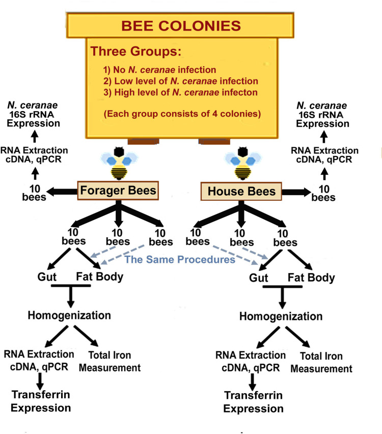
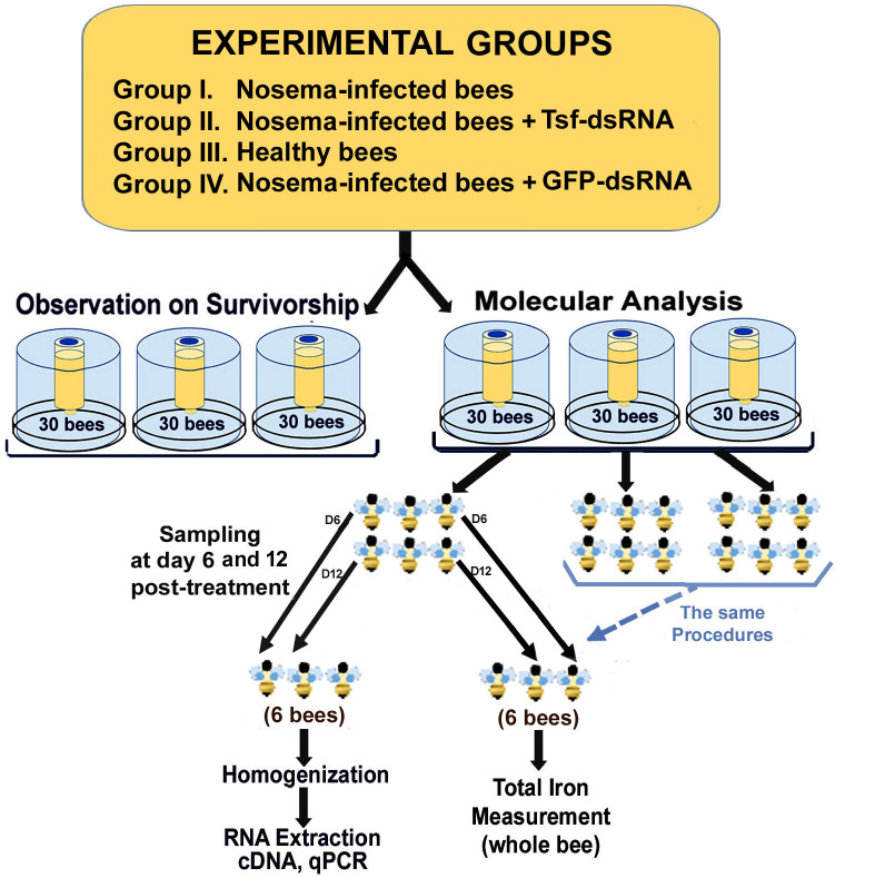
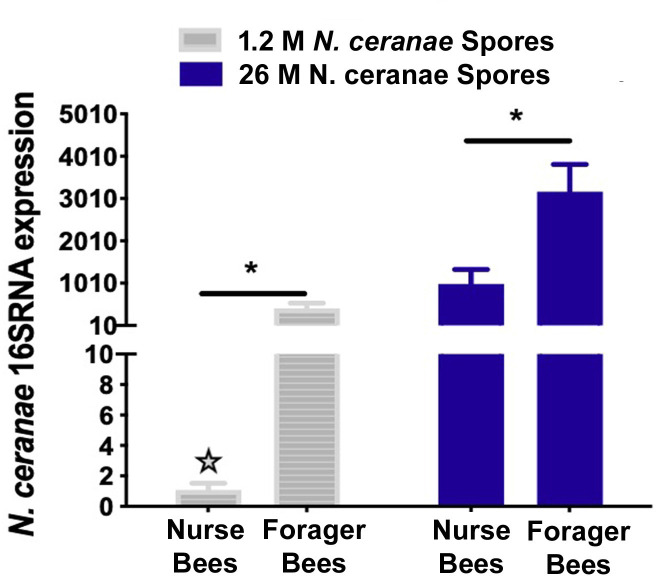
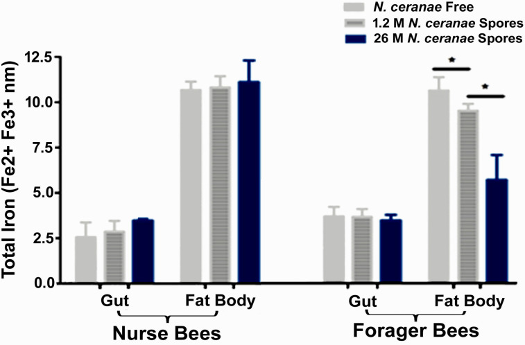
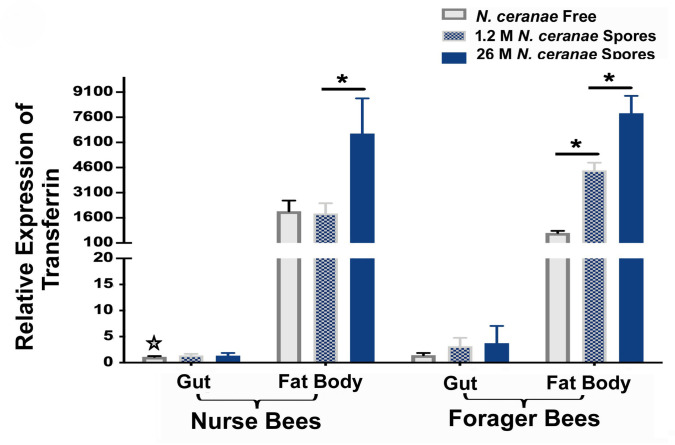
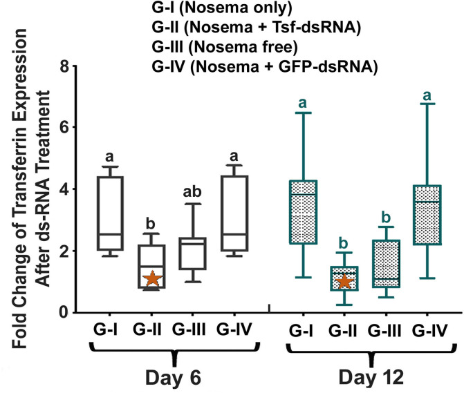
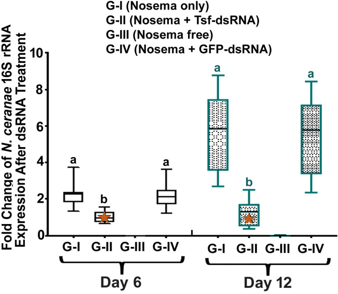
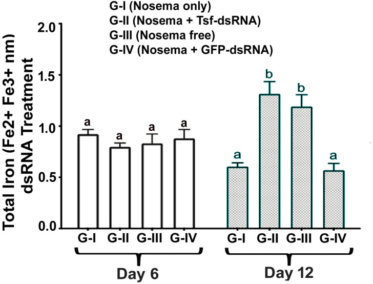
Nosemosis C, a Nosema disease caused by microsporidia parasite Nosema ceranae, is a significant disease burden of the European honey bee Apis mellifera which is one of the most economically important insect pollinators. Nevertheless, there is no effective treatment currently available for Nosema disease and the disease mechanisms underlying the pathological effects of N. ceranae infection in honey bees are poorly understood. Iron is an essential nutrient for growth and survival of hosts and pathogens alike. The iron tug-of-war between host and pathogen is a central battlefield at the host-pathogen interface which determines the outcome of an infection, however, has not been explored in honey bees. To fill the gap, we conducted a study to investigate the impact of N. ceranae infection on iron homeostasis in honey bees. The expression of transferrin, an iron binding and transporting protein that is one of the key players of iron homeostasis, in response to N. ceranae infection was analysed. Furthermore, the functional roles of transferrin in iron homeostasis and honey bee host immunity were characterized using an RNA interference (RNAi)-based method. The results showed that N. ceranae infection causes iron deficiency and upregulation of the A. mellifera transferrin (AmTsf) mRNA in honey bees, implying that higher expression of AmTsf allows N. ceranae to scavenge more iron from the host for its proliferation and survival. The suppressed expression levels of AmTsf via RNAi could lead to reduced N. ceranae transcription activity, alleviated iron loss, enhanced immunity, and improved survival of the infected bees. The intriguing multifunctionality of transferrin illustrated in this study is a significant contribution to the existing body of literature concerning iron homeostasis in insects. The uncovered functional role of transferrin on iron homeostasis, pathogen growth and honey bee’s ability to mount immune responses may hold the key for the development of novel strategies to treat or prevent diseases in honey bees.
Overview
The claims and statistics of this paper haven’t been checked yet.
Run the check — free, no account needed. It reads the open-access text and checks the sources it cites.
Integrity signals
Registry
Cites
Statistics
Figures
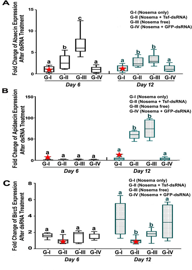
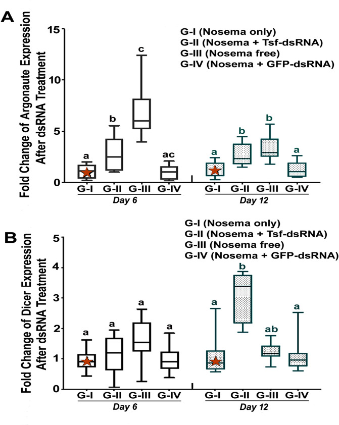
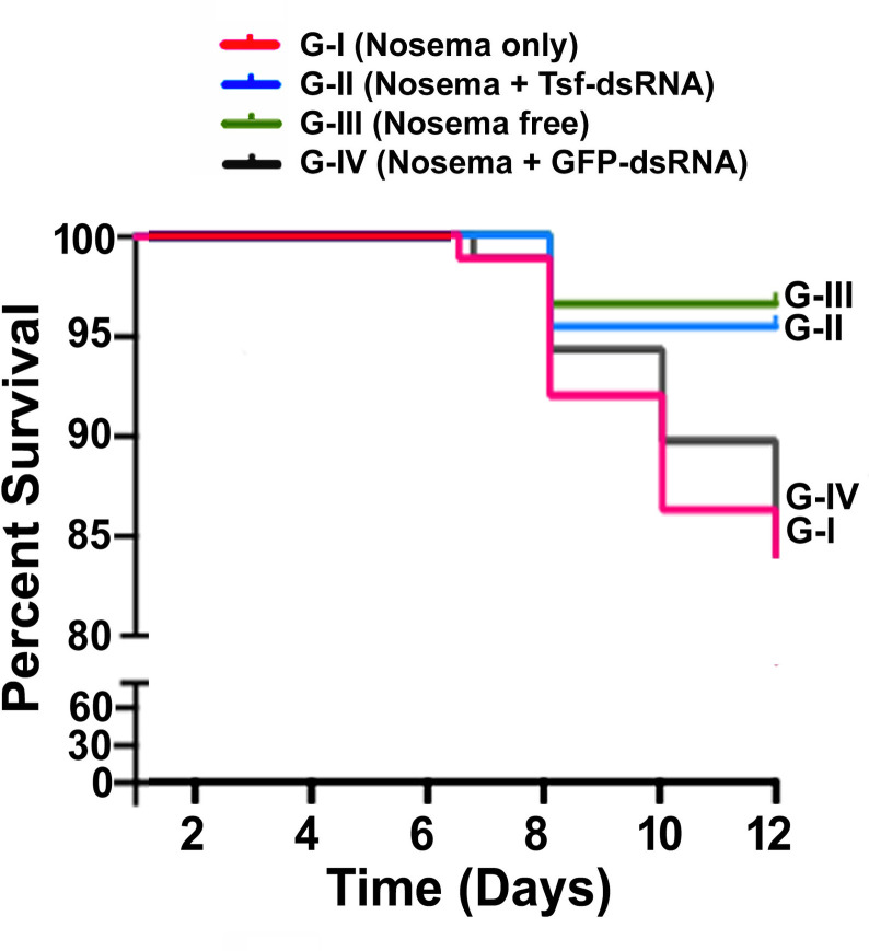
Verification
Claims the paper makes
The claims and statistics of this paper haven’t been checked yet.
Run the check — free, no account needed. It reads the open-access text and checks the sources it cites.
Claims other papers make about this one
Each is checked against this paper’s own text: does it say what the citing paper says it does?
In the citing paper
“Thus, to guarantee iron's vital nutritional benefits and avoid its harmful consequences, its acquisition, transportation, usage, and storage must be tightly controlled through a precisely calibrated process (Rodríguez‐García et al. 2021).”
From Nutritional Immunity in Wound Infection: Unveiling the Role of Dietary Elements in Host–Pathogen Interaction (2025) · cites this paper as Rodríguez‐García et al. 2021
In the citing paper
“Because transferrin has a high affinity for iron, it can keep the amount of free iron in bodily fluids low, which stops invasive bacteria from having access to iron (Rodríguez‐García et al. 2021; Barber and Elde 2014).”
From Nutritional Immunity in Wound Infection: Unveiling the Role of Dietary Elements in Host–Pathogen Interaction (2025) · cites this paper as Rodríguez‐García et al. 2021
Self-check
Percentages, sums and reported differences re-computed from the paper’s own numbers.
The self-check needs the paper’s full text.
Citations
Cites
Iron in infection and immunity
Cassat, J. E., & Skaar, E. P. (2013) · Cell Host & Microbe · doi:10.1016/j.chom.2013.04.010 · Cited by 1,214
Iron metabolism and iron disorders revisited in the hepcidin era
Camaschella, C., Nai, A., & Silvestri, L. (2020) · Haematologica · doi:10.3324/haematol.2019.232124 · Cited by 734
Mechanisms of mammalian iron homeostasis
Pantopoulos, K., Porwal, S. K., Tartakoff, A., & Devireddy, L. (2012) · Biochemistry · doi:10.1021/bi300752r · Cited by 598
The Role of Iron Regulation in Immunometabolism and Immune-Related Disease
Cronin, S. J. F., Woolf, C. J., Weiss, G., & Penninger, J. M. (2019) · Frontiers in Molecular Biosciences · doi:10.3389/fmolb.2019.00116 · Cited by 299
Sequestration and scavenging of iron in infection
Parrow, N. L., Fleming, R. E., & Minnick, M. F. (2013) · Infection and Immunity · doi:10.1128/iai.00602-13 · Cited by 261
Iron metabolism and the innate immune response to infection
Johnson, E. E., & Wessling-Resnick, M. (2012) · Microbes and Infection · doi:10.1016/j.micinf.2011.10.001 · Cited by 253
The Role of Iron in the Immune Response to Bacterial Infection
Cherayil, B. J. (2011) · Immunologic Research · doi:10.1007/s12026-010-8199-1 · Cited by 191
Research works printed but not linked: 111
- A-M Klein, Importance of pollinators in changing landscapes for world crops Proceedings of the Royal Society B: Biological Sciences, 274, 303
- NW Calderone, 2012, Insect pollinated crops, insect pollinators and US agriculture: trend analysis of aggregate data for the period 1992–2009., PLoS One., 7, e37235, 10.1371/journal.pone.0037235 · DOI
- SG Potts, 2016, Safeguarding pollinators and their values to human well-being, Nature, 540, 220, 10.1038/nature20588 · DOI · Paper page
- DL Cox-Foster, 2007, A metagenomic survey of microbes in honey bee colony collapse disorder, Science, 318, 283, 10.1126/science.1146498 · DOI
- N. Kulhanek K S, 2017, A national survey of managed honey bee 2015–2016 annual colony losses in the USA, J Apic Res, 56, 328, 10.1080/00218839.2017.1344496 · DOI
- SG Potts, 2010, Declines of managed honey bees and beekeepers in Europe., J Api Res, 49, 15, 10.3896/IBRA.1.49.1.02 · DOI
- SG Potts, 2010, Global pollinator declines: trends, impacts and drivers, Trends Ecol Evol, 25, 345, 10.1016/j.tree.2010.01.007 · DOI · Paper page
- D Vanengelsdorp, 2010, A historical review of managed honey bee populations in Europe and the United States and the factors that may affect them, J Invertebr Pathol, 103, S80, 10.1016/j.jip.2009.06.011 · DOI
- D Goulson, 2015, Bee declines driven by combined stress from parasites, pesticides, and lack of flowers, Science, 347, 1255957, 10.1126/science.1255957 · DOI
- SA Cameron, 2011, Patterns of widespread decline in North American bumble bees, Proc Natl Acad Sci U S A, 108, 662, 10.1073/pnas.1014743108 · DOI
- I. Fries, 2010, Nosema ceranae in European honey bees (Apis mellifera)., J Invertebr Pathol, 103, S73, 10.1016/j.jip.2009.06.017 · DOI
- L. Bailey, 1968, Honey bee pathology, Annu Rev Entomol, 13, 191, 10.1146/annurev.en.13.010168.001203 · DOI
- E. Zander, 1909, Tierische parasiten als krankenheitserreger bei der biene., Münchener Bienenzeitung, 31, 196
- I Fries, 1996, Nosema ceranae n. sp. (Microspora, Nosematidae), morphological and molecular characterization of a microsporidian parasite of the Asian honey bee Apis cerana (Hymenoptera, Apidae)., Europ J Protistol, 32, 356, 10.1016/S0932-4739(96)80059-9 · DOI
- M Higes, 2006, Nosema ceranae, a new microsporidian parasite in honeybees in Europe, J Invertebr Pathol, 92, 93, 10.1016/j.jip.2006.02.005 · DOI
- Y Chen, 2008, Nosema ceranae is a long-present and wide-spread microsporidian infection of the European honey bee (Apis mellifera) in the United States., J Invertebr Pathol, 97, 186, 10.1016/j.jip.2007.07.010 · DOI
- M Higes, 2009, Honeybee colony collapse due to Nosema ceranae in professional apiaries, Environ Microbiol Rep, 1, 110, 10.1111/j.1758-2229.2009.00014.x · DOI
- WF Huang, 2015, Infectivity and virulence of Nosema ceranae and Nosema apis in commercially available North American honey bees, J Invertebr Pathol, 124, 107, 10.1016/j.jip.2014.10.006 · DOI
- J Klee, 2007, Widespread dispersal of the microsporidian Nosema ceranae, an emergent pathogen of the western honey bee, Apis mellifera, J Invertebr Pathol, 96, 1, 10.1016/j.jip.2007.02.014 · DOI
- MO Milbrath, 2015, Comparative virulence and competition between Nosema apis and Nosema ceranae in honey bees (Apis mellifera)., J Invertebr Pathol, 125, 9, 10.1016/j.jip.2014.12.006 · DOI
- R Martin-Hernandez, 2012, Microsporidia infecting Apis mellifera: coexistence or competition. Is Nosema ceranae replacing Nosema apis?, Environ Microbiol, 14, 2127, 10.1111/j.1462-2920.2011.02645.x · DOI
- RJ Paxton, 2007, Nosema ceranae has infected Apis mellifera in Europe since at least 1998 and may be more virulent than Nosema apis, Apidologie, 38, 558, 10.1051/apido:2007037 · DOI
- M-HR Higes M, 2010, Nosema ceranae in Europe: an emergent type C nosemosis, Apidologie, 41, 375, 10.1051/apido/2010019 · DOI
- RJ Paxton, 2010, Does infection by Nosema ceranae cause "Colony Collapse Disorder" in honey bees (Apis mellifera)?., J Api Res, 49, 80, 10.3896/IBRA.1.49.1.11 · DOI
- R Martin-Hernandez, 2018, Nosema ceranae in Apis mellifera: a 12 years postdetection perspective, Environ Microbiol, 20, 1302, 10.1111/1462-2920.14103 · DOI
- M Hges, Nosema ceranae in Europe: an emergent type Cnosemosis, Apidologie, 41, 375, 10.1051/apido/2010019 · DOI
- HL Holt, 2016, Approaches and Challenges to Managing Nosema (Microspora: Nosematidae) Parasites in Honey Bee (Hymenoptera: Apidae) Colonies., J Econ Entomol, 109, 1487, 10.1093/jee/tow103 · DOI
- C Botias, 2013, Nosema spp. infection and its negative effects on honey bees (Apis mellifera iberiensis) at the colony level., Vet Res, 44, 25, 10.1186/1297-9716-44-25 · DOI
- BB Bailey L, 1991, Honey bee pathology.
- M. Goblirsch, 2018, Nosema ceranae diseases of honey bee (Apis mellifera)., Apidologie, 49, 131, 10.1007/s13592-017-0535-1 · DOI
- C Mayack, 2009, Energetic stress in the honeybee Apis mellifera from Nosema ceranae infection, J Invertebr Pathol, 100, 185, 10.1016/j.jip.2008.12.001 · DOI
- K Antunez, 2009, Immune suppression in the honey bee (Apis mellifera) following infection by Nosema ceranae (Microsporidia)., Environ Microbiol, 11, 2284, 10.1111/j.1462-2920.2009.01953.x · DOI
- SL Gage, 2018, Nosema ceranae parasitism impacts olfactory learning and memory and neurochemistry in honey bees (Apis mellifera)., J Exp Biol, 221
- HL Holt, 2013, Chronic parasitization by Nosema microsporidia causes global expression changes in core nutritional, metabolic and behavioral pathways in honey bee workers (Apis mellifera)., BMC Genomics, 14, 799, 10.1186/1471-2164-14-799 · DOI
- C Vidau, 2014, Differential proteomic analysis of midguts from Nosema ceranae-infected honeybees reveals manipulation of key host functions, J Invertebr Pathol, 121, 89, 10.1016/j.jip.2014.07.002 · DOI
- K Antúnez, 2009, Immune suppression in the honey bee (Apis mellifera) following infection by Nosema ceranae (Microsporidia)., Environmental Microbiology, 11, 2284, 10.1111/j.1462-2920.2009.01953.x · DOI
- V Chaimanee, 2012, Differential expression of immune genes of adult honey bee (Apis mellifera) after inoculated by Nosema ceranae., Journal of Insect Physiology, 58, 1090, 10.1016/j.jinsphys.2012.04.016 · DOI
- M Goblirsch, 2013, Physiological and behavioral changes in honey bees (Apis mellifera) induced by Nosema ceranae infection., PLoS One., 8, e58165, 10.1371/journal.pone.0058165 · DOI
- M Higes, 2013, Apoptosis in the pathogenesis of Nosema ceranae (Microsporidia: Nosematidae) in honey bees (Apis mellifera)., Environmental Microbiology Reports, 5, 530, 10.1111/1758-2229.12059 · DOI
- R Martín-Hernández, 2017, Microsporidia infection impacts the host cell's cycle and reduces host cell apoptosis, PLoS ONE, 12, 10.1371/journal.pone.0170183 · DOI
- C Costa, 2011, Negative correlation between Nosema ceranae spore loads and deformed wing virus infection levels in adult honey bee workers, J Invertebr Pathol, 108, 224, 10.1016/j.jip.2011.08.012 · DOI
- HQ Zheng, 2015, Evidence of the synergistic interaction of honey bee pathogens Nosema ceranae and Deformed wing virus, Vet Microbiol, 177, 1, 10.1016/j.vetmic.2015.02.003 · DOI
- M Higes, 2008, How natural infection by Nosema ceranae causes honeybee colony collapse, Environ Microbiol, 10, 2659, 10.1111/j.1462-2920.2008.01687.x · DOI
- D Vanengelsdorp, 2009, Colony collapse disorder: a descriptive study., PLoS One., 4, e6481, 10.1371/journal.pone.0006481 · DOI
- C Botías, 2012, The effect of induced queen replacement on Nosema spp. infection in honey bee (Apis mellifera iberiensis) colonies., Environmental microbiology, 14, 845, 10.1111/j.1462-2920.2011.02647.x · DOI
- I Muñoz, 2014, Presence of Nosema ceranae associated with honeybee queen introductions, Infection, Genetics and Evolution, 23, 161, 10.1016/j.meegid.2014.02.008 · DOI
- L. Bailey, 1955, The epidemiology and control of Nosema disease of the honey-bee, Annals of Applied Biology, 43, 379, 10.1111/j.1744-7348.1955.tb02488.x · DOI
- M Higes, 2008, Detection of infective Nosema ceranae (Microsporidia) spores in corbicular pollen of forager honeybees., Journal of Invertebrate Pathology, 97, 76, 10.1016/j.jip.2007.06.002 · DOI
- M Higes, 2009, Horizontal transmission of Nosema ceranae (Microsporidia) from worker honeybees to queens (Apis mellifera)., Environmental Microbiology Reports, 1, 495, 10.1111/j.1758-2229.2009.00052.x · DOI
- LA Malone, 2001, Effects of time, temperature, and honey on Nosema apis (Microsporidia: Nosematidae), a parasite of the honeybee, Apis mellifera (Hymenoptera: Apidae)., Journal of Invertebrate Pathology, 77, 258, 10.1006/jipa.2001.5028 · DOI
- ML Smith, 2012, The honey bee parasite nosema ceranae: Transmissible via food exchange?, PLoS ONE., 7, 10.1371/journal.pone.0043319 · DOI
- U Glavinić, 2014, Nosema ceranae DNA in honey bee haemolymph and honey bee mite Varroa destructor, Acta Veterinaria, 64, 349, 10.2478/acve-2014-0033 · DOI
- Y Mendoza, 2017, Nosema ceranae Winter Control: Study of the Effectiveness of Different Fumagillin Treatments and Consequences on the Strength of Honey Bee (Hymenoptera: Apidae) Colonies., Journal of economic entomology, 110, 1
- A Giacobino, 2016, Fumagillin control of Nosema ceranae (Microsporidia: Nosematidae) infection in honey bee (Hymenoptera: Apidae) colonies in Argentina., Veterinaria Italiana, 52, 145
- WF Huang, 2013, Nosema apis and nosema ceranae; a comparative study in the honey bee host, American Bee Journal, 153, 277
- M Higes, 2011, The stability and effectiveness of fumagillin in controlling Nosema ceranae (Microsporidia) infection in honey bees (Apis mellifera) under laboratory and field conditions., Apidologie, 42, 364, 10.1007/s13592-011-0003-2 · DOI
- GR Williams, 2008, Does fumagillin control the recently detected invasive parasite Nosema ceranae in western honey bees (Apis mellifera)?, Journal of Invertebrate Pathology, 99, 342, 10.1016/j.jip.2008.04.005 · DOI
- WF Huang, 2013, Nosema ceranae escapes fumagillin control in honey bees., PLoS Pathog., 9, e1003185, 10.1371/journal.ppat.1003185 · DOI
- LL Dunn, 2007, Iron uptake and metabolism in the new millennium, Trends in cell biology, 17, 93, 10.1016/j.tcb.2006.12.003 · DOI
- H Nichol, 2002, Iron metabolism in insects, Annual review of entomology, 47, 535, 10.1146/annurev.ento.47.091201.145237 · DOI
- GJ Anderson, 2017, Current understanding of iron homeostasis, Am J Clin Nutr, 106, 1559s, 10.3945/ajcn.117.155804 · DOI · Paper page
- HA Huebers, 1987, The physiology of transferrin and transferrin receptors, Physiol Rev, 67, 520, 10.1152/physrev.1987.67.2.520 · DOI
- JH Brock, 1989, Iron-binding proteins., Acta Paediatr Scand Suppl, 361, 31, 10.1111/apa.1989.78.s361.31 · DOI
- G Gao, 2019, Cellular Iron Metabolism and Regulation, Adv Exp Med Biol, 1173, 21, 10.1007/978-981-13-9589-5_2 · DOI
- X Tang, 2013, Iron homeostasis in insects: Insights from Drosophila studies, IUBMB Life, 65, 863, 10.1002/iub.1211 · DOI
- DL Geiser, 2012, Insect transferrins: multifunctional proteins, Biochim Biophys Acta, 1820, 437, 10.1016/j.bbagen.2011.07.011 · DOI
- G Xiao, 2019, Transferrin 1 Functions in Iron Trafficking and Genetically Interacts with Ferritin in Drosophila melanogaster, Cell Rep, 26, 748, 10.1016/j.celrep.2018.12.053 · DOI
- A Dutta, 2019, First report on transferrin in the silkworm, Antheraea mylitta, with a putative role in antioxidant defense: Insights from proteomic analysis and immunodetection, Comp Biochem Physiol B Biochem Mol Biol, 233, 23, 10.1016/j.cbpb.2019.03.010 · DOI
- B Dunkov, 2006, Insect iron binding proteins: insights from the genomes, Insect Biochem Mol Biol, 36, 300, 10.1016/j.ibmb.2006.01.007 · DOI
- T Yoshiga, 1999, Drosophila melanogaster transferrin. Cloning, deduced protein sequence, expression during the life cycle, gene localization and up-regulation on bacterial infection, Eur J Biochem, 260, 414, 10.1046/j.1432-1327.1999.00173.x · DOI
- R Kucharski, 2003, Transcriptional profiling reveals multifunctional roles for transferrin in the honeybee, Apis mellifera., J Insect Sci, 3, 27, 10.1093/jis/3.1.27 · DOI
- BY Kim, 2008, Insect transferrin functions as an antioxidant protein in a beetle larva, Comp Biochem Physiol B Biochem Mol Biol, 150, 161, 10.1016/j.cbpb.2008.02.009 · DOI
- M Hirai, 2000, A juvenile hormone-repressible transferrin-like protein from the bean bug, Riptortus clavatus: cDNA sequence analysis and protein identification during diapause and vitellogenesis, Arch Insect Biochem Physiol, 44, 17, 10.1002/(SICI)1520-6327(200005)44:1<17::AID-ARCH3>3.0.CO;2-O · DOI
- AM do Nascimento, 2004, Honey bee (Apis mellifera) transferrin-gene structure and the role of ecdysteroids in the developmental regulation of its expression., Insect Biochem Mol Biol, 34, 415, 10.1016/j.ibmb.2003.12.003 · DOI
- R Kucharski, 2003, Transcriptional profiling reveals multifunctional roles for transferrin in the honeybee, Apis mellifera, Journal of Insect Science, 3, 27, 10.1093/jis/3.1.27 · DOI
- J Li, 2019, The Phylogeny and Pathogenesis of Sacbrood Virus (SBV) Infection in European Honey Bees, Apis mellifera, Viruses, 11, 61, 10.3390/v11010061 · DOI
- JH Li, 2017, New evidence showing that the destruction of gut bacteria by antibiotic treatment could increase the honey bee's vulnerability to Nosema infection, PLoS One, 12, e0187505, 10.1371/journal.pone.0187505 · DOI
- GE Cantwell, 1970, Standard methods for counting Nosema spores, American Bee Journal, 110, 222
- JD Evans, 2009, Bee cups: Single-use cages for honey bee experiments, Journal of Apicultural Research, 48, 300, 10.1080/00218839.2009.11101548 · DOI
- SK Huang, 2014, Evaluation of cage designs and feeding regimes for honey bee (Hymenoptera: Apidae) laboratory experiments., Journal of Economic Entomology, 107, 54, 10.1603/EC13213 · DOI
- T Horn, 2010, E-RNAi: a web application for the multi-species design of RNAi reagents—2010 update, Nucleic Acids Res, 38, W332, 10.1093/nar/gkq317 · DOI
- CJ Jack, 2016, Colony Level Prevalence and Intensity of Nosema ceranae in Honey Bees (Apis mellifera L.)., PLoS One., 11, e0163522, 10.1371/journal.pone.0163522 · DOI
- MD Smart, 2012, Nosema ceranae in age cohorts of the western honey bee (Apis mellifera)., J Invertebr Pathol, 109, 148, 10.1016/j.jip.2011.09.009 · DOI
- S Wolf, 2014, So near and yet so far: harmonic radar reveals reduced homing ability of Nosema infected honeybees., PLoS One., 9, e103989, 10.1371/journal.pone.0103989 · DOI
- KE Roberts, 2014, Immunosenescence and resistance to parasite infection in the honey bee, Apis mellifera, J Invertebr Pathol, 121, 1, 10.1016/j.jip.2014.06.004 · DOI
- J Kralj, 2010, Nosema sp. influences flight behaviour of infected honeybee (Apis mellifera) foragers., Apidologie, 41, 21, 10.1051/apido/2009046 · DOI
- CJ Perry, 2015, Rapid behavioral maturation accelerates failure of stressed honey bee colonies, Proc Natl Acad Sci U S A, 112, 3427, 10.1073/pnas.1422089112 · DOI
- J Vávra, 2013, Microsporidia and 'the art of living together'., Adv Parasitol, 82, 253, 10.1016/B978-0-12-407706-5.00004-6 · DOI
- ED Weinberg, 1975, Nutritional immunity. Host's attempt to withold iron from microbial invaders, Jama, 231, 39, 10.1001/jama.1975.03240130021018 · DOI · Paper page
- M Nairz, 2010, The struggle for iron—a metal at the host-pathogen interface, Cell Microbiol, 12, 1691, 10.1111/j.1462-5822.2010.01529.x · DOI · Paper page
- H Haas, 2008, Siderophores in fungal physiology and virulence, Annu Rev Phytopathol, 46, 149, 10.1146/annurev.phyto.45.062806.094338 · DOI
- CC Philpott, 2006, Iron uptake in fungi: a system for every source, Biochim Biophys Acta, 1763, 636, 10.1016/j.bbamcr.2006.05.008 · DOI · Paper page
- A Aznar, 2014, Scavenging iron: a novel mechanism of plant immunity activation by microbial siderophores, Plant Physiol, 164, 2167, 10.1104/pp.113.233585 · DOI
- T Yoshiga, 1997, Mosquito transferrin, an acute-phase protein that is up-regulated upon infection, Proc Natl Acad Sci U S A, 94, 12337, 10.1073/pnas.94.23.12337 · DOI
- ST Ong, 2006, Iron-withholding strategy in innate immunity, Immunobiology, 211, 295, 10.1016/j.imbio.2006.02.004 · DOI · Paper page
- LM Brummett, 2017, The immune properties of Manduca sexta transferrin, Insect Biochem Mol Biol, 81, 1, 10.1016/j.ibmb.2016.12.006 · DOI
- A Fire, 1998, Potent and specific genetic interference by double-stranded RNA in Caenorhabditis elegans, Nature, 391, 806, 10.1038/35888 · DOI · Paper page
- FM Nunes, 2013, Non-Target Effects of Green Fluorescent Protein (GFP)-Derived Double-Stranded RNA (dsRNA-GFP) Used in Honey Bee RNA Interference (RNAi) Assays., Insects, 4, 90, 10.3390/insects4010090 · DOI
- R Bartoszewski, 2019, Editorial focus: understanding off-target effects as the key to successful RNAi therapy, Cell Mol Biol Lett, 24, 69, 10.1186/s11658-019-0196-3 · DOI
- DR Caffrey, 2011, siRNA off-target effects can be reduced at concentrations that match their individual potency., PLoS One., 6, e21503, 10.1371/journal.pone.0021503 · DOI
- E Maori, 2009, IAPV, a bee-affecting virus associated with Colony Collapse Disorder can be silenced by dsRNA ingestion, Insect Mol Biol, 18, 55, 10.1111/j.1365-2583.2009.00847.x · DOI
- EM Campbell, 2010, Gene-knockdown in the honey bee mite Varroa destructor by a non-invasive approach: studies on a glutathione S-transferase., Parasit Vectors., 3, 73, 10.1186/1756-3305-3-73 · DOI
- SD Desai, 2012, Reduction in deformed wing virus infection in larval and adult honey bees (Apis mellifera L.) by double-stranded RNA ingestion., Insect Mol Biol, 21, 446, 10.1111/j.1365-2583.2012.01150.x · DOI
- AJ Burnham, 2019, Scientific Advances in Controlling Nosema ceranae (Microsporidia) Infections in Honey Bees (Apis mellifera)., Front Vet Sci., 6, 79, 10.3389/fvets.2019.00079 · DOI
- N Paldi, 2010, Effective gene silencing in a microsporidian parasite associated with honeybee (Apis mellifera) colony declines., Appl Environ Microbiol, 76, 5960, 10.1128/AEM.01067-10 · DOI
- C Rodríguez-García, 2018, Nosemosis control in European honey bees, Apis mellifera, by silencing the gene encoding Nosema ceranae polar tube protein 3, J Exp Biol, 221
- Q Huang, 2019, Dicer regulates Nosema ceranae proliferation in honeybees, Insect Mol Biol, 28, 74, 10.1111/imb.12534 · DOI
- W Li, 2016, Silencing the Honey Bee (Apis mellifera) Naked Cuticle Gene (nkd) Improves Host Immune Function and Reduces Nosema ceranae Infections., Appl Environ Microbiol, 82, 6779, 10.1128/AEM.02105-16 · DOI
- L Aagaard, 2007, RNAi therapeutics: principles, prospects and challenges, Adv Drug Deliv Rev, 59, 75, 10.1016/j.addr.2007.03.005 · DOI
- JD Evans, 2006, Immune pathways and defence mechanisms in honey bees Apis mellifera, Insect Mol Biol, 15, 645, 10.1111/j.1365-2583.2006.00682.x · DOI
- F Leulier, 2006, The Drosophila inhibitor of apoptosis protein DIAP2 functions in innate immunity and is essential to resist gram-negative bacterial infection, Mol Cell Biol, 26, 7821, 10.1128/MCB.00548-06 · DOI
Cited by
Sorted by citations
Chen, C., Hu, X., He, D., He, X., & Shen, L. (2025) · Food Science & Nutrition · doi:10.1002/fsn3.70854 · Cited by 6
Metadata
- Authors
- Rodríguez-García, C., Heerman, M. C., Cook, S. C., Evans, J. D., DeGrandi-Hoffman, G., Banmeke, O., Zhang, Y., Huang, S., Hamilton, M., & Chen, Y. P.
- ORCID
- Venue
- PLOS Pathogens
- Publisher
- Public Library of Science (PLoS)
- Published
- 2021-02-18
- Volume
- 17
- Issue
- 2
- Pages
- e1009270
- ISSN
- 1553-7374
- PMC
- PMC7891791
- Cited by
- 50
- References
- 7
- Language
- en
About these checks
Registry, retraction, Cites and statistics results are computed, not judged, with one exception: a printed reference that no index lists is read once by a model before it counts as a citation error. Claims are extracted by a model and kept only when their quote appears verbatim in the text; each cited source is then checked, with a model’s help, for whether it says what the claim relies on. Those verdicts can be wrong — read the claim’s quote and the cited source before relying on one.
Statistics and arithmetic are recomputed, not judged.
Confidence bands are shown only for fields calibrated on our annotated test set. None is calibrated yet, so no band is shown.
What counts as a claim ›How we measure confidence ›How we check this ›
Are you an author, or do you see a problem with this page? Request a review
