Cited by 151Neurogenesis and neuroplasticity mechanismsAxon Guidance and Neuronal SignalingDevelopmental Biology and Gene Regulation

The DOI matches its Crossref record; 56 of the 57 references it cites were found in a registry or an index.

Layer Acquisition by Cortical GABAergic Interneurons Is Independent of Reelin Signaling

Open access
Pla, Ramón, Borrell, Víctor, Flames, Nuria, Marín, Oscar·2006·The Journal of Neuroscience

Abstract

Functioning of the cerebral cortex requires the coordinated assembly of circuits involving glutamatergic projection neurons and GABAergic interneurons. Despite their segregated origin in different regions of the telencephalon, projection neurons and interneurons born synchronically end up adopting the same cortical layer, suggesting that layer acquisition is highly coordinated for both neuronal types. The radial migration and laminar arrangement of projection neurons depends on Reelin, a secreted glycoprotein expressed near the pial surface during embryogenesis. In contrast, the mechanisms controlling layer acquisition by cortical interneurons remain essentially unknown. Here, we have used an ultrasound-guided transplantation approach to analyze the mechanisms underlying the acquisition of laminar locations by cortical interneurons. We found that layer acquisition by cortical GABAergic interneurons does not directly depend on Reelin signaling. Moreover, interneurons invade their target layers well after synchronically generated projection neurons reach their final destination. These results suggest a model in which cues provided by projection neurons guide cortical interneurons to their appropriate layer, and reveal that, at least for some neuronal types, long-range radial migration does not directly require Reelin.

Overview

The claims and statistics of this paper haven’t been checked yet.

Run the check — free, no account needed. It reads the open-access text and checks the sources it cites.

A few free checks a day · papers already verified open instantly · results are public

Integrity signals

Registry

Crossref · 10.1523/jneurosci.0245-06.2006

Cites

56 of 56 references verified0 citation errors · 0 not checkable

Statistics

Figures

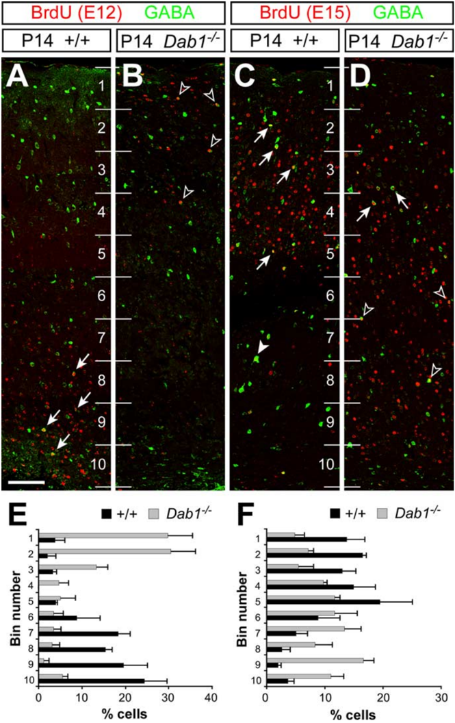
Figure1 Thelaminarpositionofcortical interneurons is inverted in Dab1 mutantmice. A-D , Confocal microscopic reconstructions of the cerebral cortex of wild-type ( A and C ) and Dab1 mutant ( B and D ) mice stained for BrdU (red) and GABA (green) after injection of BrdU at E12 ( A and B ) or E15 ( C and D ). A , B , Early-born cortical interneurons (BrdU + /GABA + ) acquire deep cortical positions in control mice (arrows), whereas they occupy superficial positions in Dab1 mutants (open arrowheads). C , D , Late-born interneurons (BrdU + /GABA + ) populate mostly superficial layers (arrows) in wild-type mice, although a few also occupy deep layers (solid arrowhead). In Dab1 mutants, the majority of late-born interneurons occupy deep positions (open arrowheads), although some populate superficial layers (arrows). Numbers identify bins for quantification. E , F , Binned quantifications of the distribution of E12 ( E ) and E15 ( F ) interneurons in the cerebral cortex of wild-type (black bars) and Dab1 mutant (gray bars) mice (average + SEM). The width of the cortex proper (layers 1- 6) was divided into 10 equal bins for laminar distribution analysis, with bin 1 at the top (marginal zone) and bin 10 at the bottom (white matter). Scale bar, 100 𝛍 m.
Figure1 Thelaminarpositionofcortical interneurons is inverted in Dab1 mutantmice. A-D , Confocal microscopic reconstructions of the cerebral cortex of wild-type ( A and C ) and Dab1 mutant ( B and D ) mice stained for BrdU (red) and GABA (green) after injection of BrdU at E12 ( A and B ) or E15 ( C and D ). A , B , Early-born cortical interneurons (BrdU + /GABA + ) acquire deep cortical positions in control mice (arrows), whereas they occupy superficial positions in Dab1 mutants (open arrowheads). C , D , Late-born interneurons (BrdU + /GABA + ) populate mostly superficial layers (arrows) in wild-type mice, although a few also occupy deep layers (solid arrowhead). In Dab1 mutants, the majority of late-born interneurons occupy deep positions (open arrowheads), although some populate superficial layers (arrows). Numbers identify bins for quantification. E , F , Binned quantifications of the distribution of E12 ( E ) and E15 ( F ) interneurons in the cerebral cortex of wild-type (black bars) and Dab1 mutant (gray bars) mice (average + SEM). The width of the cortex proper (layers 1- 6) was divided into 10 equal bins for laminar distribution analysis, with bin 1 at the top (marginal zone) and bin 10 at the bottom (white matter). Scale bar, 100 𝛍 m.CC-BY-NC-SA · From the PDF
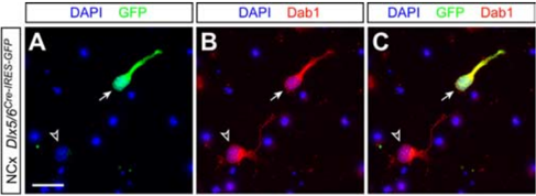
Figure 2 Expression of Dab1 in cortical interneurons during corticogenesis. Images of dissociated cultures obtained from E17.5 cortices of Dlx5/6 Cre-IRES-GFP embryos showing nuclear staining [4 ′ ,6 ′ -diamidino-2-phenylindoledihydrochloride(DAPI)]andimmunohistochemistry for GFP ( A and C ) and Dab1 ( B and C ) are shown. Scale bar, 20 𝛍 m.
Figure 2 Expression of Dab1 in cortical interneurons during corticogenesis. Images of dissociated cultures obtained from E17.5 cortices of Dlx5/6 Cre-IRES-GFP embryos showing nuclear staining [4 ′ ,6 ′ -diamidino-2-phenylindoledihydrochloride(DAPI)]andimmunohistochemistry for GFP ( A and C ) and Dab1 ( B and C ) are shown. Scale bar, 20 𝛍 m.CC-BY-NC-SA · From the PDF
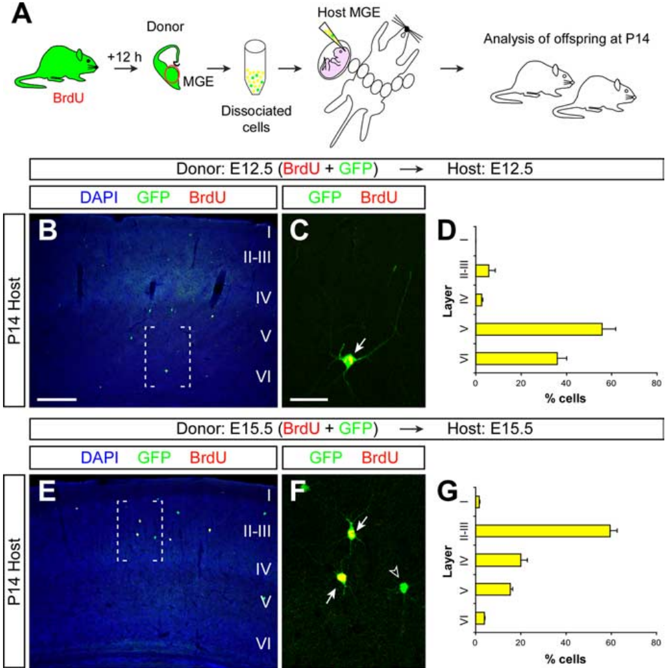
Figure 3 Cortical interneurons transplanted homotypically and isochronically in utero retain their program for laminar specificity. A , Schematic diagram of the experimental design. GFP + donor pregnant mice received a single injection of BrdU, and 12 h later the MGE was dissected from their embryos and dissociated. GFP + donorMGEcells[alsoBrdU + (yellow), or BrdU -(green)] were then injected into the MGE of age-matched host embryos. Host embryos were allowed to be born and analyzed at P14. B , E , Coronal sections through the somatosensory cortex of P14 mice showing the distribution of E12.5 ( B ) and E15.5 ( E ) GFP + MGE-derived cells after nuclear staining [4 ′ ,6 ′ -diamidino-2-phenylindole dihydrochloride (DAPI)] and immunohistochemistry for BrdU (red) and GFP (green). C , F , Magnifications of the areas boxed in B and E , respectively. Arrows show BrdU + /GFP + cells; the open arrowhead indicates an example of BrdU -/GFP + cell not included in the analysis. D , G , Quantification of the distribution of isochronically transplanted E12.5 ( D ) and E15.5 ( G ) interneurons in the P14 cortex (average + SEM). Scale bars: (in B ) B , E , 300 𝛍 m; (in C ) C , F , 50 𝛍 m.
Figure 3 Cortical interneurons transplanted homotypically and isochronically in utero retain their program for laminar specificity. A , Schematic diagram of the experimental design. GFP + donor pregnant mice received a single injection of BrdU, and 12 h later the MGE was dissected from their embryos and dissociated. GFP + donorMGEcells[alsoBrdU + (yellow), or BrdU -(green)] were then injected into the MGE of age-matched host embryos. Host embryos were allowed to be born and analyzed at P14. B , E , Coronal sections through the somatosensory cortex of P14 mice showing the distribution of E12.5 ( B ) and E15.5 ( E ) GFP + MGE-derived cells after nuclear staining [4 ′ ,6 ′ -diamidino-2-phenylindole dihydrochloride (DAPI)] and immunohistochemistry for BrdU (red) and GFP (green). C , F , Magnifications of the areas boxed in B and E , respectively. Arrows show BrdU + /GFP + cells; the open arrowhead indicates an example of BrdU -/GFP + cell not included in the analysis. D , G , Quantification of the distribution of isochronically transplanted E12.5 ( D ) and E15.5 ( G ) interneurons in the P14 cortex (average + SEM). Scale bars: (in B ) B , E , 300 𝛍 m; (in C ) C , F , 50 𝛍 m.CC-BY-NC-SA · From the PDF
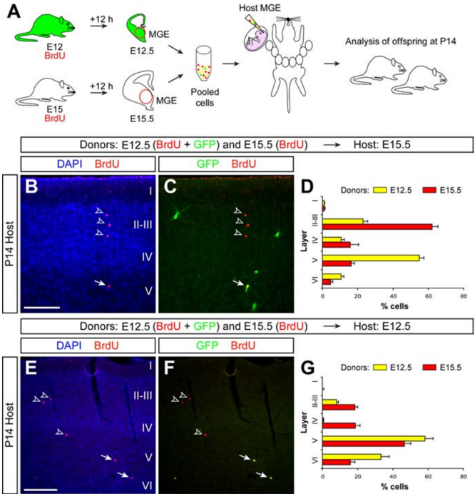
Figure 4 Cortical interneurons adopt multiple laminar fates after heterochronic transplantation. A , Schematic diagram of the experimental design. GFP + donor pregnant mice received a single injection of BrdU at E12, and wild-type donor pregnant mice received a single injection of BrdU at E15. Twelve hours after BrdU injection, the MGE was dissected from embryos and dissociated. Pooled donor MGE cells were then injected into the MGE of either E12.5 or E15.5 host embryos. Host embryos were allowed to be born and analyzed at P14. B , C , Coronal section through the somatosensory cortex of a transplanted P14 mouse showing the distribution of E12.5 (BrdU + /GFP + ) and E15.5 (BrdU + /GFP -) MGE-derived cells after nuclear staining [4 ′ ,6 ′ -diamidino-2phenylindole dihydrochloride (DAPI)] and immunohistochemistry for BrdU (red) and GFP (green). Arrows point to a BrdU + / GFP + cell; open arrowheads indicate BrdU + /GFP -cells. D , Quantification of the distribution of heterochronically transplanted E12.5 (yellow) and isochronically transplanted E15.5 (red) interneurons in the P14 cortex (average + SEM). E , F , Coronal section through the somatosensory cortex of a transplanted P14 mouse showing the distribution of E12.5 (BrdU + /GFP + ) and E15.5 (BrdU + /GFP -) MGE-derived cells after nuclear staining (DAPI) and immunohistochemistry for BrdU (red) and GFP (green). Arrows point to BrdU + /GFP + cells; open arrowheads indicate BrdU + /GFP -cells. G , Quantification of the distribution of isochronically transplanted E12.5 (yellow) and heterochronically transplanted E15.5 (red) interneurons in the P14 cortex (average + SEM). Scale bars: B , C , 100 𝛍 m; E , F , 200 𝛍 m.
Figure 4 Cortical interneurons adopt multiple laminar fates after heterochronic transplantation. A , Schematic diagram of the experimental design. GFP + donor pregnant mice received a single injection of BrdU at E12, and wild-type donor pregnant mice received a single injection of BrdU at E15. Twelve hours after BrdU injection, the MGE was dissected from embryos and dissociated. Pooled donor MGE cells were then injected into the MGE of either E12.5 or E15.5 host embryos. Host embryos were allowed to be born and analyzed at P14. B , C , Coronal section through the somatosensory cortex of a transplanted P14 mouse showing the distribution of E12.5 (BrdU + /GFP + ) and E15.5 (BrdU + /GFP -) MGE-derived cells after nuclear staining [4 ′ ,6 ′ -diamidino-2phenylindole dihydrochloride (DAPI)] and immunohistochemistry for BrdU (red) and GFP (green). Arrows point to a BrdU + / GFP + cell; open arrowheads indicate BrdU + /GFP -cells. D , Quantification of the distribution of heterochronically transplanted E12.5 (yellow) and isochronically transplanted E15.5 (red) interneurons in the P14 cortex (average + SEM). E , F , Coronal section through the somatosensory cortex of a transplanted P14 mouse showing the distribution of E12.5 (BrdU + /GFP + ) and E15.5 (BrdU + /GFP -) MGE-derived cells after nuclear staining (DAPI) and immunohistochemistry for BrdU (red) and GFP (green). Arrows point to BrdU + /GFP + cells; open arrowheads indicate BrdU + /GFP -cells. G , Quantification of the distribution of isochronically transplanted E12.5 (yellow) and heterochronically transplanted E15.5 (red) interneurons in the P14 cortex (average + SEM). Scale bars: B , C , 100 𝛍 m; E , F , 200 𝛍 m.CC-BY-NC-SA · From the PDF
Image 5
CC-BY-NC-SA · From the PDF
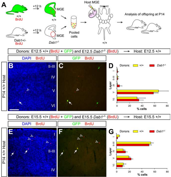
Figure 6 The laminar distribution of cortical interneurons is independent of Reelin signaling. A , Schematic diagram of the experimental design. GFP + and Dab1 + / -donor pregnant mice received a single injection of BrdU at E12 or E15. Twelve hours after BrdU injection, the MGE of Dab1 -/ -embryos was collected and dissociated and pooled together with cells obtained from the MGE of GFP + embryos. Pooled donor MGE cells were then injected into the MGE of either E12.5 or E15.5 wild-type host embryos. Host embryos were allowed to be born and analyzed at P14. B , C , Coronal section through the somatosensory cortex of a transplanted P14 mouse showing the distribution of E12.5 wild-type (BrdU + /GFP + ) and E12.5 Dab1 -/ -(BrdU + /GFP -) MGE-derived interneurons after nuclear staining [4 ′ ,6 ′ -diamidino-2-phenylindole dihydrochloride (DAPI)] and immunohistochemistryforBrdU(red)andGFP(green).Arrowspointtoawild-type(BrdU + /GFP + ) cell; open arrowheads indicate a Dab1 -/ -(BrdU + /GFP -) cell. D , Quantification of the distribution of wild-type E12.5 (yellow) and Dab1 -/ -E12.5 (red) interneurons in the P14 cortex (average + SEM). E , F , Coronal section through the somatosensory cortex of a transplanted P14 mouse showing the distribution of E15.5 wild-type (BrdU + /GFP + ) and E15.5 Dab1 -/ -(BrdU + /GFP -) MGE-derived interneurons after nuclear staining (DAPI) and immunohistochemistry for BrdU (red) and GFP (green). Arrows point to wild-type (BrdU + /GFP + ) cells; open arrowheads indicate Dab1 -/ -(BrdU + /GFP -) cells. G , Quantification of the distribution of wild-type E15.5 (yellow) and Dab1 -/ -E15.5 (red) interneurons in the P14 cortex (average + SEM). Scale bar, 200 𝛍 m.
Figure 6 The laminar distribution of cortical interneurons is independent of Reelin signaling. A , Schematic diagram of the experimental design. GFP + and Dab1 + / -donor pregnant mice received a single injection of BrdU at E12 or E15. Twelve hours after BrdU injection, the MGE of Dab1 -/ -embryos was collected and dissociated and pooled together with cells obtained from the MGE of GFP + embryos. Pooled donor MGE cells were then injected into the MGE of either E12.5 or E15.5 wild-type host embryos. Host embryos were allowed to be born and analyzed at P14. B , C , Coronal section through the somatosensory cortex of a transplanted P14 mouse showing the distribution of E12.5 wild-type (BrdU + /GFP + ) and E12.5 Dab1 -/ -(BrdU + /GFP -) MGE-derived interneurons after nuclear staining [4 ′ ,6 ′ -diamidino-2-phenylindole dihydrochloride (DAPI)] and immunohistochemistryforBrdU(red)andGFP(green).Arrowspointtoawild-type(BrdU + /GFP + ) cell; open arrowheads indicate a Dab1 -/ -(BrdU + /GFP -) cell. D , Quantification of the distribution of wild-type E12.5 (yellow) and Dab1 -/ -E12.5 (red) interneurons in the P14 cortex (average + SEM). E , F , Coronal section through the somatosensory cortex of a transplanted P14 mouse showing the distribution of E15.5 wild-type (BrdU + /GFP + ) and E15.5 Dab1 -/ -(BrdU + /GFP -) MGE-derived interneurons after nuclear staining (DAPI) and immunohistochemistry for BrdU (red) and GFP (green). Arrows point to wild-type (BrdU + /GFP + ) cells; open arrowheads indicate Dab1 -/ -(BrdU + /GFP -) cells. G , Quantification of the distribution of wild-type E15.5 (yellow) and Dab1 -/ -E15.5 (red) interneurons in the P14 cortex (average + SEM). Scale bar, 200 𝛍 m.CC-BY-NC-SA · From the PDF
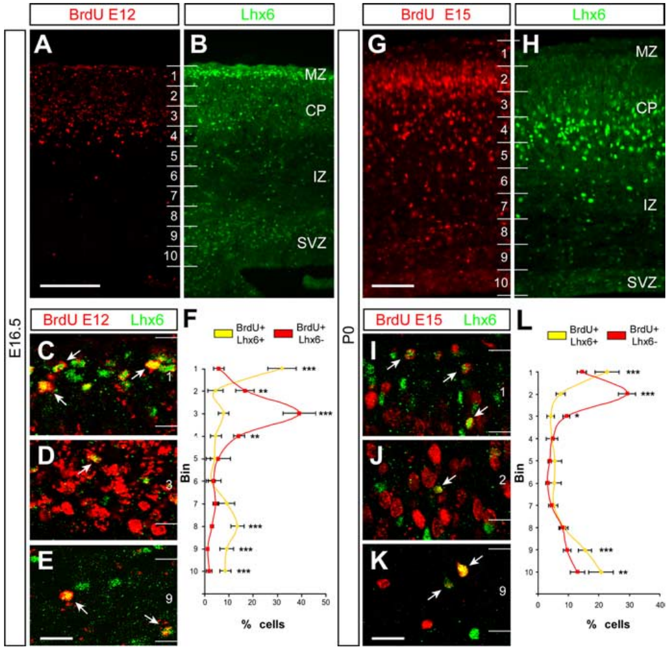
Figure 7 The timing of cortical plate invasion is different for interneurons than for projection neurons. A , B , Coronal sections through the somatosensory cortex of an E16.5 mouse embryo that received a single BrdU injection at E12, showing immunohistochemistry for BrdU ( A ) and Lhx6 ( B ). The majority of BrdU + cells occupy the cortical plate (CP), although labeled cells are scattered through all layers. In turn, Lhx6 staining is more abundant in the marginal zone (MZ), cortical plate, and subventricular zone (SVZ). Numbers indicate bins for quantification. C-E , Confocal microscopic images of the marginal zone (MZ) ( C ), the CP ( D ), and the SVZ ( E ) showing BrdU (red) and Lhx6 (green) staining at E16.5. The large majority of BrdU + /Lhx6 + cells (arrows) were found in the MZ ( C ) and SVZ ( E ). Only a small fraction of BrdU + /Lhx6 + cells were located in the CP. F , Quantification of the relative distribution of BrdU + /Lhx6 + cells (yellow) and BrdU + /Lhx6 -cells in the E16.5 cortex (average + SEM). ** p < 0.01, *** p < 0.001 ( 𝛘 2 test). G , H , Coronal section through the somatosensory cortex of a P0 mouse that received a single BrdU injection at E15, showing immunohistochemistry for BrdU ( G ) and Lhx6 ( H ). Although the majority of BrdU + cells occupy the top portion of the CP, labeled cells are scattered through all layers. Lhx6 staining is also present in all layers, but high levels of staining predominate in the bottom portion of the CP. I-K , Confocal microscopic images of the MZ ( I ), the CP ( J ), and the SVZ ( K ) showing BrdU (red) and Lhx6 (green) staining at P0. The large majority of BrdU + /Lhx6 + cells (arrows) were found in the MZ ( I ) and SVZ ( K ). Only a small fraction of BrdU + /Lhx6 + cells were located in the CP. L , Quantification of the relative distribution of BrdU + / Lhx6 + cells (yellow) and BrdU + /Lhx6 -cells in the P0 cortex (average + SEM). * p < 0.05, ** p < 0.01, *** p < 0.001 ( 𝛘 2 test). Scale bars: A , B , 150 𝛍 m; G , H , 100 𝛍 m; C-E , I-K , 40 𝛍 m.
Figure 7 The timing of cortical plate invasion is different for interneurons than for projection neurons. A , B , Coronal sections through the somatosensory cortex of an E16.5 mouse embryo that received a single BrdU injection at E12, showing immunohistochemistry for BrdU ( A ) and Lhx6 ( B ). The majority of BrdU + cells occupy the cortical plate (CP), although labeled cells are scattered through all layers. In turn, Lhx6 staining is more abundant in the marginal zone (MZ), cortical plate, and subventricular zone (SVZ). Numbers indicate bins for quantification. C-E , Confocal microscopic images of the marginal zone (MZ) ( C ), the CP ( D ), and the SVZ ( E ) showing BrdU (red) and Lhx6 (green) staining at E16.5. The large majority of BrdU + /Lhx6 + cells (arrows) were found in the MZ ( C ) and SVZ ( E ). Only a small fraction of BrdU + /Lhx6 + cells were located in the CP. F , Quantification of the relative distribution of BrdU + /Lhx6 + cells (yellow) and BrdU + /Lhx6 -cells in the E16.5 cortex (average + SEM). ** p < 0.01, *** p < 0.001 ( 𝛘 2 test). G , H , Coronal section through the somatosensory cortex of a P0 mouse that received a single BrdU injection at E15, showing immunohistochemistry for BrdU ( G ) and Lhx6 ( H ). Although the majority of BrdU + cells occupy the top portion of the CP, labeled cells are scattered through all layers. Lhx6 staining is also present in all layers, but high levels of staining predominate in the bottom portion of the CP. I-K , Confocal microscopic images of the MZ ( I ), the CP ( J ), and the SVZ ( K ) showing BrdU (red) and Lhx6 (green) staining at P0. The large majority of BrdU + /Lhx6 + cells (arrows) were found in the MZ ( I ) and SVZ ( K ). Only a small fraction of BrdU + /Lhx6 + cells were located in the CP. L , Quantification of the relative distribution of BrdU + / Lhx6 + cells (yellow) and BrdU + /Lhx6 -cells in the P0 cortex (average + SEM). * p < 0.05, ** p < 0.01, *** p < 0.001 ( 𝛘 2 test). Scale bars: A , B , 150 𝛍 m; G , H , 100 𝛍 m; C-E , I-K , 40 𝛍 m.CC-BY-NC-SA · From the PDF
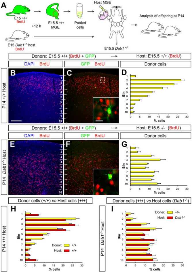
Figure 8 The laminar distribution of cortical interneurons correlates with the allocation of projection neurons. A , Schematic diagram of the experimental design. GFP + donor and Dab1 + / -host pregnant mice received a single injection of BrdU at E15. Twelve hours after BrdU injection, the MGE of GFP + embryos was collected, dissociated, and then injected into the MGE of E15.5 Dab1 + /? and Dab1 -/ -host embryos. Host embryos were allowed to be born and analyzed at P14. B , C , Coronal section through the somatosensory cortex of a transplanted P14 wild-type mouse showing the distribution of E15.5 wild-type (BrdU + /GFP + , see inset) MGE-derived interneurons and E15.5 host-derived neurons (BrdU + /GFP -) after nuclear staining [4 ′ ,6 ′ -diamidino2-phenylindole dihydrochloride (DAPI)] and immunohistochemistry for BrdU (red) and GFP (green). Numbers indicate bins for quantification. D , Quantification of the distribution of transplanted wild-type E15.5 interneurons in the P14 wild-type cortex (average + SEM). E , F , Coronal section through the somatosensory cortex of a transplanted P14 Dab1 -/ -mouse showing the distribution of E15.5 wild-type (BrdU + /GFP + , see inset) MGE-derived interneurons and E15.5 host-derived neurons (BrdU + / GFP -) after nuclear staining (DAPI) and immunohistochemistry for BrdU (red) and GFP (green). G , Quantification of the distribution of transplanted wild-type E15.5 interneurons in the P14 Dab1 -/ -cortex (average + SEM). H , I , Quantification of the relative distribution of transplanted wild-type E15.5 interneurons (yellow) in relation to host (red) wild-type ( H ) or Dab1 -/ -( I ) E15.5 neurons in the postnatal cortex (average + SEM). Scale bars: 200 𝛍 m( B , C , E , F ); 20 𝛍 m(insets in C , F ).
Figure 8 The laminar distribution of cortical interneurons correlates with the allocation of projection neurons. A , Schematic diagram of the experimental design. GFP + donor and Dab1 + / -host pregnant mice received a single injection of BrdU at E15. Twelve hours after BrdU injection, the MGE of GFP + embryos was collected, dissociated, and then injected into the MGE of E15.5 Dab1 + /? and Dab1 -/ -host embryos. Host embryos were allowed to be born and analyzed at P14. B , C , Coronal section through the somatosensory cortex of a transplanted P14 wild-type mouse showing the distribution of E15.5 wild-type (BrdU + /GFP + , see inset) MGE-derived interneurons and E15.5 host-derived neurons (BrdU + /GFP -) after nuclear staining [4 ′ ,6 ′ -diamidino2-phenylindole dihydrochloride (DAPI)] and immunohistochemistry for BrdU (red) and GFP (green). Numbers indicate bins for quantification. D , Quantification of the distribution of transplanted wild-type E15.5 interneurons in the P14 wild-type cortex (average + SEM). E , F , Coronal section through the somatosensory cortex of a transplanted P14 Dab1 -/ -mouse showing the distribution of E15.5 wild-type (BrdU + /GFP + , see inset) MGE-derived interneurons and E15.5 host-derived neurons (BrdU + / GFP -) after nuclear staining (DAPI) and immunohistochemistry for BrdU (red) and GFP (green). G , Quantification of the distribution of transplanted wild-type E15.5 interneurons in the P14 Dab1 -/ -cortex (average + SEM). H , I , Quantification of the relative distribution of transplanted wild-type E15.5 interneurons (yellow) in relation to host (red) wild-type ( H ) or Dab1 -/ -( I ) E15.5 neurons in the postnatal cortex (average + SEM). Scale bars: 200 𝛍 m( B , C , E , F ); 20 𝛍 m(insets in C , F ).CC-BY-NC-SA · From the PDF

Verification

Claims the paper makes

The claims and statistics of this paper haven’t been checked yet.

Run the check — free, no account needed. It reads the open-access text and checks the sources it cites.

A few free checks a day · papers already verified open instantly · results are public

Claims other papers make about this one

Each is checked against this paper’s own text: does it say what the citing paper says it does?

Self-check

Percentages, sums and reported differences re-computed from the paper’s own numbers.

The self-check needs the paper’s full text.

Citations

Cites

Show all 57 references

Research works printed but not linked: 2

  1. (1982), Brain Res, 256, 293
  2. (1985), Brain Res, 355, 187

Cited by

Sorted by citations

See all 151

Metadata

Authors
Pla, R., Borrell, V., Flames, N., & Marín, O.
Affiliations
Consejo Superior de Investigaciones Científicas (ES)
Venue
The Journal of Neuroscience
Published
2006-06-28
Volume
26
Issue
26
Pages
6924-6934
ISSN
  • 0270-6474 (ISSN-L)
  • 1529-2401
OpenAlex
W2042566086
Cited by
151
References
57
Citations by year
2026: 4 · 2025: 4 · 2024: 6 · 2023: 4 · 2022: 4 · 2021: 7 · 2020: 8 · 2019: 10 · 2018: 8 · 2017: 13 · 2015: 7 · 2014: 10 · 2013: 9 · 2012: 11
FWCI
2.17
Type
journal article
Language
en
Open access
Locations
  • Journal of Neuroscience (publishedVersion, cc-by-nc-sa)
  • PubMed Central (publishedVersion)
  • PubMed (publishedVersion)
  • DIGITAL.CSIC (Spanish National Research Council (CSIC)) (submittedVersion, other-oa)
Funders
  • National Alliance for Research on Schizophrenia and Depression
  • European Commission (005139)
  • Ministerio de Economía y Competitividad
Topics
  • Life Sciences › Neuroscience › Developmental Neuroscience › Neurogenesis and neuroplasticity mechanisms
  • Life Sciences › Neuroscience › Cellular and Molecular Neuroscience › Axon Guidance and Neuronal Signaling
  • Life Sciences › Biochemistry, Genetics and Molecular Biology › Molecular Biology › Developmental Biology and Gene Regulation
Keywords
GABAergic interneurons, Reelin signaling, radial migration, cortical lamination, projection neurons, interneuron migration, telencephalon, embryonic brain development
MeSH terms
Reelin Protein, Animals, Animals, Newborn, Cell Movement, Cerebral Cortex, Embryo, Mammalian, gamma-Aminobutyric Acid, Interneurons, Mice, Transgenic, Nerve Tissue Proteins, Synaptic Transmission, Neurons, Serine Endopeptidases, Time Factors, Signal Transduction, Cell Adhesion Molecules, Neuronal, Extracellular Matrix Proteins, Mice, Knockout, Mice

About these checks

Registry, retraction, Cites and statistics results are computed, not judged, with one exception: a printed reference that no index lists is read once by a model before it counts as a citation error. Claims are extracted by a model and kept only when their quote appears verbatim in the text; each cited source is then checked, with a model’s help, for whether it says what the claim relies on. Those verdicts can be wrong — read the claim’s quote and the cited source before relying on one.

Statistics and arithmetic are recomputed, not judged.

Confidence bands are shown only for fields calibrated on our annotated test set. None is calibrated yet, so no band is shown.

What counts as a claim ›How we measure confidence ›How we check this ›

Are you an author, or do you see a problem with this page? Request a review