

The DOI matches its Crossref record; 2 of the 2 references it cites were found in a registry or an index.
Tyrosine 394 Is Phosphorylated in Alzheimer's Paired Helical Filament Tau and in Fetal Tau with c-Abl as the Candidate Tyrosine Kinase
Abstract
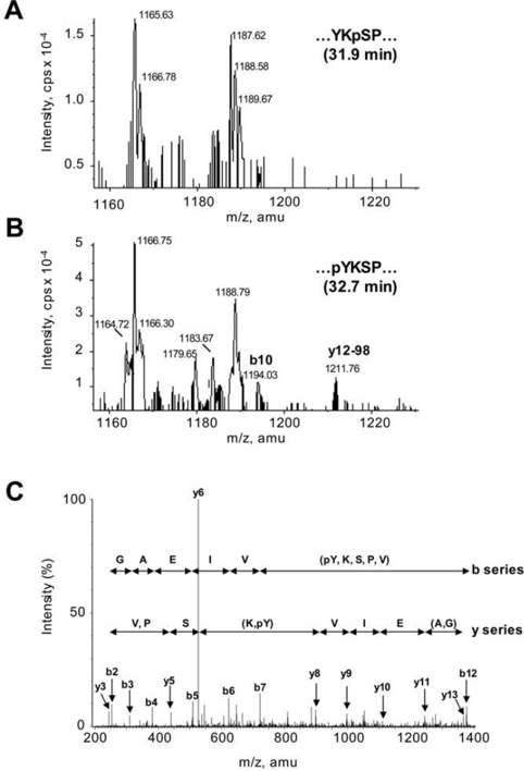
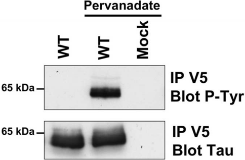
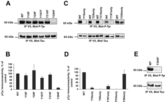
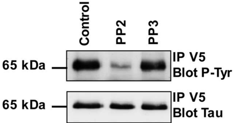
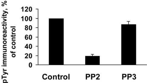
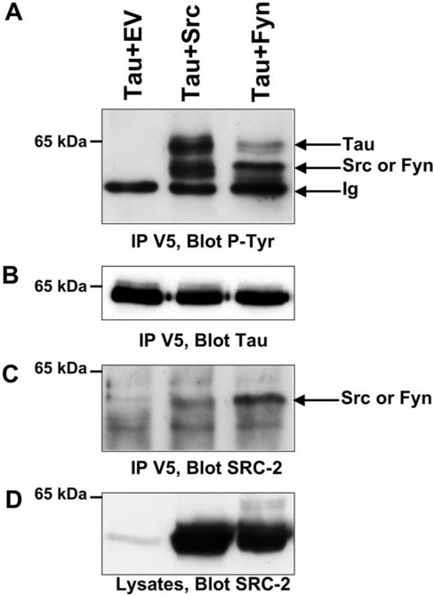
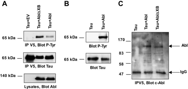
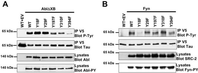
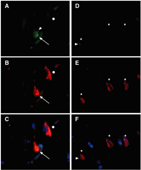
Tau is a major microtubule-associated protein of axons and is also the principal component of the paired helical filaments (PHFs) that comprise the neurofibrillary tangles found in Alzheimer's disease and other tauopathies. Besides phosphorylation of tau on serine and threonine residues in both normal tau and tau from neurofibrillary tangles, Tyr-18 was reported to be a site of phosphorylation by the Src-family kinase Fyn. We examined whether tyrosine residues other than Tyr-18 are phosphorylated in tau and whether other tyrosine kinases might phosphorylate tau. Using mass spectrometry, we positively identified phosphorylated Tyr-394 in PHF-tau from an Alzheimer brain and in human fetal brain tau. When wild-type human tau was transfected into fibroblasts or neuroblastoma cells, treatment with pervanadate caused tau to become phosphorylated on tyrosine by endogenous kinases. By replacing each of the five tyrosines in tau with phenylalanine, we identified Tyr-394 as the major site of tyrosine phosphorylation in tau. Tyrosine phosphorylation of tau was inhibited by PP2 (4-amino-5-(4-chlorophenyl-7-(t-butyl)pyrazolo[3,4-d]pyrimidine), which is known to inhibit Src-family kinases and c-Abl. Cotransfection of tau and kinases showed that Tyr-18 was the major site for Fyn phosphorylation, but Tyr-394 was the main residue for Abl.In vitro, Abl phosphorylated tau directly. Abl could be coprecipitated with tau and was present in pretangle neurons in brain sections from Alzheimer cases. These results show that phosphorylation of tau on Tyr-394 is a physiological event that is potentially part of a signal relay and suggest that Abl could have a pathogenic role in Alzheimer's disease.
Overview
The claims and statistics of this paper haven’t been checked yet.
Run the check — free, no account needed. It reads the open-access text and checks the sources it cites.
Integrity signals
Registry
Cites
Statistics
Figures
Verification
Claims the paper makes
The claims and statistics of this paper haven’t been checked yet.
Run the check — free, no account needed. It reads the open-access text and checks the sources it cites.
Claims other papers make about this one
Each is checked against this paper’s own text: does it say what the citing paper says it does?
In the citing paper
“Earlier work showed that fetal tau was highly phosphorylated during development, but the precise mechanisms and functions of this process are still unknown32,80–82.”
From Multi-omic phenotyping of MAPT V337M neurons reveals early changes in axonogenesis and tau phosphorylation (2026) · cites this paper as [32]
Self-check
Percentages, sums and reported differences re-computed from the paper’s own numbers.
The self-check needs the paper’s full text.
Citations
Cites
Association of missense and 5′-splice-site mutations in tau with the inherited dementia FTDP-17
Hutton, M., Lendon, C. L., Rizzu, P., Baker, M., Froelich, S., Houlden, H., Pickering-Brown, S., Chakraverty, S., Isaacs, A., Grover, A., et al. (1998) · Nature · doi:10.1038/31508 · Cited by 3,498
Maas, T., Eidenmüller, J., & Brandt, R. (2000) · Journal of Biological Chemistry · doi:10.1074/jbc.m000389200 · Cited by 226
Research works printed but not linked: 65
- 10.1016/j.nbd.2004.06.007 · DOI · Paper page
- 2001, Biochem Soc Symp, 67, 73, 10.1042/bss0670073 · DOI
- 10.1074/jbc.272.20.12922 · DOI
- 10.1016/0896-6273(93)90279-Z · DOI · Paper page
- 10.1083/jcb.131.5.1327 · DOI
- 10.1111/j.1471-4159.1993.tb03298.x · DOI
- 10.1016/S0165-0173(00)00019-9 · DOI
- 1992, Mol Cell Biol, 12, 1864
- Delacourte A, Buée L (2000) Tau pathology: a marker of neurodegenerative disorders. Curr Opin Neurol 371-376. · DOI
- 10.1006/jmbi.1997.1073 · DOI
- Derkinderen P, Girault JA (1997) Protein tyrosine phosphorylation. In: Post translational modifications: techniques and methods. (Boulton AA, Baker GB, Hemmings HC, eds), pp 251-274. Totowa, NJ: Humana. · DOI
- 10.1042/0264-6021:3570759 · DOI
- 10.1016/0197-0186(93)90139-V · DOI
- 10.1016/S0014-5793(97)00483-3 · DOI
- 10.1016/0076-6879(91)01043-2 · DOI · Paper page
- 10.1046/j.1471-4159.1998.71062465.x · DOI
- 10.1074/jbc.271.2.695 · DOI · Paper page
- 10.1038/nrm1280 · DOI
- 10.1016/0092-8674(95)90405-0 · DOI
- 10.1074/jbc.272.2.843 · DOI
- 10.1074/jbc.271.31.18342 · DOI
- 1989, EMBO J, 8, 449, 10.1002/j.1460-2075.1989.tb03397.x · DOI
- 10.1097/00001756-199801050-00014 · DOI
- 10.1523/JNEUROSCI.1264-04.2004 · DOI
- 2002, J Neurosci, 22, 698, 10.1523/JNEUROSCI.22-03-00698.2002 · DOI · Paper page
- 10.1016/S0896-6273(00)80646-7 · DOI
- 10.1038/227680a0 · DOI · Paper page
- 1998, J Cell Sci, 111, 3167, 10.1242/jcs.111.21.3167 · DOI
- 10.1523/JNEUROSCI.4162-03.2004 · DOI
- 10.1007/s004010100435 · DOI
- 10.1074/jbc.273.34.21901 · DOI
- 10.1016/S1074-5521(99)80118-5 · DOI
- 10.1016/S0306-4522(96)00577-5 · DOI
- 10.1093/emboj/20.1.27 · DOI
- 10.1046/j.1432-1327.2001.02024.x · DOI
- 10.1016/j.conb.2003.08.002 · DOI
- 10.1074/jbc.270.2.823 · DOI · Paper page
- 10.1016/0014-5793(94)00702-0 · DOI
- 1996, J Neurochem, 67, 1622, 10.1046/j.1471-4159.1996.67041622.x · DOI
- 10.1016/S0896-6273(03)00259-9 · DOI
- 10.1093/nar/gkg584 · DOI
- 10.1016/S0065-230X(02)85003-5 · DOI
- 10.1101/gad.13.18.2400 · DOI
- 10.1002/ana.410430617 · DOI · Paper page
- 10.1073/pnas.092136199 · DOI · Paper page
- 10.1046/j.1471-4159.1997.68041736.x · DOI
- 10.1046/j.1471-4159.2000.0741587.x · DOI
- 10.1002/bms.1200111109 · DOI
- 10.1385/MB:20:1:107 · DOI
- 10.1111/j.1365-2990.1994.tb00977.x · DOI
- 10.1016/S0968-0004(00)89103-3 · DOI
- 10.1074/jbc.274.36.25490 · DOI
- 10.1016/S0166-2236(98)01337-X · DOI · Paper page
- 10.1074/jbc.M006219200 · DOI
- 10.1074/jbc.M209321200 · DOI
- 10.1021/jm970124v · DOI
- 1992, Cancer Treat Res, 63, 167
- 10.1016/S0962-8924(99)01549-4 · DOI
- 2004, Leuk Res, 28, S21, 10.1016/j.leukres.2003.10.005 · DOI
- 10.1083/jcb.124.3.325 · DOI
- 10.1182/blood-2002-01-0288 · DOI
- 2002, J Neurosci, 22, 10, 10.1523/JNEUROSCI.22-01-00010.2002 · DOI · Paper page
- 10.1083/jcb.200110014 · DOI
- 2002, Comb Chem High Throughput Screen, 5, 83, 10.2174/1386207023330516 · DOI
- 10.1016/S0896-6273(00)81200-3 · DOI
Cited by
Roles of tau protein in health and disease
Guo, T., Noble, W., & Hanger, D. P. (2017) · Acta Neuropathologica · doi:10.1007/s00401-017-1707-9 · Cited by 1,003
Self-citationTau protein kinases: Involvement in Alzheimer's disease
Martin, L., Latypova, X., Wilson, C. M., Magnaudeix, A., Perrin, M.-L., Yardin, C., & Terro, F. (2013) · Ageing Research Reviews · doi:10.1016/j.arr.2012.06.003 · Cited by 631
Arendt, T., Stieler, J. T., & Holzer, M. (2016) · Brain Research Bulletin · doi:10.1016/j.brainresbull.2016.08.018 · Cited by 609
The role of tau in neurodegeneration
Gendron, T. F., & Petrucelli, L. (2009) · Molecular Neurodegeneration · doi:10.1186/1750-1326-4-13 · Cited by 478
The importance of tau phosphorylation for neurodegenerative diseases
Noble, W., Hanger, D. P., Miller, C. C. J., & Lovestone, S. (2013) · Frontiers in Neurology · doi:10.3389/fneur.2013.00083 · Cited by 475
Self-citationAlquezar, C., Arya, S., & Kao, A. W. (2021) · Frontiers in Neurology · doi:10.3389/fneur.2020.595532 · Cited by 367
Biochemistry of Tau in Alzheimer’s disease and related neurological disorders
Sergeant, N., Bretteville, A., Hamdane, M., Caillet-Boudin, M.-L., Grognet, P., Bombois, S., Blum, D., Delacourte, A., Pasquier, F., Vanmechelen, E., et al. (2008) · Expert Review of Proteomics · doi:10.1586/14789450.5.2.207 · Cited by 300
Structural Principles of Tau and the Paired Helical Filaments of Alzheimer's Disease
Mandelkow, E., Von Bergen, M., Biernat, J., & Mandelkow, E. (2007) · Brain Pathology · doi:10.1111/j.1750-3639.2007.00053.x · Cited by 243
A role for tau at the synapse in Alzheimer’s disease pathogenesis
Pooler, A. M., Noble, W., & Hanger, D. P. (2014) · Neuropharmacology · doi:10.1016/j.neuropharm.2013.09.018 · Cited by 203
Self-citationBasheer, N., Smolek, T., Hassan, I., Liu, F., Iqbal, K., Zilka, N., & Novak, P. (2023) · Molecular Psychiatry · doi:10.1038/s41380-023-02113-z · Cited by 159
Lonskaya, I., Hebron, M. L., Desforges, N. M., Schachter, J. B., & Moussa, C. E.-H. (2014) · Journal of Molecular Medicine · doi:10.1007/s00109-013-1112-3 · Cited by 152
Early axonopathy preceding neurofibrillary tangles in mutant tau transgenic mice
Leroy, K., Bretteville, A., Schindowski, K., Gilissen, E., Authelet, M., De Decker, R., Yilmaz, Z., Buée, L., & Brion, J.-P. (2007) · The American Journal of Pathology · doi:10.2353/ajpath.2007.070345 · Cited by 135
Self-citationThomas, S. N., Funk, K. E., Wan, Y., Liao, Z., Davies, P., Kuret, J., & Yang, A. J. (2012) · Acta Neuropathologica · doi:10.1007/s00401-011-0893-0 · Cited by 123
c-Abl tyrosine kinase modulates tau pathology and Cdk5 phosphorylation in AD transgenic mice
Cancino, G. I., Perez de Arce, K., Castro, P. U., Toledo, E. M., von Bernhardi, R., & Alvarez, A. R. (2011) · Neurobiology of Aging · doi:10.1016/j.neurobiolaging.2009.07.007 · Cited by 110
Targeting tau: clinical trials and novel therapeutic approaches
VandeVrede, L., Boxer, A. L., & Polydoro, M. (2020) · Neuroscience Letters · doi:10.1016/j.neulet.2020.134919 · Cited by 90
Altered subcellular distribution of c-Abl in Alzheimer’s disease
Jing, Z., Caltagarone, J., & Bowser, R. (2009) · Journal of Alzheimer s Disease · doi:10.3233/jad-2009-1062 · Cited by 83
Oxidative stress, DNA damage, and c-Abl signaling: At the crossroad in neurodegenerative diseases?
Gonfloni, S., Maiani, E., Di Bartolomeo, C., Diederich, M., & Cesareni, G. (2012) · International Journal of Cell Biology · doi:10.1155/2012/683097 · Cited by 63
Targeting dementias through cancer kinases inhibition
Fagiani, F., Lanni, C., Racchi, M., & Govoni, S. (2020) · Alzheimer's & Dementia: Translational Research & Clinical Interventions · doi:10.1002/trc2.12044 · Cited by 43
Mohl, G. A., Dixon, G., Marzette, E., McKetney, J., Samelson, A. J., Serras, C. P., Jin, J., Ariqat, N., Keys, A., Chavira, C., et al. (2026) · npj Dementia · doi:10.1038/s44400-026-00076-w · Cited by 5
Metadata
- Authors
- Derkinderen, P., Scales, T. M. E., Hanger, D. P., Leung, K.-Y., Byers, H. L., Ward, M. A., Lenz, C., Price, C., Bird, I. N., Perera, T., Kellie, S., Williamson, R., Noble, W., Van Etten, R. A., Leroy, K., Brion, J.-P., Reynolds, C. H., & Anderton, B. H.
- Venue
- The Journal of Neuroscience
- Publisher
- Society for Neuroscience
- Published
- 2005-07-13
- Volume
- 25
- Issue
- 28
- Pages
- 6584-6593
- ISSN
- 0270-6474
- 1529-2401
- Cited by
- 169
- References
- 2
- Language
- en
About these checks
Registry, retraction, Cites and statistics results are computed, not judged, with one exception: a printed reference that no index lists is read once by a model before it counts as a citation error. Claims are extracted by a model and kept only when their quote appears verbatim in the text; each cited source is then checked, with a model’s help, for whether it says what the claim relies on. Those verdicts can be wrong — read the claim’s quote and the cited source before relying on one.
Statistics and arithmetic are recomputed, not judged.
Confidence bands are shown only for fields calibrated on our annotated test set. None is calibrated yet, so no band is shown.
What counts as a claim ›How we measure confidence ›How we check this ›
Are you an author, or do you see a problem with this page? Request a review
Data sources
Bibliographic data: Semantic Scholar, OpenAlex, Crossref.
Contains information from the Semantic Scholar Academic Graph Datasets, made available under the ODC Attribution License (ODC-BY 1.0).
Semantic Scholar’s data is described in: Kinney, R., et al. (2023). The Semantic Scholar Open Data Platform. arXiv:2301.10140.
