

The DOI matches its Crossref record; 32 of the 33 references it cites were found in a registry or an index.
β-Amyloid Monomers Are Neuroprotective
Open accessAbstract
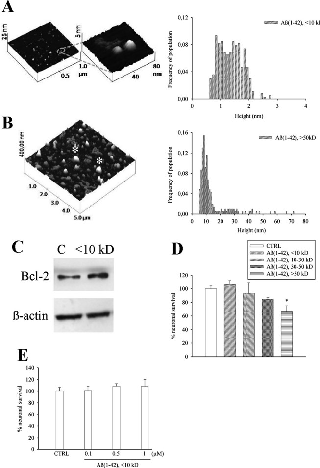
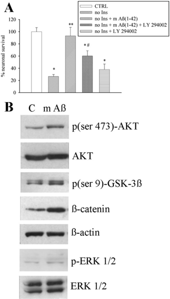
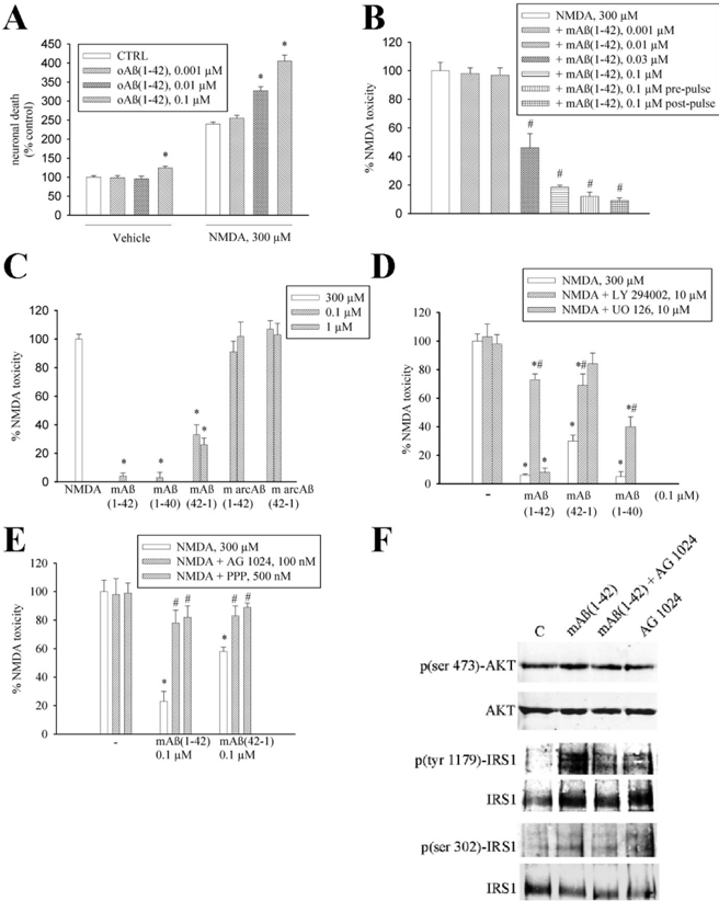
The 42-aa-long beta-amyloid protein--Abeta(1-42)--is thought to play a central role in the pathogenesis of Alzheimer's disease (AD) (Walsh and Selkoe, 2007). Data from AD brain (Shankar et al., 2008), transgenic APP (amyloid precursor protein)-overexpressing mice (Lesné et al., 2006), and neuronal cultures treated with synthetic Abeta peptides (Lambert et al., 1998) indicate that self-association of Abeta(1-42) monomers into soluble oligomers is required for neurotoxicity. The function of monomeric Abeta(1-42) is unknown. The evidence that Abeta(1-42) is present in the brain and CSF of normal individuals suggests that the peptide is physiologically active (Shoji, 2002). Here we show that synthetic Abeta(1-42) monomers support the survival of developing neurons under conditions of trophic deprivation and protect mature neurons against excitotoxic death, a process that contributes to the overall neurodegeneration associated with AD. The neuroprotective action of Abeta(1-42) monomers was mediated by the activation of the PI-3-K (phosphatidylinositol-3-kinase) pathway, and involved the stimulation of IGF-1 (insulin-like growth factor-1) receptors and/or other receptors of the insulin superfamily. Interestingly, monomers of Abeta(1-42) carrying the Arctic mutation (E22G) associated with familiar AD (Nilsberth et al., 2001) were not neuroprotective. We suggest that pathological aggregation of Abeta(1-42) may also cause neurodegeneration by depriving neurons of the protective activity of Abeta(1-42) monomers. This "loss-of-function" hypothesis of neuronal death should be taken into consideration when designing therapies aimed at reducing Abeta burden.
Overview
The claims and statistics of this paper haven’t been checked yet.
Run the check — free, no account needed. It reads the open-access text and checks the sources it cites.
Integrity signals
Registry
Cites
Statistics
Figures
Verification
Claims the paper makes
The claims and statistics of this paper haven’t been checked yet.
Run the check — free, no account needed. It reads the open-access text and checks the sources it cites.
Claims other papers make about this one
Each is checked against this paper’s own text: does it say what the citing paper says it does?
In the citing paper
“Relative levels of Aβ42 to Aβ40 appear to influence the mechanism of oligomerization, with higher levels of Aβ40 in normal individuals likely impeding formation of stable toxic oligomers (Giuffrida et al., 2009).”
From The Aβ oligomer hypothesis for synapse failure and memory loss in Alzheimer’s disease (2011) · cites this paper as Giuffrida et al., 2009
Self-check
Percentages, sums and reported differences re-computed from the paper’s own numbers.
The self-check needs the paper’s full text.
Citations
Cites
1 retracted
RETRACTED ARTICLE: A specific amyloid-β protein assembly in the brain impairs memory
Lesné, S., Koh, M. T., Kotilinek, L., Kayed, R., Glabe, C. G., Yang, A., Gallagher, M., & Ashe, K. H. (2006) · Nature · doi:10.1038/nature04533 · Cited by 2,769 · cited 2×
“In Tg2576 mice expressing a human APP (amyloid precursor protein) variant linked to AD, a 56 kDa soluble A assembly (A *56) disrupts memory (Lesné et al., 2006).”
BackgroundRetractedThe Bcl-2 Protein Family: Arbiters of Cell Survival
Adams, J. M., & Cory, S. (1998) · Science · doi:10.1126/science.281.5381.1322 · Cited by 5,334
Walsh, D. M., Klyubin, I., Fadeeva, J. V., Cullen, W. K., Anwyl, R., Wolfe, M. S., Rowan, M. J., & Selkoe, D. J. (2002) · Nature · doi:10.1038/416535a · Cited by 4,291
Shankar, G. M., Li, S., Mehta, T. H., Garcia-Munoz, A., Shepardson, N. E., Smith, I., Brett, F. M., Farrell, M. A., Rowan, M. J., Lemere, C. A., et al. (2008) · Nature Medicine · doi:10.1038/nm1782 · Cited by 3,685
Diffusible, nonfibrillar ligands derived from Aβ 1–42 are potent central nervous system neurotoxins
Lambert, M. P., Barlow, A. K., Chromy, B. A., Edwards, C., Freed, R., Liosatos, M., Morgan, T. E., Rozovsky, I., Trommer, B., Viola, K. L., et al. (1998) · Proceedings of the National Academy of Sciences · doi:10.1073/pnas.95.11.6448 · Cited by 3,506
Vlahos, C. J., Matter, W. F., Hui, K. Y., & Brown, R. F. (1994) · Journal of Biological Chemistry · doi:10.1016/s0021-9258(17)37680-9 · Cited by 3,248
Identification of a Novel Inhibitor of Mitogen-activated Protein Kinase Kinase
Favata, M. F., Horiuchi, K. Y., Manos, E. J., Daulerio, A. J., Stradley, D. A., Feeser, W. S., Dyk, D. E. V., Pitts, W. J., Earl, R. A., Hobbs, F. W., et al. (1998) · Journal of Biological Chemistry · doi:10.1074/jbc.273.29.18623 · Cited by 3,016
Aβ Oligomers – a decade of discovery
Walsh, D. M., & Selkoe, D. J. (2007) · Journal of Neurochemistry · doi:10.1111/j.1471-4159.2006.04426.x · Cited by 1,938
PI3K: Downstream AKTion Blocks Apoptosis
Franke, T. F., Kaplan, D. R., & Cantley, L. C. (1997) · Cell · doi:10.1016/s0092-8674(00)81883-8 · Cited by 1,665
APP Processing and Synaptic Function
Kamenetz, F., Tomita, T., Hsieh, H., Seabrook, G., Borchelt, D., Iwatsubo, T., Sisodia, S., & Malinow, R. (2003) · Neuron · doi:10.1016/s0896-6273(03)00124-7 · Cited by 1,644
Cited by
Sorted by citations
Items 401–423 of 423
The Role of c-Abl in Alzheimer’s Disease: Guilty or not Guilty?
Li, W., Wang, X., Liu, J., & Chen, Y. (2025) · Cellular and Molecular Neurobiology · doi:10.1007/s10571-025-01650-1
Rofo, F., Buijs, J., Falk, R., Honek, K., Lannfelt, L., Lilja, A. M., Metzendorf, N. G., Gustavsson, T., Sehlin, D., Söderberg, L., et al. (2021) · Figshare · doi:10.6084/m9.figshare.16689644
Barger, S. W., & Moerman, A. (2026) · Frontiers in Molecular Neuroscience · doi:10.3389/fnmol.2026.1781541
Man, V. H., He, X., Niu, T., Cai, L., Han, F., Nguyen, P. H., & Wang, J. (2026) · ACS Chemical Neuroscience · doi:10.1021/acschemneuro.6c00003
Rôle du peptide amyloïde dans les maladies neurodégénératives
Combes, M. (2018) · theses.fr (ABES) · doi:10.70675/a219ef4dzbca0z4272z8bf2z74080a1976e5 · Cited by 1
Spontaneous Insertion of Aβ42 Dimers but Not Monomers into a Cholesterol-Rich Lipid Bilayer
Pandey, R., Ruggiero, T., Andrews, B., & Urbanc, B. (2026) · ACS Chemical Neuroscience · doi:10.1021/acschemneuro.5c00942
Basile, S., Schipper, F., Zimbone, S., Vermorgen, S., Bellia, F., Parisi, C., Sortino, S., Walter, J., Giuffrida, M. L., Rozemüller, A. J. M., et al. (2026) · Research Square · doi:10.21203/rs.3.rs-10769225/v1
Self-citationLyagin, I. (2026) · Moscow University Chemistry Bulletin · doi:10.3103/s0027131426700276
Modulating Calcium Signaling by Protein Design and Analysis of Calcium Binding Proteins
Zhuo, Y. (2022) · Digital Archive @ GSU · doi:10.57709/4865017
Metadata
- Authors
- Giuffrida, M. L., Caraci, F., Pignataro, B., Cataldo, S., De Bona, P., Bruno, V., Molinaro, G., Pappalardo, G., Messina, A., Palmigiano, A., Garozzo, D., Nicoletti, F., Rizzarelli, E., & Copani, A.
- Affiliations
- University of Catania (IT)
- University of Palermo (IT)
- Istituto Neurologico Mediterraneo (IT)
- Institute of Biostructure and Bioimaging (IT)
- National Research Council (IT)
- ORCID
- Venue
- The Journal of Neuroscience
- Publisher
- Society for Neuroscience
- Published
- 2009-08-26
- Volume
- 29
- Issue
- 34
- Pages
- 10582-10587
- ISSN
- 0270-6474 (ISSN-L)
- 1529-2401
- PMC
- PMC6665714
- OpenAlex
- W1991988242
- Cited by
- 421
- References
- 33
- Citations by year
- 2026: 10 · 2025: 19 · 2024: 30 · 2023: 27 · 2022: 21 · 2021: 34 · 2020: 23 · 2019: 21 · 2018: 27 · 2017: 30 · 2016: 22 · 2015: 23 · 2014: 31 · 2013: 23 · 2012: 18
- FWCI
- 17.73
- Type
- journal article
- Language
- en
- Open access
- hybrid
- doi.org
- Licence
- Locations
- Journal of Neuroscience (publishedVersion, cc-by-nc-sa)
- Europe PMC (PubMed Central) (publishedVersion)
- PubMed Central (publishedVersion)
- PubMed (publishedVersion)
- Nova Science Publishers (Nova Science Publishers, Inc.) (submittedVersion, other-oa)
- IRIS Research product catalog (Sapienza University of Rome) (submittedVersion)
- Funders
- Università di Catania
- Topics
- Life Sciences › Neuroscience › Cellular and Molecular Neuroscience › Alzheimer's disease research and treatments
- Life Sciences › Neuroscience › Neurology › Neuroinflammation and Neurodegeneration Mechanisms
- Health Sciences › Medicine › Psychiatry and Mental health › Dementia and Cognitive Impairment Research
- Keywords
- amyloid-beta 42, neuroprotection, Alzheimer's disease, excitotoxicity, IGF-1 receptor, Arctic mutation, amyloid fibril formation, neurodegeneration
- MeSH terms
- Analysis of Variance, Animals, Butadienes, Cells, Cultured, Cerebral Cortex, Dose-Response Relationship, Drug, Embryo, Mammalian, Enzyme Inhibitors, Neurons, Nitriles, Peptide Fragments, Podophyllotoxin, N-Methylaspartate, Amyloid beta-Peptides, Cell Death, Excitatory Amino Acid Agonists, Neuroprotective Agents, Tyrphostins, Rats
About these checks
Registry, retraction, Cites and statistics results are computed, not judged, with one exception: a printed reference that no index lists is read once by a model before it counts as a citation error. Claims are extracted by a model and kept only when their quote appears verbatim in the text; each cited source is then checked, with a model’s help, for whether it says what the claim relies on. Those verdicts can be wrong — read the claim’s quote and the cited source before relying on one.
Statistics and arithmetic are recomputed, not judged.
Confidence bands are shown only for fields calibrated on our annotated test set. None is calibrated yet, so no band is shown.
What counts as a claim ›How we measure confidence ›How we check this ›
Are you an author, or do you see a problem with this page? Request a review
