

The DOI matches its Crossref record; 32 of the 33 references it cites were found in a registry or an index.
β-Amyloid Monomers Are Neuroprotective
Open accessAbstract
The 42-aa-long beta-amyloid protein--Abeta(1-42)--is thought to play a central role in the pathogenesis of Alzheimer's disease (AD) (Walsh and Selkoe, 2007). Data from AD brain (Shankar et al., 2008), transgenic APP (amyloid precursor protein)-overexpressing mice (Lesné et al., 2006), and neuronal cultures treated with synthetic Abeta peptides (Lambert et al., 1998) indicate that self-association of Abeta(1-42) monomers into soluble oligomers is required for neurotoxicity. The function of monomeric Abeta(1-42) is unknown. The evidence that Abeta(1-42) is present in the brain and CSF of normal individuals suggests that the peptide is physiologically active (Shoji, 2002). Here we show that synthetic Abeta(1-42) monomers support the survival of developing neurons under conditions of trophic deprivation and protect mature neurons against excitotoxic death, a process that contributes to the overall neurodegeneration associated with AD. The neuroprotective action of Abeta(1-42) monomers was mediated by the activation of the PI-3-K (phosphatidylinositol-3-kinase) pathway, and involved the stimulation of IGF-1 (insulin-like growth factor-1) receptors and/or other receptors of the insulin superfamily. Interestingly, monomers of Abeta(1-42) carrying the Arctic mutation (E22G) associated with familiar AD (Nilsberth et al., 2001) were not neuroprotective. We suggest that pathological aggregation of Abeta(1-42) may also cause neurodegeneration by depriving neurons of the protective activity of Abeta(1-42) monomers. This "loss-of-function" hypothesis of neuronal death should be taken into consideration when designing therapies aimed at reducing Abeta burden.
Overview
The claims and statistics of this paper haven’t been checked yet.
Run the check — free, no account needed. It reads the open-access text and checks the sources it cites.
Integrity signals
Registry
Cites
Statistics
Figures
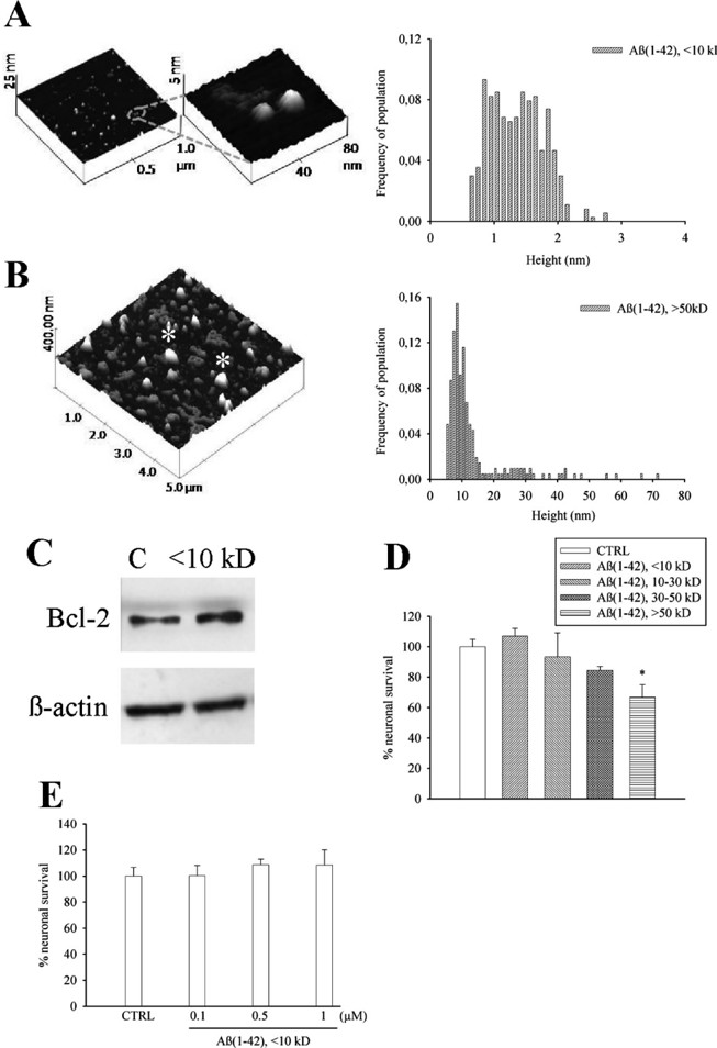
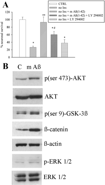
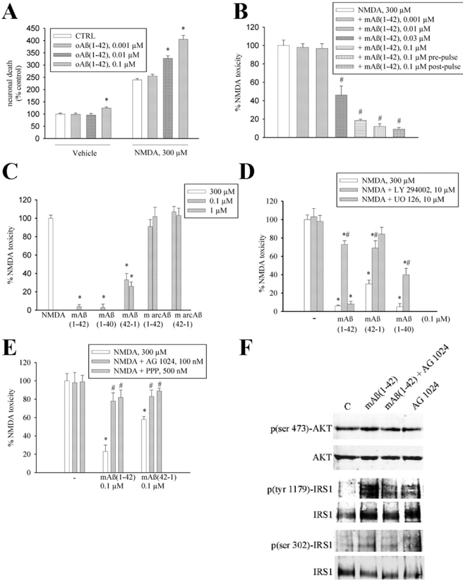
Verification
Claims the paper makes
The claims and statistics of this paper haven’t been checked yet.
Run the check — free, no account needed. It reads the open-access text and checks the sources it cites.
Claims other papers make about this one
Each is checked against this paper’s own text: does it say what the citing paper says it does?
In the citing paper
“Relative levels of Aβ42 to Aβ40 appear to influence the mechanism of oligomerization, with higher levels of Aβ40 in normal individuals likely impeding formation of stable toxic oligomers (Giuffrida et al., 2009).”
From The Aβ oligomer hypothesis for synapse failure and memory loss in Alzheimer’s disease (2011) · cites this paper as Giuffrida et al., 2009
Self-check
Percentages, sums and reported differences re-computed from the paper’s own numbers.
The self-check needs the paper’s full text.
Citations
Cites
1 retracted
RETRACTED ARTICLE: A specific amyloid-β protein assembly in the brain impairs memory
Lesné, S., Koh, M. T., Kotilinek, L., Kayed, R., Glabe, C. G., Yang, A., Gallagher, M., & Ashe, K. H. (2006) · Nature · doi:10.1038/nature04533 · Cited by 2,769 · cited 2×
“In Tg2576 mice expressing a human APP (amyloid precursor protein) variant linked to AD, a 56 kDa soluble A assembly (A *56) disrupts memory (Lesné et al., 2006).”
BackgroundRetractedThe Bcl-2 Protein Family: Arbiters of Cell Survival
Adams, J. M., & Cory, S. (1998) · Science · doi:10.1126/science.281.5381.1322 · Cited by 5,334
Walsh, D. M., Klyubin, I., Fadeeva, J. V., Cullen, W. K., Anwyl, R., Wolfe, M. S., Rowan, M. J., & Selkoe, D. J. (2002) · Nature · doi:10.1038/416535a · Cited by 4,291
Shankar, G. M., Li, S., Mehta, T. H., Garcia-Munoz, A., Shepardson, N. E., Smith, I., Brett, F. M., Farrell, M. A., Rowan, M. J., Lemere, C. A., et al. (2008) · Nature Medicine · doi:10.1038/nm1782 · Cited by 3,685
Diffusible, nonfibrillar ligands derived from Aβ 1–42 are potent central nervous system neurotoxins
Lambert, M. P., Barlow, A. K., Chromy, B. A., Edwards, C., Freed, R., Liosatos, M., Morgan, T. E., Rozovsky, I., Trommer, B., Viola, K. L., et al. (1998) · Proceedings of the National Academy of Sciences · doi:10.1073/pnas.95.11.6448 · Cited by 3,506
Vlahos, C. J., Matter, W. F., Hui, K. Y., & Brown, R. F. (1994) · Journal of Biological Chemistry · doi:10.1016/s0021-9258(17)37680-9 · Cited by 3,248
Identification of a Novel Inhibitor of Mitogen-activated Protein Kinase Kinase
Favata, M. F., Horiuchi, K. Y., Manos, E. J., Daulerio, A. J., Stradley, D. A., Feeser, W. S., Dyk, D. E. V., Pitts, W. J., Earl, R. A., Hobbs, F. W., et al. (1998) · Journal of Biological Chemistry · doi:10.1074/jbc.273.29.18623 · Cited by 3,016
Aβ Oligomers – a decade of discovery
Walsh, D. M., & Selkoe, D. J. (2007) · Journal of Neurochemistry · doi:10.1111/j.1471-4159.2006.04426.x · Cited by 1,938
PI3K: Downstream AKTion Blocks Apoptosis
Franke, T. F., Kaplan, D. R., & Cantley, L. C. (1997) · Cell · doi:10.1016/s0092-8674(00)81883-8 · Cited by 1,665
APP Processing and Synaptic Function
Kamenetz, F., Tomita, T., Hsieh, H., Seabrook, G., Borchelt, D., Iwatsubo, T., Sisodia, S., & Malinow, R. (2003) · Neuron · doi:10.1016/s0896-6273(03)00124-7 · Cited by 1,644
Cited by
Sorted by citations
Alzheimer Mechanisms and Therapeutic Strategies
Huang, Y., & Mucke, L. (2012) · Cell · doi:10.1016/j.cell.2012.02.040 · Cited by 1,758
Miranda, M., Morici, J. F., Zanoni, M. B., & Bekinschtein, P. (2019) · Frontiers in Cellular Neuroscience · doi:10.3389/fncel.2019.00363 · Cited by 1,416
Danysz, W., & Parsons, C. G. (2012) · British Journal of Pharmacology · doi:10.1111/j.1476-5381.2012.02057.x · Cited by 500
Copper in the brain and Alzheimer’s disease
Hung, Y. H., Bush, A. I., & Cherny, R. A. (2009) · JBIC Journal of Biological Inorganic Chemistry · doi:10.1007/s00775-009-0600-y · Cited by 503
Amyloid toxicity in Alzheimer’s disease
Reiss, A. B., Arain, H. A., Stecker, M. M., Siegart, N. M., & Kasselman, L. J. (2018) · Reviews in the Neurosciences · doi:10.1515/revneuro-2017-0063 · Cited by 447
The Aβ oligomer hypothesis for synapse failure and memory loss in Alzheimer’s disease
Ferreira, S. T., & Klein, W. L. (2011) · Neurobiology of Learning and Memory · doi:10.1016/j.nlm.2011.08.003 · Cited by 442
Calcium Signaling and Amyloid Toxicity in Alzheimer Disease
Demuro, A., Parker, I., & Stutzmann, G. E. (2010) · Journal of Biological Chemistry · doi:10.1074/jbc.r109.080895 · Cited by 426
Structural and kinetic basis for the selectivity of aducanumab for aggregated forms of amyloid-β
Arndt, J. W., Qian, F., Smith, B. A., Quan, C., Kilambi, K. P., Bush, M. W., Walz, T., Pepinsky, R. B., Bussière, T., Hamann, S., et al. (2018) · Scientific Reports · doi:10.1038/s41598-018-24501-0 · Cited by 369
A Pathologic Cascade Leading to Synaptic Dysfunction in -Synuclein-Induced Neurodegeneration
Scott, D. A., Tabarean, I. V., Tang, Y., Cartier, A. E., Masliah, E., & Roy, S. (2010) · Journal of Neuroscience · doi:10.1523/jneurosci.1091-10.2010 · Cited by 365
Salomone, S., Caraci, F., Leggio, G. M., Fedotova, J., & Drago, F. (2011) · British Journal of Clinical Pharmacology · doi:10.1111/j.1365-2125.2011.04134.x · Cited by 335
Self-citationImmunotherapeutic Approaches for Alzheimer’s Disease
Wisniewski, T., & Goñi, F. (2015) · Neuron · doi:10.1016/j.neuron.2014.12.064 · Cited by 284
Diógenes, M. J., Dias, R. B., Rombo, D. M., Miranda, H. V., Maiolino, F., Guerreiro, P. S., Näsström, T., Franquelim, H. G., Oliveira, L. M. A., Castanho, M. A. R. B., et al. (2012) · Journal of Neuroscience · doi:10.1523/jneurosci.0234-12.2012 · Cited by 274
Gao, N., Sun, H., Dong, K., Ren, J., Duan, T., Xu, C., & Qu, X. (2014) · Nature Communications · doi:10.1038/ncomms4422 · Cited by 263
Amyloid-β as a Modulator of Synaptic Plasticity
Parihar, M. S., & Brewer, G. J. (2010) · Journal of Alzheimer s Disease · doi:10.3233/jad-2010-101020 · Cited by 264
A 3D human brain–like tissue model of herpes-induced Alzheimer’s disease
Cairns, D. M., Rouleau, N., Parker, R. N., Walsh, K. G., Gehrke, L., & Kaplan, D. L. (2020) · Science Advances · doi:10.1126/sciadv.aay8828 · Cited by 247
Rajasekhar, K., Chakrabarti, M., & Govindaraju, T. (2015) · Chemical Communications · doi:10.1039/c5cc05264e · Cited by 243
Functions of Aβ, sAPPα and sAPPβ : similarities and differences
Chasseigneaux, S., & Allinquant, B. (2011) · Journal of Neurochemistry · doi:10.1111/j.1471-4159.2011.07584.x · Cited by 278
Nie, Q., Du, X., & Geng, M. (2011) · Acta Pharmacologica Sinica · doi:10.1038/aps.2011.14 · Cited by 241
Use of curcumin in diagnosis, prevention, and treatment of Alzheimer's disease
Zhang, K., Chen, M., Du, Z.-Y., Zheng, X., Li, D., & Zhou, R. (2018) · Neural Regeneration Research · doi:10.4103/1673-5374.230303 · Cited by 243
McNay, E. C., & Recknagel, A. K. (2011) · Neurobiology of Learning and Memory · doi:10.1016/j.nlm.2011.08.005 · Cited by 233
Goyal, D., Shuaib, S., Mann, S., & Goyal, B. (2017) · ACS Combinatorial Science · doi:10.1021/acscombsci.6b00116 · Cited by 232
Rushworth, J. V., & Hooper, N. M. (2010) · International Journal of Alzheimer s Disease · doi:10.4061/2011/603052 · Cited by 234
The duality of amyloid-β: its role in normal and Alzheimer’s disease states
Azargoonjahromi, A. (2024) · Molecular Brain · doi:10.1186/s13041-024-01118-1 · Cited by 214
Goure, W. F., Krafft, G. A., Jerecic, J., & Hefti, F. (2014) · Alzheimer's Research & Therapy · doi:10.1186/alzrt272 · Cited by 190
Nature of the Amyloid-β Monomer and the Monomer-Oligomer Equilibrium
Nag, S., Sarkar, B., Bandyopadhyay, A., Sahoo, B., Sreenivasan, V. K. A., Kombrabail, M. H., Chandrakesan, M., & Maiti, S. (2011) · Journal of Biological Chemistry · doi:10.1074/jbc.m110.199885 · Cited by 191
Jiménez, S., Torres, M., Vizuete, M., Sanchez-Varo, R. M., Sánchez‐Mejías, E., Trujillo‐Estrada, L., Carmona‐Cuenca, I., Caballero, C., Ruano, D., Gutiérrez, A., et al. (2011) · Journal of Biological Chemistry · doi:10.1074/jbc.m110.209718 · Cited by 190
Exosomes neutralize synaptic-plasticity-disrupting activity of Aβ assemblies in vivo
An, K., Klyubin, I. V., Kim, Y., Jung, J. H., Mably, A. J., O’Dowd, S., Lynch, T., Kanmert, D., Lemere, C. A., Finan, G. M., et al. (2013) · Molecular Brain · doi:10.1186/1756-6606-6-47 · Cited by 196
Amyloid Beta: Multiple Mechanisms of Toxicity and Only Some Protective Effects?
Carrillo‐Mora, P., Luna, R., & Colín-Barenque, L. (2014) · Oxidative Medicine and Cellular Longevity · doi:10.1155/2014/795375 · Cited by 187
Amyloid beta deregulates astroglial mGluR5‐mediated calcium signaling via calcineurin and Nf‐kB
Lim, D., Iyer, A., Ronco, V., Grolla, A. A., Canonico, P. L., Aronica, E., & Genazzani, A. A. (2013) · Glia · doi:10.1002/glia.22502 · Cited by 169
Toward a comprehensive neurobiology of IGF‐I
Torres‐Alemán, I. (2010) · Developmental Neurobiology · doi:10.1002/dneu.20778 · Cited by 177
Chen, X., & Mobley, W. C. (2019) · Frontiers in Neuroscience · doi:10.3389/fnins.2019.00446 · Cited by 179
Grassi, S., Giussani, P., Mauri, L., Prioni, S., Sonnino, S., & Prinetti, A. (2019) · Journal of Lipid Research · doi:10.1194/jlr.tr119000427 · Cited by 174
Chami, L., & Checler, F. (2012) · Molecular Neurodegeneration · doi:10.1186/1750-1326-7-52 · Cited by 168
Nygaard, H. B., Wagner, A. F., Bowen, G. S., Good, S. P., MacAvoy, M. G., Strittmatter, K. A., Kaufman, A. C., Rosenberg, B. J., Sekine-Konno, T., Varma, P., et al. (2015) · Alzheimer s Research & Therapy · doi:10.1186/s13195-015-0119-0 · Cited by 168
Intraneuronal Aβ detection in 5xFAD mice by a new Aβ-specific antibody
Youmans, K. L., Tai, L. M., Kanekiyo, T., Stine Jr, W. B., Michon, S.-C., Nwabuisi-Heath, E., Manelli, A. M., Fu, Y., Riordan, S., Eimer, W. A., et al. (2012) · Molecular Neurodegeneration · doi:10.1186/1750-1326-7-8 · Cited by 164
Rammes, G., Hasenjäger, A., Sroka-Saidi, K., Deussing, J. M., & Parsons, C. G. (2011) · Neuropharmacology · doi:10.1016/j.neuropharm.2011.01.051 · Cited by 163
Alifragis, P., & Marsh, J. (2018) · Neural Regeneration Research · doi:10.4103/1673-5374.230276 · Cited by 156
Man, V. H., Nguyen, P. H., & Derreumaux, P. (2017) · The Journal of Physical Chemistry B · doi:10.1021/acs.jpcb.7b04689 · Cited by 152
Alzheimer’s disease: How metal ions define β-amyloid function
Kepp, K. P. (2017) · Coordination Chemistry Reviews · doi:10.1016/j.ccr.2017.05.007 · Cited by 154
Paula-Lima, A. C., Adasme, T., SanMartín, C. D., Sebollela, A. S., Hetz, C. A., Carrasco, M. A., Ferreira, S. T., & Hidalgo, C. (2010) · Antioxidants and Redox Signaling · doi:10.1089/ars.2010.3287 · Cited by 141
Alzheimer’s disease due to loss of function: A new synthesis of the available data
Kepp, K. P. (2016) · Progress in Neurobiology · doi:10.1016/j.pneurobio.2016.06.004 · Cited by 123
Cenini, G., Dowling, A. L. S., Beckett, T. L., Barone, E., Mancuso, C., Murphy, M. P., LeVine, H., Lott, I. T., Schmitt, F. A., Butterfield, D. A., et al. (2011) · Biochimica et Biophysica Acta (BBA) - Molecular Basis of Disease · doi:10.1016/j.bbadis.2011.10.001 · Cited by 126
Amyloid-β Peptide: Dr. Jekyll or Mr. Hyde?
Puzzo, D., & Arancio, O. (2012) · Journal of Alzheimer s Disease · doi:10.3233/jad-2012-129033 · Cited by 134
Amyloid‐β Oligomers: Possible Roles as Key Neurotoxins in Alzheimer's Disease
Lublin, A. L., & Gandy, S. (2010) · Mount Sinai Journal of Medicine A Journal of Translational and Personalized Medicine · doi:10.1002/msj.20160 · Cited by 116
Wang, Y., Xia, Z., Xu, J., Wang, Y., Hou, L., Qiu, Y. L., & Chen, H. (2011) · Neuropharmacology · doi:10.1016/j.neuropharm.2011.09.016 · Cited by 119
Hemoglobin-derived Peptides as Novel Type of Bioactive Signaling Molecules
Gomes, I., Dale, C. S., Casten, K., Geigner, M. A., Gozzo, F. C., Ferro, E. S., Heimann, A. S., & Devi, L. A. (2010) · The AAPS Journal · doi:10.1208/s12248-010-9217-x · Cited by 122
Caraci, F., Molinaro, G., Battaglia, G., Giuffrida, M. L., Riozzi, B., Traficante, A., Bruno, V., Cannella, M., Merlo, S., Wang, X.-S., et al. (2010) · Molecular Pharmacology · doi:10.1124/mol.110.067488 · Cited by 121
Self-citationSingle-Particle Characterization of Aβ Oligomers in Solution
Yusko, E. C., Prangkio, P., Sept, D., Rollings, R. C., Li, J., & Mayer, M. (2012) · ACS Nano · doi:10.1021/nn300542q · Cited by 122
Role of Cholesterol in APP Metabolism and Its Significance in Alzheimer’s Disease Pathogenesis
Maulik, M., Westaway, D. A., Jhamandas, J. H., & Kar, S. (2012) · Molecular Neurobiology · doi:10.1007/s12035-012-8337-y · Cited by 121
Dysfunction of TGF-β1 signaling in Alzheimer’s disease: perspectives for neuroprotection
Caraci, F., Spampinato, S. F., Sortino, M. A., Bosco, P., Battaglia, G., Bruno, V., Drago, F., Nicoletti, F., & Copani, A. (2011) · Cell and Tissue Research · doi:10.1007/s00441-011-1230-6 · Cited by 116
Self-citationCaruso, G., Caraci, F., & Jolivet, R. B. (2019) · Progress in Neurobiology · doi:10.1016/j.pneurobio.2018.12.004 · Cited by 110
CSF N-glycoproteomics for early diagnosis in Alzheimer's disease
Palmigiano, A., Barone, R., Sturiale, L., Sanfilippo, C., Bua, R. O., Romeo, D. A., Messina, A., Capuana, M. L., Maci, T., Le Pira, F., et al. (2016) · Journal of Proteomics · doi:10.1016/j.jprot.2015.10.006 · Cited by 107
Self-citationThe impact of curcumin and its modified formulations on Alzheimer's disease
Farkhondeh, T., Samarghandian, S., Pourbagher‐Shahri, A. M., & Sedaghat, M. (2019) · Journal of Cellular Physiology · doi:10.1002/jcp.28411 · Cited by 110
Grolla, A. A., Sim, J. A., Lim, D., Rodriguez, J. J., Genazzani, A. A., & Verkhratsky, A. (2013) · Cell Death and Disease · doi:10.1038/cddis.2013.145 · Cited by 106
The Development of New Therapeutics for Alzheimer's Disease
Carter, M. D., Simms, G. A., & Weaver, D. F. (2010) · Clinical Pharmacology & Therapeutics · doi:10.1038/clpt.2010.165 · Cited by 107
The Beta Amyloid Dysfunction (BAD) Hypothesis for Alzheimer’s Disease
Hillen, H. (2019) · Frontiers in Neuroscience · doi:10.3389/fnins.2019.01154 · Cited by 106
Advances in development of fluorescent probes for detecting amyloid-β aggregates
Xu, M., Ren, W., Tang, X.-C., Hu, Y., & Zhang, H. (2016) · Acta Pharmacologica Sinica · doi:10.1038/aps.2015.155 · Cited by 101
Anti-Amyloid Therapies for Alzheimer’s Disease and the Amyloid Cascade Hypothesis
Fedele, E. (2023) · International Journal of Molecular Sciences · doi:10.3390/ijms241914499 · Cited by 102
Alzheimer’s disease Aβ assemblies mediating rapid disruption of synaptic plasticity and memory
Klyubin, I. V., Cullen, W. K., Hu, N., & Rowan, M. J. (2012) · Molecular Brain · doi:10.1186/1756-6606-5-25 · Cited by 105
Immunotherapy for Alzheimer's disease
Wısnıewskı, T., & Goñi, F. (2014) · Biochemical Pharmacology · doi:10.1016/j.bcp.2013.12.020 · Cited by 103
Chadwick, W., Mitchell, N., Caroll, J., Zhou, Y., Park, S.-S., Wang, L., Becker, K. G., Zhang, Y., Lehrmann, E., Wood, W. H., et al. (2011) · PLoS ONE · doi:10.1371/journal.pone.0021660 · Cited by 100
Mullane, K., & Williams, M. (2018) · Biochemical Pharmacology · doi:10.1016/j.bcp.2018.09.026 · Cited by 98
Wang, D., Fu, Q., Zhou, Y., Xu, B. Y., Shi, Q., Igwe, B., Matt, L., Hell, J. W., Wisely, E. V., Oddo, S., et al. (2013) · Journal of Biological Chemistry · doi:10.1074/jbc.m112.415141 · Cited by 101
Anti-Viral Properties of Amyloid-β Peptides
Bourgade, K., Dupuis, G., Frost, E. H., & Fülöp, T. (2016) · Journal of Alzheimer s Disease · doi:10.3233/jad-160517 · Cited by 97
Aβ leads to Ca2+ signaling alterations and transcriptional changes in glial cells
Grolla, A. A., Fakhfouri, G., Balzaretti, G., Marcello, E. E., Gardoni, F., Canonico, P. L., Luca, M. D., Genazzani, A. A., & Lim, D. (2012) · Neurobiology of Aging · doi:10.1016/j.neurobiolaging.2012.05.005 · Cited by 93
The Monomer State of Beta-Amyloid: Where the Alzheimer's Disease Protein Meets Physiology
Giuffrida, M. L., Caraci, F., Bona, P. D., Pappalardo, G., Nicoletti, F., Rizzarelli, E., & Copani, A. (2010) · Reviews in the Neurosciences · doi:10.1515/revneuro.2010.21.2.83 · Cited by 94
Self-citationCopper imbalance in Alzheimer’s disease: Convergence of the chemistry and the clinic
Kepp, K. P., & Squitti, R. (2019) · Coordination Chemistry Reviews · doi:10.1016/j.ccr.2019.06.018 · Cited by 93
Amyloid-β-induced Synapse Damage Is Mediated via Cross-linkage of Cellular Prion Proteins
Bate, C., & Williams, A. (2011) · Journal of Biological Chemistry · doi:10.1074/jbc.m111.248724 · Cited by 93
Enantiomeric Aβ peptides inhibit the fluid shear stress response of PIEZO1
Maneshi, M. M., Ziegler, L. M., Sachs, F., Hua, S. Z., & Gottlieb, P. A. (2018) · Scientific Reports · doi:10.1038/s41598-018-32572-2 · Cited by 85
Chasseigneaux, S., Dinc, L., Rose, C., Chabret, C., Coulpier, F., Topilko, P., Mauger, G., & Allinquant, B. (2011) · PLoS ONE · doi:10.1371/journal.pone.0016301 · Cited by 89
The missing link in the amyloid cascade of Alzheimer’s disease – Metal ions
Tiiman, A., Palumaa, P., & Tõugu, V. (2013) · Neurochemistry International · doi:10.1016/j.neuint.2013.01.023 · Cited by 92
Kim, S. H., Fraser, P. E., Westaway, D. A., George‐Hyslop, P. S., Ehrlich, M. E., & Gandy, S. (2010) · Journal of Neuroscience · doi:10.1523/jneurosci.4717-09.2010 · Cited by 89
Prangkio, P., Yusko, E. C., Sept, D., Yang, J., & Mayer, M. (2012) · PLoS ONE · doi:10.1371/journal.pone.0047261 · Cited by 88
Cognitive outcomes in trials of two BACE inhibitors in Alzheimer's disease
Wessels, A. M., Lines, C., Stern, R. A., Kost, J., Voss, T., Mozley, L. H., Furtek, C., Mukai, Y., Aisen, P. S., Cummings, J. L., et al. (2020) · Alzheimer's & Dementia · doi:10.1002/alz.12164 · Cited by 86
Attanasio, F., Convertino, M., Magno, A., Caflisch, A., Corazza, A., Haridas, H., Esposito, G., Cataldo, S., Pignataro, B., Milardi, D., et al. (2013) · ChemBioChem · doi:10.1002/cbic.201200704 · Cited by 84
Self-citationTorrisi, S. A., Geraci, F., Tropea, M. R., Grasso, M., Caruso, G., Fidilio, A., Musso, N., Sanfilippo, G., Tascedda, F., Palmeri, A., et al. (2019) · Frontiers in Pharmacology · doi:10.3389/fphar.2019.00693 · Cited by 90
Self-citationCapping of Aβ42 Oligomers by Small Molecule Inhibitors
Fu, Z., Aucoin, D., Ahmed, M., Ziliox, M., Nostrand, W. E. V., & Smith, S. O. (2014) · Biochemistry · doi:10.1021/bi500910b · Cited by 87
Distinct Morphologies for Amyloid Beta Protein Monomer: Aβ1–40, Aβ1–42, and Aβ1–40(D23N)
Côté, S., Derreumaux, P., & Mousseau, N. (2011) · Journal of Chemical Theory and Computation · doi:10.1021/ct1006967 · Cited by 85
Preclinical Alzheimer disease: Brain oxidative stress, Aβ peptide and proteomics
Aluise, C. D., Sowell, R. A., Beckett, T. L., Murphy, M. P., Cai, J., Pierce, W. M. J., Markesbery, W. R., & Butterfield, D. A. (2010) · Neurobiology of Disease · doi:10.1016/j.nbd.2010.04.011 · Cited by 83
Murine models of Alzheimer's disease and their use in developing immunotherapies
Wısnıewskı, T., & Sigurdsson, E. M. (2010) · Biochimica et Biophysica Acta (BBA) - Molecular Basis of Disease · doi:10.1016/j.bbadis.2010.05.004 · Cited by 84
Zimbone, S., Monaco, I., Gianì, F., Pandini, G., Copani, A. G., Giuffrida, M. L., & Rizzarelli, E. (2018) · Aging Cell · doi:10.1111/acel.12684 · Cited by 79
Self-citationKitiyanant, N., Kitiyanant, Y., Svendsen, C. N., & Thangnipon, W. (2011) · Neurochemical Research · doi:10.1007/s11064-011-0592-1 · Cited by 76
Anti-Aggregating Effect of the Naturally Occurring Dipeptide Carnosine on Aβ1-42 Fibril Formation
Aloisi, A., Barca, A., Romano, A., Guerrieri, S., Storelli, C., Rinaldi, R., & Verri, T. (2013) · PLoS ONE · doi:10.1371/journal.pone.0068159 · Cited by 75
Inflammasome activation in traumatic brain injury and Alzheimer's disease
Johnson, N. H., Vaccari, J. P. D. R., Bramlett, H. M., Keane, R. W., & Dietrich, W. D. (2022) · Translational research · doi:10.1016/j.trsl.2022.08.014 · Cited by 74
HIV-1 protein-mediated amyloidogenesis in rat hippocampal cell cultures
Aksenov, M. Y., Aksenova, M. V., Mactutus, C. F., & Booze, R. M. (2010) · Neuroscience Letters · doi:10.1016/j.neulet.2010.03.073 · Cited by 73
Lipopolysaccharide endotoxemia induces amyloid-β and p-tau formation in the rat brain.
Wang, L., Wu, Q., Kirk, R. A., Horn, K. P., Salem, A. E., Hoffman, J. M., Yap, J. T., Sonnen, J. A., Towner, R. A., Bozza, F. A., et al. (2018) · Europe PMC (PubMed Central) · Cited by 74
Mechanisms of AD neurodegeneration may be independent of Aβ and its derivatives
Robakis, N. K. R. K. (2010) · Neurobiology of Aging · doi:10.1016/j.neurobiolaging.2010.05.022 · Cited by 72
Gulisano, W., Melone, M., Puma, D. D. L., Tropea, M. R., Palmeri, A., Arancio, O., Grassi, C., Conti, F., & Puzzo, D. (2018) · Neurobiology of Aging · doi:10.1016/j.neurobiolaging.2018.06.025 · Cited by 72
Granzotto, A., Vissel, B., & Sensi, S. L. (2024) · eLife · doi:10.7554/elife.90633 · Cited by 71
MAPK, β-amyloid and synaptic dysfunction: the role of RAGE
Origlia, N., Arancio, O., Domenici, L., & Yan, S. S. (2009) · Expert Review of Neurotherapeutics · doi:10.1586/ern.09.107 · Cited by 71
Torres, M., Price, S. L., Fiol-deRoque, M. A., Marcilla-Etxenike, A., Ahyayauch, H., Barceló‐Coblijn, G., Terés, S., Katsouri, L., Ordinas, M., López, D. J., et al. (2014) · Biochimica et Biophysica Acta (BBA) - Biomembranes · doi:10.1016/j.bbamem.2013.12.016 · Cited by 68
Sugiura, Y., Ikeda, K., & Nakano, M. (2015) · Langmuir · doi:10.1021/acs.langmuir.5b03332 · Cited by 69
Peptides as Potential Therapeutics for Alzheimer’s Disease
Ribarič, S. (2018) · Molecules · doi:10.3390/molecules23020283 · Cited by 69
Karisetty, B. C., Bhatnagar, A., Armour, E. M., Beaver, M., Zhang, H., & Elefant, F. (2020) · Frontiers in Molecular Neuroscience · doi:10.3389/fnmol.2020.577622 · Cited by 75
Mukhopadhyay, S., & Banerjee, D. (2021) · Journal of Alzheimer s Disease · doi:10.3233/jad-215065 · Cited by 66
Imbimbo, B. P., Ippati, S., Watling, M., & Imbimbo, C. (2022) · Pharmacological Research · doi:10.1016/j.phrs.2022.106631 · Cited by 69
Belluti, F., Rampa, A., Gobbi, S., & Bisi, A. (2013) · Expert Opinion on Therapeutic Patents · doi:10.1517/13543776.2013.772983 · Cited by 68
The functional neurophysiology of the amyloid precursor protein (APP) processing pathway
Randall, A. D., Witton, J., Booth, C., Hynes-Allen, A. M., & Brown, J. T. (2010) · Neuropharmacology · doi:10.1016/j.neuropharm.2010.02.011 · Cited by 66
Self-Assembly and Anti-Amyloid Cytotoxicity Activity of Amyloid beta Peptide Derivatives
Castelletto, V., Ryumin, P., Cramer, R., Hamley, I. W., Taylor, M., Allsop, D., Reza, M., Ruokolainen, J., Arnold, T., Hermida‐Merino, D., et al. (2017) · Scientific Reports · doi:10.1038/srep43637 · Cited by 65
Zheng, Q., Liu, H., Yu, W., Dong, Y., Zhou, L., Deng, W., & Hua, F. (2023) · Brain and Behavior · doi:10.1002/brb3.3136 · Cited by 61
Metadata
- Authors
- Giuffrida, M. L., Caraci, F., Pignataro, B., Cataldo, S., De Bona, P., Bruno, V., Molinaro, G., Pappalardo, G., Messina, A., Palmigiano, A., Garozzo, D., Nicoletti, F., Rizzarelli, E., & Copani, A.
- Affiliations
- University of Catania (IT)
- University of Palermo (IT)
- Istituto Neurologico Mediterraneo (IT)
- Institute of Biostructure and Bioimaging (IT)
- National Research Council (IT)
- ORCID
- Venue
- The Journal of Neuroscience
- Publisher
- Society for Neuroscience
- Published
- 2009-08-26
- Volume
- 29
- Issue
- 34
- Pages
- 10582-10587
- ISSN
- 0270-6474 (ISSN-L)
- 1529-2401
- PMC
- PMC6665714
- OpenAlex
- W1991988242
- Cited by
- 421
- References
- 33
- Citations by year
- 2026: 10 · 2025: 19 · 2024: 30 · 2023: 27 · 2022: 21 · 2021: 34 · 2020: 23 · 2019: 21 · 2018: 27 · 2017: 30 · 2016: 22 · 2015: 23 · 2014: 31 · 2013: 23 · 2012: 18
- FWCI
- 17.73
- Type
- journal article
- Language
- en
- Open access
- hybrid
- doi.org
- Licence
- Locations
- Journal of Neuroscience (publishedVersion, cc-by-nc-sa)
- Europe PMC (PubMed Central) (publishedVersion)
- PubMed Central (publishedVersion)
- PubMed (publishedVersion)
- Nova Science Publishers (Nova Science Publishers, Inc.) (submittedVersion, other-oa)
- IRIS Research product catalog (Sapienza University of Rome) (submittedVersion)
- Funders
- Università di Catania
- Topics
- Life Sciences › Neuroscience › Cellular and Molecular Neuroscience › Alzheimer's disease research and treatments
- Life Sciences › Neuroscience › Neurology › Neuroinflammation and Neurodegeneration Mechanisms
- Health Sciences › Medicine › Psychiatry and Mental health › Dementia and Cognitive Impairment Research
- Keywords
- amyloid-beta 42, neuroprotection, Alzheimer's disease, excitotoxicity, IGF-1 receptor, Arctic mutation, amyloid fibril formation, neurodegeneration
- MeSH terms
- Analysis of Variance, Animals, Butadienes, Cells, Cultured, Cerebral Cortex, Dose-Response Relationship, Drug, Embryo, Mammalian, Enzyme Inhibitors, Neurons, Nitriles, Peptide Fragments, Podophyllotoxin, N-Methylaspartate, Amyloid beta-Peptides, Cell Death, Excitatory Amino Acid Agonists, Neuroprotective Agents, Tyrphostins, Rats
About these checks
Registry, retraction, Cites and statistics results are computed, not judged, with one exception: a printed reference that no index lists is read once by a model before it counts as a citation error. Claims are extracted by a model and kept only when their quote appears verbatim in the text; each cited source is then checked, with a model’s help, for whether it says what the claim relies on. Those verdicts can be wrong — read the claim’s quote and the cited source before relying on one.
Statistics and arithmetic are recomputed, not judged.
Confidence bands are shown only for fields calibrated on our annotated test set. None is calibrated yet, so no band is shown.
What counts as a claim ›How we measure confidence ›How we check this ›
Are you an author, or do you see a problem with this page? Request a review
