

The DOI matches its Crossref record; 65 of the 66 references it cites were found in a registry or an index.
Dose-Dependent Effects of Theta Burst rTMS on Cortical Excitability and Resting-State Connectivity of the Human Motor System
Open accessAbstract
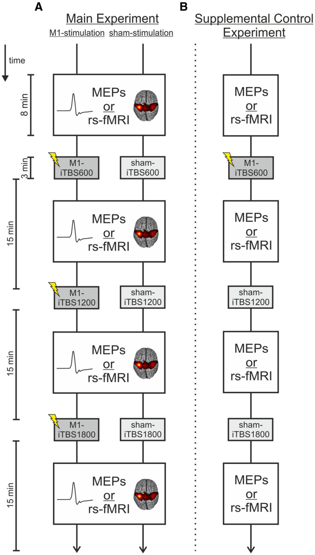
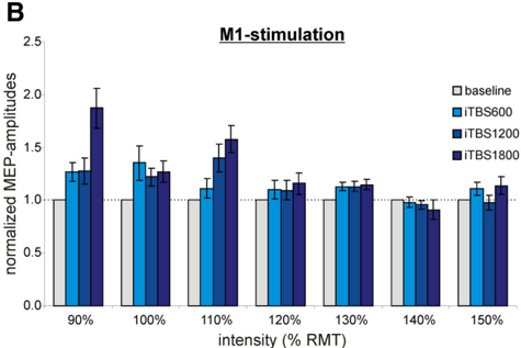
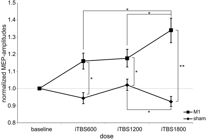
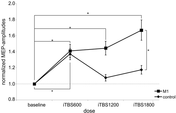
Theta burst stimulation (TBS), a specific protocol of repetitive transcranial magnetic stimulation (rTMS), induces changes in cortical excitability that last beyond stimulation. TBS-induced aftereffects, however, vary between subjects, and the mechanisms underlying these aftereffects to date remain poorly understood. Therefore, the purpose of this study was to investigate whether increasing the number of pulses of intermittent TBS (iTBS) (1) increases cortical excitability as measured by motor-evoked potentials (MEPs) and (2) alters functional connectivity measured using resting-state fMRI, in a dose-dependent manner. Sixteen healthy, human subjects received three serially applied iTBS blocks of 600 pulses over the primary motor cortex (M1 stimulation) and the parieto-occipital vertex (sham stimulation) to test for dose-dependent iTBS effects on cortical excitability and functional connectivity (four sessions in total). iTBS over M1 increased MEP amplitudes compared with sham stimulation after each stimulation block. Although the increase in MEP amplitudes did not differ between the first and second block of M1 stimulation, we observed a significant increase after three blocks (1800 pulses). Furthermore, iTBS enhanced resting-state functional connectivity between the stimulated M1 and premotor regions in both hemispheres. Functional connectivity between M1 and ipsilateral dorsal premotor cortex further increased dose-dependently after 1800 pulses of iTBS over M1. However, no correlation between changes in MEP amplitudes and functional connectivity was detected. In summary, our data show that increasing the number of iTBS stimulation blocks results in dose-dependent effects at the local level (cortical excitability) as well as at a systems level (functional connectivity) with a dose-dependent enhancement of dorsal premotor cortex-M1 connectivity.
Overview
The claims and statistics of this paper haven’t been checked yet.
Run the check — free, no account needed. It reads the open-access text and checks the sources it cites.
Integrity signals
Registry
Cites
Statistics
Figures
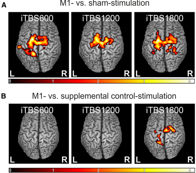
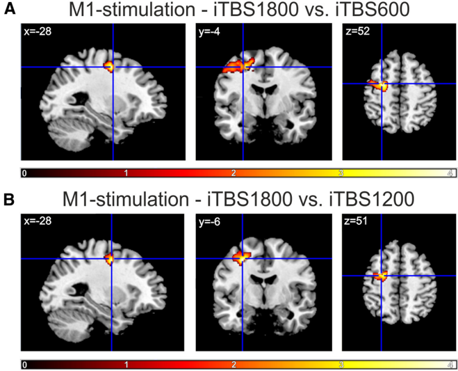
Verification
Claims the paper makes
The claims and statistics of this paper haven’t been checked yet.
Run the check — free, no account needed. It reads the open-access text and checks the sources it cites.
Claims other papers make about this one
Each is checked against this paper’s own text: does it say what the citing paper says it does?
The source backs part of the claim, not all of it — or backs it for a different population, condition or comparison.
In the citing paper
“Studies demonstrate that this number of pulses results in an optimal “window” of cortical excitability, and additional pulses may have an inhibitory effect [59-61].”
From Frontal Pole Neuromodulation for Impulsivity and Suicidality in Veterans With Mild Traumatic Brain Injury and Common Co-Occurring Mental Health Conditions: Protocol for a Pilot Randomized Controlled Trial (2024) · cites this paper as [61]
Experimental — accuracy not yet measuredThe cited source says what the claim attributes to it.
In the citing paper
“It has also been suggested that iTBS is dose-dependent and repeated application may be of benefit (Nettekoven et al., 2014).”
From Investigation of neurobiological responses to theta burst stimulation during recovery from mild traumatic brain injury (mTBI) (2023) · cites this paper as Nettekoven et al., 2014
Self-check
Percentages, sums and reported differences re-computed from the paper’s own numbers.
The self-check needs the paper’s full text.
Citations
Cites
The assessment and analysis of handedness: The Edinburgh inventory
Oldfield, R. C. (1971) · Neuropsychologia · doi:10.1016/0028-3932(71)90067-4 · Cited by 35,900
Ashburner, J., & Friston, K. J. (2005) · NeuroImage · doi:10.1016/j.neuroimage.2005.02.018 · Cited by 7,507
A mechanism for cognitive dynamics: neuronal communication through neuronal coherence.
Fries, P. (2005) · Trends in Cognitive Sciences · doi:10.1016/j.tics.2005.08.011 · Cited by 4,284
Theta Burst Stimulation of the Human Motor Cortex
Huang, Y.-Z., Edwards, M. J., Rounis, E., Bhatia, K. P., & Rothwell, J. C. (2005) · Neuron · doi:10.1016/j.neuron.2004.12.033 · Cited by 4,158
A new SPM toolbox for combining probabilistic cytoarchitectonic maps and functional imaging data
Eickhoff, S. B., Stephan, K. E., Mohlberg, H., Grefkes, C., Fink, G. R., Amunts, K., & Zilles, K. (2005) · NeuroImage · doi:10.1016/j.neuroimage.2004.12.034 · Cited by 4,033
Self-citationCorticocortical inhibition in human motor cortex.
Kujirai, T., Caramia, M. D., Rothwell, J. C., Day, B. L., Thompson, P. D., Ferbert, A., Wroe, S. J., Asselman, P., & MARSDEN, C. D. (1993) · The Journal of Physiology · doi:10.1113/jphysiol.1993.sp019912 · Cited by 3,132
Bienenstock, E., Cooper, L., & Munro, P. (1982) · The Journal of Neuroscience · doi:10.1523/jneurosci.02-01-00032.1982 · Cited by 2,896
Wassermann, E. M. (1998) · Electroencephalography and Clinical Neurophysiology/Evoked Potentials Section · doi:10.1016/s0168-5597(97)00096-8 · Cited by 2,337
Satterthwaite, T. D., Elliott, M. A., Gerraty, R. T., Ruparel, K., Loughead, J., Calkins, M. E., Eickhoff, S. B., Hakonarson, H. H., Gur, R. C., Gur, R. E., et al. (2012) · NeuroImage · doi:10.1016/j.neuroimage.2012.08.052 · Cited by 1,982
Self-citationThe plastic human brain cortex
Pascual-Leone, A., Amedi, A., Fregni, F., & Merabet, L. B. (2005) · Annual Review of Neuroscience · doi:10.1146/annurev.neuro.27.070203.144216 · Cited by 1,929
Cited by
Sorted by citations
Stanford Accelerated Intelligent Neuromodulation Therapy for Treatment-Resistant Depression
Cole, E. J., Stimpson, K. H., Bentzley, B. S., Gulser, M., Cherian, K., Tischler, C., Nejad, R., Pankow, H., Choi, E., Aaron, H., et al. (2020) · American Journal of Psychiatry · doi:10.1176/appi.ajp.2019.19070720 · Cited by 802
Stanford Neuromodulation Therapy (SNT): A Double-Blind Randomized Controlled Trial
Cole, E. J., Phillips, A. L., Bentzley, B. S., Stimpson, K. H., Nejad, R., Barmak, F., Veerapal, C., Khan, N., Cherian, K., Felber, E., et al. (2022) · American Journal of Psychiatry · doi:10.1176/appi.ajp.2021.20101429 · Cited by 795
Ten years of theta burst stimulation in humans: Established knowledge, unknowns and prospects
Suppa, A., Huang, Y.-Z., Funke, K., Ridding, M. C., Cheeran, B., Di Lazzaro, V., Ziemann, U., & Rothwell, J. C. (2016) · Brain Stimulation · doi:10.1016/j.brs.2016.01.006 · Cited by 609
Recovery from stroke: current concepts and future perspectives
Grefkes, C., & Fink, G. R. (2020) · Neurological Research and Practice · doi:10.1186/s42466-020-00060-6 · Cited by 563
Self-citationPlasticity induced by non-invasive transcranial brain stimulation: a position paper
Huang, Y.-Z., Lu, M.-K., Antal, A., Classen, J., Nitsche, M., Ziemann, U., Ridding, M., Hamada, M., Ugawa, Y., Jaberzadeh, S., et al. (2017) · Clinical Neurophysiology · doi:10.1016/j.clinph.2017.09.007 · Cited by 401
Metadata
- Authors
- Nettekoven, C., Volz, L. J., Kutscha, M., Pool, E.-M., Rehme, A. K., Eickhoff, S. B., Fink, G. R., & Grefkes, C.
- Affiliations
- Max Planck Institute for Metabolism Research (DE)
- Forschungszentrum Jülich (DE)
- ORCID
- Venue
- The Journal of Neuroscience
- Publisher
- Society for Neuroscience
- Published
- 2014-05-14
- Volume
- 34
- Issue
- 20
- Pages
- 6849-6859
- ISSN
- 0270-6474 (ISSN-L)
- 1529-2401
- PMC
- PMC4019799
- OpenAlex
- W2077740203
- Cited by
- 264
- References
- 66
- Citations by year
- 2026: 25 · 2025: 30 · 2024: 27 · 2023: 26 · 2022: 26 · 2021: 29 · 2020: 29 · 2019: 15 · 2018: 16 · 2017: 14 · 2016: 12 · 2015: 9 · 2014: 6
- FWCI
- 7.12
- Type
- journal article
- Language
- en
- Open access
- hybrid
- doi.org
- Licence
- Locations
- Journal of Neuroscience (publishedVersion, cc-by-nc-sa)
- Europe PMC (PubMed Central) (publishedVersion)
- PubMed Central (publishedVersion)
- PubMed (publishedVersion)
- Kölner Universitäts PublikationsServer (Universität zu Köln) (publishedVersion)
- Max Planck Institute for Plasma Physics (submittedVersion)
- MPG.PuRe (Max Planck Society) (submittedVersion)
- Funders
- Deutsche Forschungsgemeinschaft (LA 3071/3‐1, KFO219-TP8, 816/4-1, 3285/2-1, GR 3285/2‐1, 3071/3-1, KFO-219, EI 816/4–1)
- National Institutes of Health (R01-MH074457-01A1, MH074457)
- National Institute of Mental Health (R01MH074457)
- Topics
- Life Sciences › Neuroscience › Neurology › Transcranial Magnetic Stimulation Studies
- Life Sciences › Neuroscience › Cellular and Molecular Neuroscience › Neuroscience and Neuropharmacology Research
- Health Sciences › Medicine › Clinical Neurology › Deep Brain Stimulation and Movement Disorders
- Keywords
- theta burst stimulation, intermittent theta burst stimulation, repetitive transcranial magnetic stimulation, dose-dependent effects, cortical excitability, motor evoked potentials, resting-state functional connectivity, primary motor cortex, dorsal premotor cortex, motor system, aftereffects
- MeSH terms
- Adult, Female, Humans, Male, Motor Cortex, Nerve Net, Neuronal Plasticity, Theta Rhythm, Single-Blind Method, Cross-Over Studies, Evoked Potentials, Motor, Transcranial Magnetic Stimulation
About these checks
Registry, retraction, Cites and statistics results are computed, not judged, with one exception: a printed reference that no index lists is read once by a model before it counts as a citation error. Claims are extracted by a model and kept only when their quote appears verbatim in the text; each cited source is then checked, with a model’s help, for whether it says what the claim relies on. Those verdicts can be wrong — read the claim’s quote and the cited source before relying on one.
Statistics and arithmetic are recomputed, not judged.
Confidence bands are shown only for fields calibrated on our annotated test set. None is calibrated yet, so no band is shown.
What counts as a claim ›How we measure confidence ›How we check this ›
Are you an author, or do you see a problem with this page? Request a review
