

The DOI matches its Crossref record; 42 of the 42 references it cites were found in a registry or an index.
Long non-coding RNA PVT1 encapsulated in bone marrow mesenchymal stem cell-derived exosomes promotes osteosarcoma growth and metastasis by stabilizing ERG and sponging miR-183-5p
Open accessAbstract
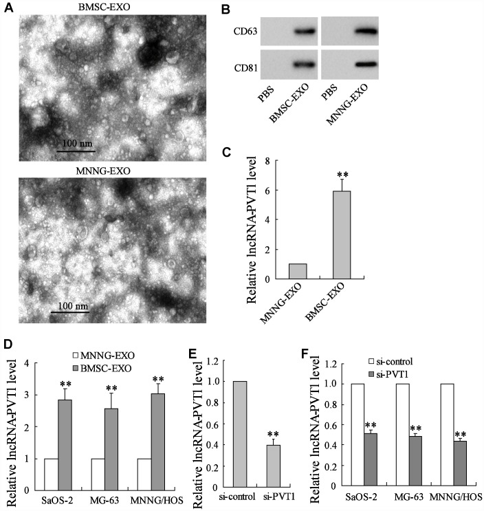
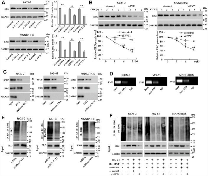
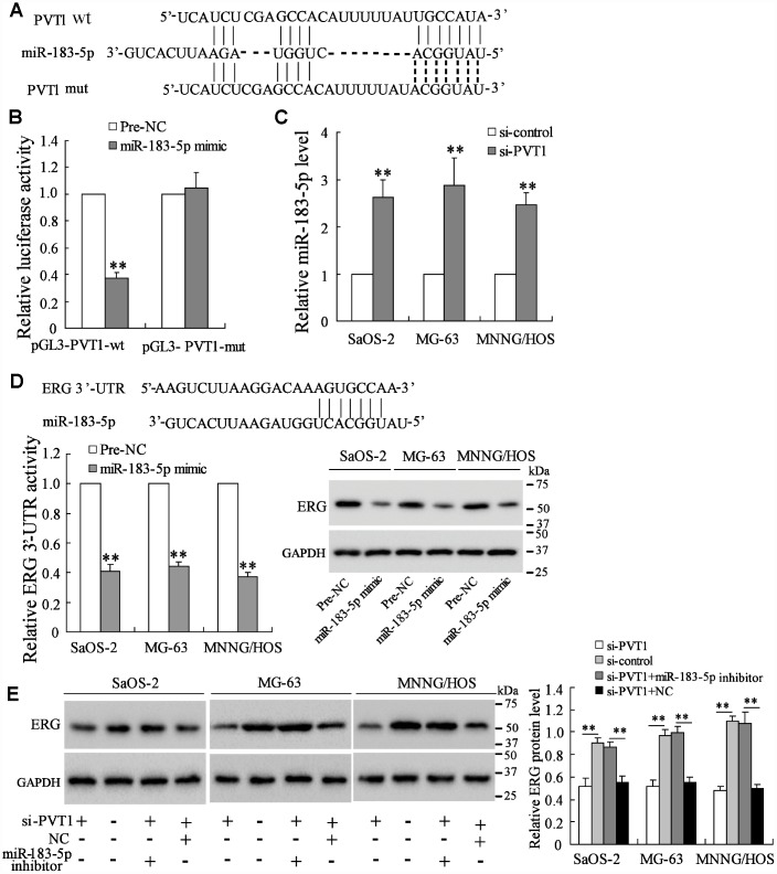
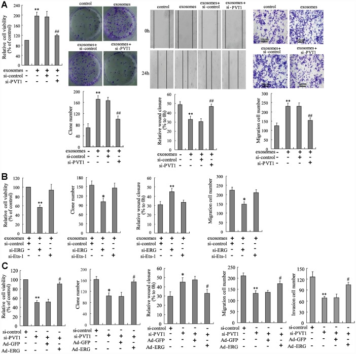
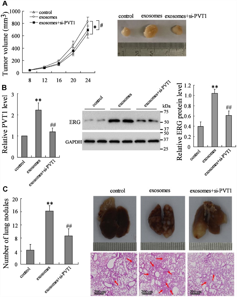
Exosomes secreted by bone marrow mesenchymal stem cells (BMSCs) promote osteosarcoma cell proliferation and migration, while the underlying mechanism remains unknown. Since the long non-coding RNA PVT1 has been reported to be upregulated in osteosarcoma cells and contributes to its growth and metastasis, we aim to investigate whether BMSC-derived exosomes promote osteosarcoma growth and metastasis via transporting PVT1 into osteosarcoma cells. The PVT1 expression in BMSC-derived exosomes was markedly higher than that in osteosarcoma cell-derived exosomes. The co-culturing of BMSC-derived exosomes and osteosarcoma cells (Saos-2, MG-63, and MNNG/HOS cell lines) significantly raised PVT1 expression of osteosarcoma cells. The direct binding between PVT1 and the oncogenic protein ERG was confirmed using RNA immunoprecipitation and RNA pull-down assays, and the transported PVT1 promotes osteosarcoma cell proliferation and migration via inhibiting degradation and ubiquitination of ERG. PVT1 also increased ERG expression through sponging miR-183-5p. In summary, our findings indicated that BMSC-derived exosomes encapsulate PVTl and transport it into osteosarcoma cells, and the transported PVT1 promotes tumor growth and metastasis by inhibiting ubiquitination and promoting expression of ERG in osteosarcoma cells. These data provide a novel insight into the mechanism of BMSC-derived exosomes in affecting osteosarcoma progression.
Overview
The claims and statistics of this paper haven’t been checked yet.
Run the check — free, no account needed. It reads the open-access text and checks the sources it cites.
Integrity signals
Registry
Cites
Statistics
Figures
Verification
Claims the paper makes
The claims and statistics of this paper haven’t been checked yet.
Run the check — free, no account needed. It reads the open-access text and checks the sources it cites.
Claims other papers make about this one
Each is checked against this paper’s own text: does it say what the citing paper says it does?
The cited source says what the claim attributes to it.
In the citing paper
“In another study, co-cultures of BM-MSC-derived exosomes with Saos-2, MG-63, and MNNG/HOS OS cells lines led to the increased expression of PVT1, a long, noncoding RNA that contributes to OS growth and metastasis [121].”
From Mesenchymal Stem/Stromal Cells: Immunomodulatory and Bone Regeneration Potential after Tumor Excision in Osteosarcoma Patients (2023) · cites this paper as [121]
Self-check
Percentages, sums and reported differences re-computed from the paper’s own numbers.
The self-check needs the paper’s full text.
Citations
Cites
3 retracted
Chen, J., Yu, Y. H., Li, H., Hu, Q., Chen, X., He, Y., Xue, C., Ren, F., Ren, Z., Li, J., et al. (2019) · Molecular Cancer · doi:10.1186/s12943-019-0947-9 · Cited by 389
Self-citationRetractedHuang, F., Chen, W., Peng, J., Li, Y., Zhuang, Y., Zhu, Z., Shao, C., Yang, W., Yao, H., & Zhang, S. (2018) · Molecular Cancer · doi:10.1186/s12943-018-0845-6 · Cited by 134
RetractedWang, D., & Hu, Y. (2018) · Molecular Therapy — Nucleic Acids · doi:10.1016/j.omtn.2018.12.006 · Cited by 82
RetractedCommunication by Extracellular Vesicles: Where We Are and Where We Need to Go
Tkach, M., & Théry, C. (2016) · Cell · doi:10.1016/j.cell.2016.01.043 · Cited by 3,444
Endogenous microRNA sponges: evidence and controversy
Thomson, D. W., & Dinger, M. E. (2016) · Nature Reviews Genetics · doi:10.1038/nrg.2016.20 · Cited by 2,185
The biology and function of exosomes in cancer
Kalluri, R. (2016) · Journal of Clinical Investigation · doi:10.1172/jci81135 · Cited by 1,880
Human mesenchymal stem cells exert potent antitumorigenic effects in a model of Kaposi's sarcoma
Khakoo, A. Y., Pati, S., Anderson, S. A., Reid, W., Elshal, M. F., Rovira, I. I., Nguyen, A. T., Malide, D., Combs, C. A., Hall, G., et al. (2006) · The Journal of Experimental Medicine · doi:10.1084/jem.20051921 · Cited by 747
Hypoxic BMSC-derived exosomal miRNAs promote metastasis of lung cancer cells via STAT3-induced EMT
Zhang, X., Sai, B., Wang, F., Wang, L., Wang, Y., Zheng, L., Li, G., Tang, J., & Xiang, J. (2019) · Molecular Cancer · doi:10.1186/s12943-019-0959-5 · Cited by 560
Long Noncoding RNA in Cancer: Wiring Signaling Circuitry
Lin, C., & Yang, L. (2018) · Trends in Cell Biology · doi:10.1016/j.tcb.2017.11.008 · Cited by 488
MVP-mediated exosomal sorting of miR-193a promotes colon cancer progression
Teng, Y., Ren, Y., Hu, X., Mu, J., Samykutty, A., Zhuang, X., Deng, Z., Kumar, A., Zhang, L., Merchant, M. L., et al. (2017) · Nature Communications · doi:10.1038/ncomms14448 · Cited by 408
Cited by
Sorted by citations
Therapeutic roles of mesenchymal stem cell-derived extracellular vesicles in cancer
Weng, Z., Zhang, B., Wu, C., Yu, F., Han, B. R., Li, B., & Li, L. (2021) · Journal of Hematology & Oncology · doi:10.1186/s13045-021-01141-y · Cited by 420
Sheng, G., Gao, Y., Yong, Y., & Wu, H. (2021) · Frontiers in Oncology · doi:10.3389/fonc.2021.780264 · Cited by 215
Zhang, H., Wang, J., Ren, T., Huang, Y., Liang, X., Yu, Y., Wang, W., Niu, J., & Guo, W. (2020) · Cancer Letters · doi:10.1016/j.canlet.2020.07.008 · Cited by 136
Self-citationRegulation of bone homeostasis: signaling pathways and therapeutic targets
Wu, Z., Li, W., Jiang, K., Lin, Z., Qian, C., Wu, M., Xia, Y., Li, N., Zhang, H., Xiao, H.-X., et al. (2024) · MedComm · doi:10.1002/mco2.657 · Cited by 129
lncRNA PVT1: a novel oncogene in multiple cancers
Li, R., Wang, X., Zhu, C., & Wang, K. (2022) · Cellular & Molecular Biology Letters · doi:10.1186/s11658-022-00385-x · Cited by 85
Yuan, Y., Wang, J., Zhang, Y., Li, L., Reza, A. M. M. T., & Gurunathan, S. (2023) · International Journal of Nanomedicine · doi:10.2147/ijn.s407029 · Cited by 85
Self-citationZeng, Z., & Xie, H. (2022) · PubMed Central · doi:10.12336/biomatertransl.2022.03.002 · Cited by 74
Exosomal Long Non-coding RNAs: Emerging Players in the Tumor Microenvironment
Pathania, A. S., & Challagundla, K. B. (2020) · Molecular Therapy — Nucleic Acids · doi:10.1016/j.omtn.2020.09.039 · Cited by 71
New perspective into mesenchymal stem cells: Molecular mechanisms regulating osteosarcoma
Chang, X., Ma, Z., Zhu, G., Lu, Y., & Yang, J. (2021) · Journal of Bone Oncology · doi:10.1016/j.jbo.2021.100372 · Cited by 70
Mesenchymal Stem Cells and Extracellular Vesicles in Osteosarcoma Pathogenesis and Therapy
Sarhadi, V. K., Daddali, R., & Seppänen-Kaijansinkko, R. (2021) · International Journal of Molecular Sciences · doi:10.3390/ijms222011035 · Cited by 63
The basic characteristics of extracellular vesicles and their potential application in bone sarcomas
Li, S. (2021) · Journal of Nanobiotechnology · doi:10.1186/s12951-021-01028-7 · Cited by 63
Wang, X., Li, Z., Cui, Y., Cui, X., Chen, C., & Wang, Z. (2021) · Frontiers in Cell and Developmental Biology · doi:10.3389/fcell.2021.644380 · Cited by 60
Self-citationNie, H., Liao, Z., Wang, Y., Zhou, J., He, X., & Ou, C. (2020) · Genes & Diseases · doi:10.1016/j.gendis.2020.12.004 · Cited by 58
Effects of microenvironment and biological behavior on the paracrine function of stem cells
Xue, Z., Liao, Y., & Li, Y. (2023) · Genes & Diseases · doi:10.1016/j.gendis.2023.03.013 · Cited by 61
Bone Cell Exosomes and Emerging Strategies in Bone Engineering
Vig, S., & Fernandes, M. H. (2022) · Biomedicines · doi:10.3390/biomedicines10040767 · Cited by 52
Han, S., Li, G., Jia, M., Zhao, Y., He, C., Huang, M., Jiang, L., Wu, M., Yang, J., Ji, X., et al. (2021) · Frontiers in Molecular Biosciences · doi:10.3389/fmolb.2021.743013 · Cited by 51
Angiogenesis is promoted by exosomal DPP4 derived from 5-fluorouracil-resistant colon cancer cells
Zheng, X., Liu, J., Li, X., Tian, R., Shang, K., Dong, X., & Cao, B. (2020) · Cancer Letters · doi:10.1016/j.canlet.2020.10.009 · Cited by 52
Cancer Stem Cells as a Source of Drug Resistance in Bone Sarcomas
Menéndez, S. T., Gallego, B., Murillo, D., Rodríguez, A., & Rodrı́guez, R. (2021) · Journal of Clinical Medicine · doi:10.3390/jcm10122621 · Cited by 48
Davodabadi, F., Mirinejad, S., Malik, S., Dhasmana, A., Ulucan‐Karnak, F., Sargazi, S., Sargazi, S., Fathi‐karkan, S., & Rahdar, A. (2024) · Nanoscale · doi:10.1039/d3nr05656b · Cited by 47
Zhang, W., Liu, Y., Jiang, J. D., Tang, Y., Tang, Y., & Liang, X. (2020) · Cancer Science · doi:10.1111/cas.14494 · Cited by 47
Recent Advances in Nanoplatforms for the Treatment of Osteosarcoma
Wu, K., Yu, B., Li, D., Tian, Y., Liu, Y., & Jiang, J. (2022) · Frontiers in Oncology · doi:10.3389/fonc.2022.805978 · Cited by 39
Exosome-based miRNA delivery: Transforming cancer treatment with mesenchymal stem cells
Balaraman, A. K., Babu, M. A., Afzal, M., Sanghvi, G. V., Rekha, M. M., Gupta, S., Rana, M., Ali, H., Goyal, K., Subramaniyan, V., et al. (2025) · Regenerative Therapy · doi:10.1016/j.reth.2025.01.019 · Cited by 46
Chen, M., Zhang, R., Lu, L., Du, J., Chen, C., Ding, K., Wei, X., Zhang, G., Huang, Y., & Hou, J. (2020) · Aging · doi:10.18632/aging.202203 · Cited by 42
Progress of Mesenchymal Stem Cell-Derived Exosomes in Tissue Repair
Zhao, G., Ge, Y., Zhang, C., Zhang, L., Xu, J., Qi, L., & Li, W. (2020) · Current Pharmaceutical Design · doi:10.2174/1381612826666200420144805 · Cited by 41
The Roles of ceRNAs-Mediated Autophagy in Cancer Chemoresistance and Metastasis
Zhang, H., & Lü, B. (2020) · Cancers · doi:10.3390/cancers12102926 · Cited by 41
Use of Stem Cell Extracellular Vesicles as a “Holistic” Approach to CNS Repair
Branscome, H., Paul, S., Yin, D., El‐Hage, N., Agbottah, E. T., Zadeh, M. A., Liotta, L. A., & Kashanchi, F. (2020) · Frontiers in Cell and Developmental Biology · doi:10.3389/fcell.2020.00455 · Cited by 37
Zhang, N., Li, L., Luo, J., Tan, J., Hu, W., Li, Z., Wang, X., & Ye, T. (2021) · Archives of Biochemistry and Biophysics · doi:10.1016/j.abb.2021.108965 · Cited by 37
RetractedExosomal non-coding RNAs have a significant effect on tumor metastasis
Wang, D., Zhang, W., Zhang, C., Wang, L., Chen, H., & Xu, J. (2022) · Molecular Therapy — Nucleic Acids · doi:10.1016/j.omtn.2022.05.034 · Cited by 40
Application of Bioprinting in Ophthalmology
Wang, Y., Wang, J., Ji, Z., Yan, W., Zhao, H. L., Huang, W., & Liu, H. (2022) · International Journal of Bioprinting · doi:10.18063/ijb.v8i2.552 · Cited by 35
Self-citationYe, H., Hu, X., Wen, Y., Tu, C., Hornicek, F. J., Duan, Z., & Min, L. (2022) · Journal of Nanobiotechnology · doi:10.1186/s12951-022-01609-0 · Cited by 35
Extracellular vesicles: A new diagnostic biomarker and targeted drug in osteosarcoma
Gao, X., Gao, B., & Li, S. (2022) · Frontiers in Immunology · doi:10.3389/fimmu.2022.1002742 · Cited by 34
Zhu, G., Xia, Y., Zhao, Z., Li, A., Li, H., & Xiao, T. (2022) · Cancer Cell International · doi:10.1186/s12935-022-02746-0 · Cited by 34
Liang, W., Lu, Y., Pan, X., Zeng, Y., Zheng, W., Li, Y., Nie, Y., Li, D., & Wang, D. (2022) · Pharmacogenomics and Personalized Medicine · doi:10.2147/pgpm.s384901 · Cited by 31
Role of extracellular vesicles in osteosarcoma
Yang, Q., Liu, J., Wu, B., Wang, X., Jiang, Y., & Zhu, D. (2022) · International Journal of Medical Sciences · doi:10.7150/ijms.74137 · Cited by 31
Functions and therapeutic potentials of exosomes in osteosarcoma
Yue, J., Chen, Z., Xu, X., & Li, S. (2022) · Acta Materia Medica · doi:10.15212/amm-2022-0024 · Cited by 29
Emerging Roles of Mesenchymal Stem/Stromal-Cell-Derived Extracellular Vesicles in Cancer Therapy
Nicodemou, A., Bernatova, S., Čeháková, M., & Danišovič, Ľ. (2023) · Pharmaceutics · doi:10.3390/pharmaceutics15051453 · Cited by 29
DDX11-AS1 contributes to osteosarcoma progression via stabilizing DDX11
Zhang, H., Lin, J., Chen, J., Gu, W., Mao, Y., Wang, H., Zhang, Y., & Liu, W. (2020) · Life Sciences · doi:10.1016/j.lfs.2020.117392 · Cited by 28
Liu, R., Ju, C., Zhang, F., Tang, X., Yan, J., Sun, J., Lv, B., Guo, Y., Liang, Y., Lv, X., et al. (2020) · Biochemical and Biophysical Research Communications · doi:10.1016/j.bbrc.2020.08.056 · Cited by 26
Osteosarcoma-derived exosomal miR-501-3p promotes osteoclastogenesis and aggravates bone loss
Lin, L., Wang, H., Guo, W., He, E., Huang, K., & Zhao, Q. (2021) · Cellular Signalling · doi:10.1016/j.cellsig.2021.109935 · Cited by 29
lncRNA MELTF-AS1 facilitates osteosarcoma metastasis by modulating MMP14 expression
Ding, L., Liu, T., Qu, Y., Kang, Z., Guo, L., Zhang, H., Jiang, J., Qu, F., Ge, W., & Zhang, S. (2021) · Molecular Therapy — Nucleic Acids · doi:10.1016/j.omtn.2021.08.022 · Cited by 26
Yang, L., Huang, X., Guo, H., Wang, L., Yang, W., Wu, W., Jing, D., & Shao, Z. (2021) · Frontiers in Cell and Developmental Biology · doi:10.3389/fcell.2021.737314 · Cited by 26
Zeng, J., Peng, Y., Wang, D., Ayesha, K., & Chen, S. (2023) · Frontiers in Cell and Developmental Biology · doi:10.3389/fcell.2023.1123065 · Cited by 31
Li, S., Han, Y., Lu, M., Liu, Z., Jin, J., Guo, Q., Wang, Y., & Liu, H. (2022) · Journal of Oral Pathology and Medicine · doi:10.1111/jop.13321 · Cited by 24
Long non-coding RNA PVT1: A promising chemotherapy and radiotherapy sensitizer
Yao, W., Li, S., Liu, R., Jiang, M., Gao, L., Lu, Y., Liang, X., & Zhang, H. (2022) · Frontiers in Oncology · doi:10.3389/fonc.2022.959208 · Cited by 23
The potential roles of exosomal noncoding RNAs in osteosarcoma
Li, S., & Wang, X. (2020) · Journal of Cellular Physiology · doi:10.1002/jcp.30101 · Cited by 22
Cao, F., Li, Z., Ding, W., Yan, L., & Zhao, Q. (2021) · Journal of Immunology Research · doi:10.1155/2021/1994328 · Cited by 21
Liu, S.-C., Cao, Y., Chen, L., Kang, R., Huang, Z., & Xin-sheng, L. (2022) · Cancer Biology & Therapy · doi:10.1080/15384047.2022.2102360 · Cited by 21
Chen, W., Lin, W., Yu, N., Zhang, L., Wu, Z., Chen, Y., Li, Z., Gong, F., Li, N., Chen, X., et al. (2022) · International Journal of Nanomedicine · doi:10.2147/ijn.s379917 · Cited by 21
Zhou, Y. L., Dong, Y., Zhang, A., Wu, J., & Sun, Q. (2023) · Frontiers in Bioengineering and Biotechnology · doi:10.3389/fbioe.2023.1214190 · Cited by 21
The Biological Roles of Exosomal Long Non-Coding RNAs in Cancers
Da, M., Jiang, H., Xie, Y., Jin, W., & Han, S. (2021) · OncoTargets and Therapy · doi:10.2147/ott.s281175 · Cited by 21
Feng, D., Li, Z., Yang, L., Liang, H., He, H., Liu, L., & Zhang, W. (2022) · Oxidative Medicine and Cellular Longevity · doi:10.1155/2022/8825784 · Cited by 20
Martins‐Neves, S. R., Sampaio-Ribeiro, G., & Gomes, C. M. F. (2022) · International Journal of Molecular Sciences · doi:10.3390/ijms231911416 · Cited by 22
Liao, Y., Yi, Q., He, J., Huang, D., Xiong, J., Sun, W., & Sun, W. (2023) · Bioengineered · doi:10.1080/21655979.2022.2161711 · Cited by 19
Wan, D., Qu, Y., Zhang, L., Ai, S., & Cheng, L. (2020) · OncoTargets and Therapy · doi:10.2147/ott.s266435 · Cited by 19
Baron, M., Drohat, P., Crawford, B., Hornicek, F. J., Best, T. M., & Kouroupis, D. (2023) · Bioengineering · doi:10.3390/bioengineering10101187 · Cited by 19
Sun, Z., Tan, Z., Peng, C., & Yi, W. (2021) · Journal of Gastrointestinal Surgery · doi:10.1007/s11605-021-05160-5 · Cited by 18
Farahzadi, R., Fathi, E., Vandghanooni, S., & Valipour, B. (2024) · Biochimica et Biophysica Acta (BBA) - Reviews on Cancer · doi:10.1016/j.bbcan.2024.189177 · Cited by 19
Wu, C., Li, Z., Feng, G., Wang, L., Xie, J., Jin, Y., Wang, L., & Liu, S. (2021) · Bioengineered · doi:10.1080/21655979.2021.1987092 · Cited by 18
He, H., Ding, M., Li, T., Zhao, W., Zhang, L., Yin, P., & Zhang, W. (2022) · Laboratory Investigation · doi:10.1038/s41374-021-00691-6 · Cited by 18
Self-citationGoyal, A., Afzal, M., Goyal, K., Ganesan, S., Kumari, M., Sunitha, S., Dash, A., Saini, S., Rana, M., Gupta, G., et al. (2025) · Regenerative Therapy · doi:10.1016/j.reth.2025.03.006 · Cited by 17
Guo, M., Li, D., Feng, Y., Li, M., & Yang, B. (2021) · Journal of Leukocyte Biology · doi:10.1002/jlb.1mia0321-164r · Cited by 17
Taheri, M., Tehrani, H. A., Dehghani, S., Rajabzadeh, A., Alibolandi, M., Zamani, N., Arefian, E., & Ramezani, M. (2024) · Cytokine & Growth Factor Reviews · doi:10.1016/j.cytogfr.2024.01.004 · Cited by 17
Li, G., Feng, J., Huang, S., & Li, Q. (2024) · Frontiers in Bioscience-Landmark · doi:10.31083/j.fbl2906207 · Cited by 16
Luongo, M., Laurenziello, P., Cesta, G., Bochicchio, A. M., Omer, L. C., Falco, G., Milone, M. R., Cibarelli, F., Russi, S., & Laurino, S. (2024) · Molecular Cancer · doi:10.1186/s12943-024-02083-y · Cited by 16
Shie, M., Huang, S., Chen, Y., Chen, M.-C., Pan, C., Chen, C., Lin, Y.-H., Yu, M., Kan, K., Chiu, S., et al. (2025) · Nature Communications · doi:10.1038/s41467-025-66451-y · Cited by 18
Noncoding RNA PVT1 in osteosarcoma: The roles of lncRNA PVT1 and circPVT1
Wu, T., Ji, Z., Lin, H., Wei, B., Xie, G., Ji, G., Fu, S., Huang, W., & Liu, H. (2022) · Cell Death Discovery · doi:10.1038/s41420-022-01192-1 · Cited by 15
Luo, Y., Liu, C., Kirby, J. A., Chu, C., Zang, D., & Chen, J. (2025) · Cancers · doi:10.3390/cancers17132186 · Cited by 15
Keremu, A., Aila, P., Tusun, A., Abulikemu, M., & Zou, X. (2022) · European Journal of Histochemistry · doi:10.4081/ejh.2022.3394 · Cited by 15
Roles of lncRNAs in childhood cancer: Current landscape and future perspectives
Liu, F., Xiong, Q.-W., Wang, J., & Peng, W. (2023) · Frontiers in Oncology · doi:10.3389/fonc.2023.1060107 · Cited by 15
Self-citationExosomes in osteosarcoma research and preclinical practice.
Zhang, X., Zhang, R., Su, X., Qi, J., Hu, Y., Shi, J.-T., Zhang, K., Wang, K.-P., & Zhou, H.-Y. (2021) · PubMed Central · Cited by 16
Ding, B., Lou, W., Fan, W., & Pan, J. (2023) · Environmental Toxicology · doi:10.1002/tox.23746 · Cited by 14
The Role of Ubiquitination in Osteosarcoma Development and Therapies
Mao, P., Feng, Z., Liu, Y., Zhang, K., Zhao, G., Lei, Z., Di, T., & Zhang, H. (2024) · Biomolecules · doi:10.3390/biom14070791 · Cited by 14
Yang, M., Zhou, W., Han, X. L., Xu, M., Wang, Z., Shi, M., Shi, Y., & Yu, Y. (2024) · Journal of Cellular and Molecular Medicine · doi:10.1111/jcmm.70115 · Cited by 12
LncRNA FAM181A-AS1 promotes gliomagenesis by sponging miR-129-5p and upregulating ZRANB2
Jiang, X., & Chen, D. (2020) · Aging · doi:10.18632/aging.103391 · Cited by 13
Huang, H., Zhong, P., Zhang, J., Chen, X., Chen, J., Tian, L., & Wu, Q. (2022) · Bioengineered · doi:10.1080/21655979.2022.2037379 · Cited by 13
The Roles of Exosomes in Metastasis of Sarcoma: From Biomarkers to Therapeutic Targets
Tan, L., Wang, Y., Hu, X., & Min, L. (2023) · Biomolecules · doi:10.3390/biom13030456 · Cited by 12
Exosomes—Promising Carriers for Regulatory Therapy in Oncology
Jóźwicka, T. M., Erdmańska, P. M., Stachowicz-Karpińska, A., Olkiewicz, M., & Jóźwicki, W. (2024) · Cancers · doi:10.3390/cancers16050923 · Cited by 13
Emerging roles of long non-coding RNAs in osteosarcoma
Liao, X., Wei, R., Zhou, J., Wu, K., & Li, J. (2024) · Frontiers in Molecular Biosciences · doi:10.3389/fmolb.2024.1327459 · Cited by 14
Self-citationLncRNAs Are Key Regulators of Transcription Factor-Mediated Endothelial Stress Responses
Lam, F., Leisegang, M. S., & Brandes, R. P. (2024) · International Journal of Molecular Sciences · doi:10.3390/ijms25179726 · Cited by 13
Shirani, N., Abdi, N., Chehelgerdi, M., Yaghoobi, H., & Chehelgerdi, M. (2025) · Frontiers in Cell and Developmental Biology · doi:10.3389/fcell.2025.1485422 · Cited by 14
Chen, J., Miao, W., Yang, S., Yin, M., Zhao, J., & Song, D. (2020) · Frontiers in Oncology · doi:10.3389/fonc.2020.563255 · Cited by 12
Emerging roles of mesenchymal stem cell-derived exosomes in gastrointestinal cancers
Wang, N., Pei, B., Yuan, X., Yi, C., Ocansey, D. K. W., Qian, H., & Mao, F. (2022) · Frontiers in Bioengineering and Biotechnology · doi:10.3389/fbioe.2022.1019459 · Cited by 12
Sun, Z., Li, X., Shi, Y., & Yao, Y. (2024) · Cancer Reports · doi:10.1002/cnr2.2085 · Cited by 11
The role of tumor-derived exosomal LncRNA in tumor metastasis
Yu, Z., Fu, J., Mantareva, V. N., Blažević, I., Wu, Y., Wen, D., Battulga, T., Wang, Y.-Q., & Zhang, J. (2025) · Cancer Gene Therapy · doi:10.1038/s41417-024-00852-x · Cited by 10
Self-citationLong Non-Coding RNAs as Competitive Endogenous RNAs in Osteosarcoma
Кушлинский, Н. Е., Fridman, M. V., & Александровна, Б. Э. (2020) · Molecular Biology · doi:10.1134/s0026893320050052 · Cited by 10
Li, M., Lin, C., & Cai, Z. (2023) · Translational Oncology · doi:10.1016/j.tranon.2023.101639 · Cited by 9
Ming, X., Lu, Y., Huang, H., Zheng, J., Wang, T., Li, Z., Yu, X., & Xiong, L. (2024) · Functional & Integrative Genomics · doi:10.1007/s10142-024-01506-9 · Cited by 9
Afkhami, H., Yarahmadi, A., Bostani, S., Yarian, N., Haddad, M. S., Lesani, S. S., Aghaei, S. S., & Zolfaghari, M. R. (2024) · Discover Oncology · doi:10.1007/s12672-024-01590-0 · Cited by 10
Jamil, N. Y., Nawrooz, M. S., Bishoyi, A. K., Ballal, S., Singh, A., Krithiga, T., Panigrahi, R., Bakhtiyarovna, B. Z., Taher, S. G., Alwan, M., et al. (2025) · Investigational New Drugs · doi:10.1007/s10637-025-01551-6 · Cited by 9
Therapeutic effects and perspective of stem cell extracellular vesicles in aging and cancer
Liu, Y., Cheng, Y., Wang, S., Su, Y., Liu, Y., Li, C., Jin, L., Wan, Q., Sang, X., & Wang, Z. (2020) · Journal of Cellular Physiology · doi:10.1002/jcp.30212 · Cited by 9
Mesenchymal stem cells elicits Anti-PD1 immunotherapy by targeted delivery of CX3CL1
Liu, J., Ma, X., Liu, C., Yang, C. H., Li, B., Zhang, W., Zeng, R., Chen, Q., Zhang, Y., & Hu, S. (2023) · Frontiers in Pharmacology · doi:10.3389/fphar.2023.1136614 · Cited by 7
Progress of extracellular vesicles-based system for tumor therapy
Wang, F., Yin, L., & Hu, Y. (2025) · Journal of Controlled Release · doi:10.1016/j.jconrel.2025.02.066 · Cited by 7
Liu, X., Ye, J., Guo, W., & Wang, J. (2024) · Frontiers in Cell and Developmental Biology · doi:10.3389/fcell.2024.1473044 · Cited by 6
Self-citationInhibition of miR-615-3p enhances dentinogenesis in scaps via PVT1-mediated mitochondrial regulation
Yang, H., Huang, Y., Song, J., Han, X. Y., Yuan, F., & Fan, Z. (2025) · Stem Cell Research & Therapy · doi:10.1186/s13287-025-04528-7 · Cited by 5
Huang, L., Xiong, J., Fu, J., Zhou, Z., Yu, H., Xu, J., Wu, L. L., & Cao, K. (2022) · Journal of Clinical and Translational Research · doi:10.18053/jctres.08.202206.015 · Cited by 5
Chen, C. C., & Benavente, C. A. (2024) · International Journal of Molecular Sciences · doi:10.3390/ijms25010568 · Cited by 7
lncRNA LINC00960 promotes apoptosis by sponging ubiquitin ligase Nrdp1-targeting miR-183-5p
Yang, H., Jiang, T.-X., Fan, L., & Qiu, X. (2023) · Acta Biochimica et Biophysica Sinica · doi:10.3724/abbs.2023005 · Cited by 4
Ye, J., He, H., Chen, S., Ren, Y., Guo, W., & Jin, Z. (2022) · International Journal of Clinical Oncology · doi:10.1007/s10147-022-02245-2 · Cited by 3
Wu, W., & He, J. (2023) · Journal of Cancer Research and Clinical Oncology · doi:10.1007/s00432-023-05273-1 · Cited by 3
Role of non-coding RNA in exosomes for the diagnosis and treatment of osteosarcoma
Liu, X., Wang, Y., Wang, C., Wang, X., Tang, G., Xiong, Z., & Zhou, W. (2024) · Frontiers in Oncology · doi:10.3389/fonc.2024.1469833 · Cited by 3
Metadata
- Authors
- Zhao, W., Qin, P., Zhang, D., Cui, X., Gao, J., Yu, Z., Chai, Y., Wang, J., & Li, J.
- Affiliations
- First Affiliated Hospital of Zhengzhou University (CN)
- Corresponding author
- Li, Juan
- Venue
- Aging
- Publisher
- Impact Journals, LLC
- Published
- 2019-11-07
- Volume
- 11
- Issue
- 21
- Pages
- 9581-9596
- ISSN
- 1945-4589 (ISSN-L)
- PMC
- PMC6874467
- OpenAlex
- W2984884798
- Cited by
- 115
- References
- 42
- Citations by year
- 2026: 5 · 2025: 14 · 2024: 18 · 2023: 16 · 2022: 26 · 2021: 19 · 2020: 17
- FWCI
- 5.13
- Type
- journal article
- Language
- en
- Open access
- green
- www.ncbi.nlm.nih.gov
- Licence
- cc-by
- Locations
- Aging (publishedVersion)
- PubMed Central (publishedVersion, cc-by)
- PubMed (publishedVersion)
- Topics
- Life Sciences › Biochemistry, Genetics and Molecular Biology › Molecular Biology › Extracellular vesicles in disease
- Life Sciences › Biochemistry, Genetics and Molecular Biology › Cancer Research › Cancer-related molecular mechanisms research
- Life Sciences › Biochemistry, Genetics and Molecular Biology › Molecular Biology › Circular RNAs in diseases
- Keywords
- bone marrow mesenchymal stem cells, exosomes, osteosarcoma, ERG, miR-183-5p, tumor growth, metastasis, protein stabilization, ubiquitination, tumor microenvironment, cell proliferation, cell migration
- MeSH terms
- Transcriptional Regulator ERG, Animals, Bone Neoplasms, Humans, Mice, Inbred BALB C, Mice, Nude, Neoplasm Metastasis, Neoplasm Transplantation, Osteosarcoma, MicroRNAs, Cell Line, Tumor, Exosomes, Primary Cell Culture, RNA, Long Noncoding
About these checks
Registry, retraction, Cites and statistics results are computed, not judged, with one exception: a printed reference that no index lists is read once by a model before it counts as a citation error. Claims are extracted by a model and kept only when their quote appears verbatim in the text; each cited source is then checked, with a model’s help, for whether it says what the claim relies on. Those verdicts can be wrong — read the claim’s quote and the cited source before relying on one.
Statistics and arithmetic are recomputed, not judged.
Confidence bands are shown only for fields calibrated on our annotated test set. None is calibrated yet, so no band is shown.
What counts as a claim ›How we measure confidence ›How we check this ›
Are you an author, or do you see a problem with this page? Request a review
