

The DOI matches its Crossref record; 89 of the 90 references it cites were found in a registry or an index.
Baseline MRI Predictors of Conversion from MCI to Probable AD in the ADNI Cohort
Open accessAbstract
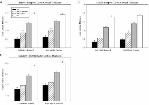
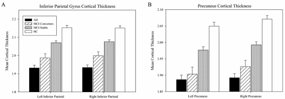
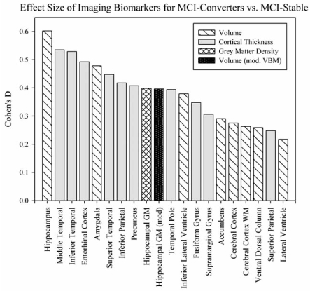
The Alzheimer's Disease Neuroimaging Initiative (ADNI) is a multi-center study assessing neuroimaging in diagnosis and longitudinal monitoring. Amnestic Mild Cognitive Impairment (MCI) often represents a prodromal form of dementia, conferring a 10-15% annual risk of converting to probable AD. We analyzed baseline 1.5T MRI scans in 693 participants from the ADNI cohort divided into four groups by baseline diagnosis and one year MCI to probable AD conversion status to identify neuroimaging phenotypes associated with MCI and AD and potential predictive markers of imminent conversion. MP-RAGE scans were analyzed using publicly available voxel-based morphometry (VBM) and automated parcellation methods. Measures included global and hippocampal grey matter (GM) density, hippocampal and amygdalar volumes, and cortical thickness values from entorhinal cortex and other temporal and parietal lobe regions. The overall pattern of structural MRI changes in MCI (n=339) and AD (n=148) compared to healthy controls (HC, n=206) was similar to prior findings in smaller samples. MCI-Converters (n=62) demonstrated a very similar pattern of atrophic changes to the AD group up to a year before meeting clinical criteria for AD. Finally, a comparison of effect sizes for contrasts between the MCI-Converters and MCI-Stable (n=277) groups on MRI metrics indicated that degree of neurodegeneration of medial temporal structures was the best antecedent MRI marker of imminent conversion, with decreased hippocampal volume (left > right) being the most robust. Validation of imaging biomarkers is important as they can help enrich clinical trials of disease modifying agents by identifying individuals at highest risk for progression to AD.
Overview
The claims and statistics of this paper haven’t been checked yet.
Run the check — free, no account needed. It reads the open-access text and checks the sources it cites.
Integrity signals
Registry
Cites
Statistics
Figures
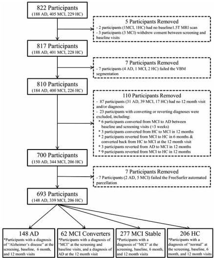
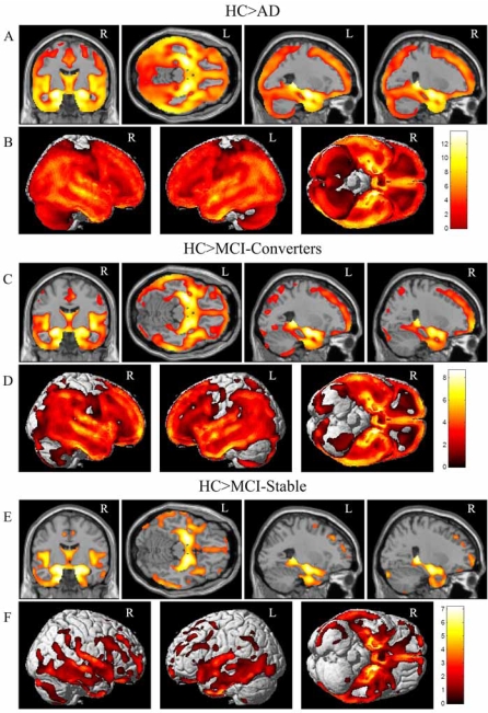
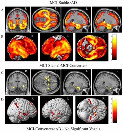
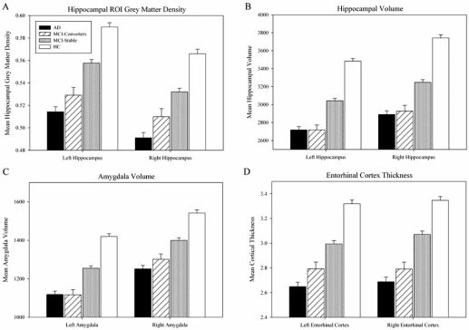
Verification
Claims the paper makes
The claims and statistics of this paper haven’t been checked yet.
Run the check — free, no account needed. It reads the open-access text and checks the sources it cites.
Claims other papers make about this one
Each is checked against this paper’s own text: does it say what the citing paper says it does?
In the citing paper
“they reliably predict cognitive decline [222]”
From Genetic architecture of Alzheimer's disease (2020) · cites this paper as [222]
In the citing paper
“a large number of novel loci and genes have been nominated by these analyses [222, 229–231]”
From Genetic architecture of Alzheimer's disease (2020) · cites this paper as [222]
Self-check
Percentages, sums and reported differences re-computed from the paper’s own numbers.
The self-check needs the paper’s full text.
Citations
Cites
Folstein, M. F., Folstein, S. E., & McHugh, P. R. (1975) · Journal of Psychiatric Research · doi:10.1016/0022-3956(75)90026-6 · Cited by 88,282
Cortical Surface-Based Analysis
Dale, A. M., Fischl, B., & Sereno, M. I. (1999) · NeuroImage · doi:10.1006/nimg.1998.0395 · Cited by 10,910
The Clinical Dementia Rating (CDR): Current version and scoring rules
Morris, J. C. (1993) · Neurology · doi:10.1212/wnl.43.11.2412-a · Cited by 9,882
Mild Cognitive Impairment: Clinical Characterization and Outcome
Petersen, R. C., Smith, G. E., Waring, S. C., Ivnik, R. J., Tangalos, E. G., & Kokmen, E. (1999) · Archives of Neurology · doi:10.1001/archneur.56.3.303 · Cited by 8,980
Voxel-Based Morphometry—The Methods
Ashburner, J., & Friston, K. J. (2000) · NeuroImage · doi:10.1006/nimg.2000.0582 · Cited by 8,676
Fischl, B., Salat, D. H., Busa, E., Albert, M., Dieterich, M., Haselgrove, C., van der Kouwe, A., Killiany, R., Kennedy, D., Klaveness, S., et al. (2002) · Neuron · doi:10.1016/s0896-6273(02)00569-x · Cited by 8,610
Mild cognitive impairment as a diagnostic entity
Petersen, R. C. (2004) · Journal of Internal Medicine · doi:10.1111/j.1365-2796.2004.01388.x · Cited by 7,864
Sheikh (1986) · doi:10.1300/j018v05n01_09 · Cited by 7,236
Cortical Surface-Based Analysis
Fischl, B., Sereno, M. I., & Dale, A. M. (1999) · NeuroImage · doi:10.1006/nimg.1998.0396 · Cited by 6,147
Measuring the thickness of the human cerebral cortex from magnetic resonance images
Fischl, B. R., & Dale, A. M. (2000) · Proceedings of the National Academy of Sciences · doi:10.1073/pnas.200033797 · Cited by 5,995
Research works printed but not linked: 1
- [35]Dickerson BC Goncharova I Sullivan MP Forchetti C Wilson RS Bennett DA MRI-derived entorhinal and hippocampal atrophy in incipient and very mild Alzheimer's disease Neurobiol Aging 2001 22 747 754 11705634
Cited by
Sorted by citations
Items 501–538 of 538
Lee, J., & Park, J. (2020) · The Journal of Korean Society of Occupational Therapy · doi:10.14519/kjot.2020.28.1.07
Cognitive Biomarker Prioritization in Alzheimer's Disease using Brain Morphometric DataPreprint
Peng, B., Yao, X., Risacher, S. L., Saykin, A. J., Shen, L., & Ning, X. (2020) · arXiv (Cornell University) · doi:10.48550/arxiv.2002.07699
Self-citationSaccades in Alzheimer’s disease: an MRI study.
Wright, S. L. (2011) · Otago University Research Archive (University of Otago)
Representative Functional Connectivity Learning for Multiple Clinical groups in Alzheimer's DiseasePreprint
Zhang, L., Yu, X., Lyu, Y., Wang, L., & Zhu, D. (2021) · arXiv (Cornell University) · doi:10.48550/arxiv.2106.07622
da Costa, R. Q. M. (2020) · Digital Library of Theses and Dissertations (Universidade de São Paulo) · doi:10.11606/t.5.2020.tde-15092021-155025
Voxel-based Morphometry Analysis for Healthy Aging and Cognitive Function
Mizuno, S., & Maesawa, S. (2020) · Higher Brain Function Research · doi:10.2496/hbfr.40.377
Experiencias educativas en el Parque Nacional de Doñana
España, M. C. C., Ortiz, E. M. L., & Giner, M. I. C. (1997) · LA Referencia (Red Federada de Repositorios Institucionales de Publicaciones Científicas)
Neuroimaging Approaches for Elderly Studies
Styliadis, C., Kartsidis, P. E., & Paraskevopoulos, E. (2016) · IGI Global eBooks · doi:10.4018/978-1-5225-0571-6.ch067
Liu, S. (2017) · Springer theses · doi:10.1007/978-981-10-3533-3_1
Meyer, B. J. (2018) · MDPI eBooks · doi:10.3390/books978-3-03842-972-2
Functional MRI Studies of Memory in Aging, Mild Cognitive Impairment, and Alzheimer’s Disease
Risacher, S. L., Wishart, H. A., & Saykin, A. J. (2014) · Springer eBooks · doi:10.1007/978-1-4939-1995-6_10
Self-citationPharmacodynamic Evaluation: CNS Methodologies
Hughes, L., Trad, M., Boyer, S., Lee, D., & Yin, W. (2017) · Springer eBooks · doi:10.1007/978-3-319-56637-5_32-1
Liu, S. (2017) · Springer theses · doi:10.1007/978-981-10-3533-3_2
Misquitta, K., Rosen, H. J., & Tartaglia, M. C. (2016) · Cambridge University Press eBooks · doi:10.1017/9781139924771.004
Li, Q., Ren, A., Cui, L., Sun, X., Zhong, Y., Wang, B., Miao, Y., & Guo, Q. (2020) · doi:10.21203/rs.3.rs-55458/v1
Adaptive Prior Correction in Alzheimer’s Disease Spatio-Temporal Modeling via Multi-task Learning
Chang, X., Zhou, M., Yang, Y., & Yang, P. (2024) · Communications in computer and information science · doi:10.1007/978-3-031-52216-1_6
Kamal, F., Morrison, C., Oliver, M. D., & Dadar, M. (2024) · medRxiv · doi:10.1101/2024.02.28.24303519
Ratnakaran, B., Jan, D., Raelly-Muze, L., Owens, T., & Wang, S. (2024) · Springer eBooks · doi:10.1007/978-3-031-55711-8_7
Comparative Analysis of Biomarkers for Alzheimer’s disease
Navale, G. J., Bhise, A., & Wagh, K. (2024) · IEEE Pune Section International Conference (PuneCon) · doi:10.1109/punecon63413.2024.10895704
ONDRUS, M. G., & Cribben, I. (2025) · bioRxiv (Cold Spring Harbor Laboratory) · doi:10.1101/2025.05.16.654552
Neurological aging and neurodegenerative disease in humans and great apes
Edler, M. K., Hearn, C. J., Guevara, E. E., Mhatre‐Winters, I., Varghese, M., Freire‐Cobo, C., Sherwood, C. C., Richardson, J. R., Raghanti, M. A., & Hof, P. R. (2025) · Elsevier eBooks · doi:10.1016/b978-0-443-27380-3.00074-9
Xie, L., Das, S. R., Wisse, L. E. M., Ittyerah, R., de Florès, R., Shaw, L. M., Yushkevich, P. A., & Wolk, D. A. (2023) · Figshare · doi:10.6084/m9.figshare.22625699
Peng, B., Yao, X., Risacher, S. L., Saykin, A. J., Shen, L., & Ning, X. (2020) · Figshare · doi:10.6084/m9.figshare.13323701
Self-citationShin, Y., Oh, K., & Suk, H. (2025) · SSRN Electronic Journal · doi:10.2139/ssrn.5332732
Sex-dependent grey matter atrophy in Alzheimer’s disease progression
Mukherjee, C., Bajaj, S., Adhikari, B. M., & Dhamala, M. (2026) · Brain Communications · doi:10.1093/braincomms/fcag103
Chen, L., Wu, Z., Zhao, F., Wang, Y. P., Lin, W., Wang, L., & Li, G. (2026) · Computerized Medical Imaging and Graphics · doi:10.1016/j.compmedimag.2026.102779
Jones, G. H., Suchting, R., Courtes, A. C., Shahani, L., Lavagnino, L., Leung, E., da Costa, S. C., de Sousa, R. T., Beal, L. L., Zanetti, M. V., et al. (2026) · Psychiatry Research Neuroimaging · doi:10.1016/j.pscychresns.2026.112257
Advancing Strain‐Specific Immunotherapy for Alzheimer's Disease and Tauopathies
Priya, E. S., Vasanth, S., Taj, T., Ameen, M. P. M., Danam, S. S., & Kumar, M. V. (2026) · Wiley eBooks · doi:10.1002/9781394336241.ch3
Structural Brain Biomarkers for Identifying Progressive Amnestic Mild Cognitive ImpairmentPreprint
Cherkasov, N. S., Tomyshev, A. S., Гаяровна, А. Е., Dudina, A. N., Fedorova, Y. B., Пономарева, Е. В., Селезнева, Н. Д., Bozhko, O. V., Si, G., Колыхалов, И. В., et al. (2026) · medRxiv · doi:10.64898/2026.07.27.26359003
Watanabe, N., Ogawa, A., Osada, T., Adachi, Y., Shirokoshi, T., Kodama, H., Oshima, Y., Tanaka, S., Kaga, H., Tamura, Y., et al. (2026) · bioRxiv (Cold Spring Harbor Laboratory) · doi:10.64898/2026.08.04.742662
Feng, C., Zhang, T., Liu, X., Liu, Z., Zhao, Y., & Zhang, P. (2026) · Biology · doi:10.3390/biology15161423
Oh, K., Shin, Y., & Suk, H. (2026) · Pattern Recognition · doi:10.1016/j.patcog.2026.114719
Gender bias in the diagnosis of Alzheimer's DiseasePreprint
Neufang, S., Akhrif, A., Beyan, O., & Initiative, N. (2026) · Research Square · doi:10.21203/rs.3.rs-10738736/v1
Mouiha, A., Taiebine, M., & Hilaly, J. E. (2026) · Journal of Education and Health Promotion · doi:10.4103/jehp.jehp_1279_25
Seçgin, Y., Yenigül, H., Karagöz, C. K., Sağlam, Ö. C., Acer, N., & Initiative, T. F. L. D. N. (2026) · Clinical Anatomy · doi:10.1002/ca.70212
Metadata
- Authors
- Risacher, S., Saykin, A., Wes, J., Shen, L., Firpi, H., & McDonald, B.
- Affiliations
- Indiana University School of Medicine (US)
- Indiana University Bloomington (US)
- Corresponding author
- Saykin, Andrew
- Venue
- Current Alzheimer Research
- Publisher
- Bentham Science Publishers Ltd.
- Published
- 2009-08-01
- Volume
- 6
- Issue
- 4
- Pages
- 347-361
- ISSN
- 1567-2050 (ISSN-L)
- PMC
- PMC2764863
- OpenAlex
- W2128422359
- Cited by
- 538
- References
- 90
- Citations by year
- 2026: 18 · 2025: 11 · 2024: 21 · 2023: 23 · 2022: 20 · 2021: 29 · 2020: 39 · 2019: 36 · 2018: 44 · 2017: 38 · 2016: 41 · 2015: 29 · 2014: 31 · 2013: 46 · 2012: 33
- FWCI
- 22.68
- Type
- journal article
- Language
- en
- Open access
- bronze
- doi.org
- Locations
- Current Alzheimer Research (publishedVersion)
- PubMed Central (publishedVersion, cc-by)
- PubMed (publishedVersion)
- IUScholarWorks (Indiana University) (submittedVersion, cc-by)
- Funders
- Brigham Young University
- National Institutes of Health (5u01ag024904-10)
- School of Medicine, Indiana University
- National Institute on Aging (U01-AG-024904, P30AG010133, R01 AG19771, R01AG019771, U19AG010483, P30 AG-10133)
- National Institute of Biomedical Imaging and Bioengineering (R03EB008674)
- Topics
- Health Sciences › Medicine › Psychiatry and Mental health › Dementia and Cognitive Impairment Research
- Life Sciences › Neuroscience › Cellular and Molecular Neuroscience › Alzheimer's disease research and treatments
- Health Sciences › Medicine › Oncology › Cancer-related cognitive impairment studies
- Keywords
- Alzheimer's disease, amnestic mild cognitive impairment, neuroimaging biomarkers, hippocampal volume, gray matter density, amygdala, entorhinal cortex, cortical thickness, medial temporal lobe atrophy, structural MRI, voxel-based morphometry, MPRAGE, disease progression prediction
- MeSH terms
- Aged, Aged, 80 and over, Alzheimer Disease, Analysis of Variance, Atrophy, Brain Mapping, Cerebral Cortex, Cognition Disorders, Female, Hippocampus, Humans, Image Interpretation, Computer-Assisted, Image Processing, Computer-Assisted, Magnetic Resonance Imaging, Male, Middle Aged, Neuropsychological Tests, Organ Size, Predictive Value of Tests, Multivariate Analysis, Disease Progression
About these checks
Registry, retraction, Cites and statistics results are computed, not judged, with one exception: a printed reference that no index lists is read once by a model before it counts as a citation error. Claims are extracted by a model and kept only when their quote appears verbatim in the text; each cited source is then checked, with a model’s help, for whether it says what the claim relies on. Those verdicts can be wrong — read the claim’s quote and the cited source before relying on one.
Statistics and arithmetic are recomputed, not judged.
Confidence bands are shown only for fields calibrated on our annotated test set. None is calibrated yet, so no band is shown.
What counts as a claim ›How we measure confidence ›How we check this ›
Are you an author, or do you see a problem with this page? Request a review
