

The DOI matches its Crossref record; 89 of the 90 references it cites were found in a registry or an index.
Baseline MRI Predictors of Conversion from MCI to Probable AD in the ADNI Cohort
Open accessAbstract
The Alzheimer's Disease Neuroimaging Initiative (ADNI) is a multi-center study assessing neuroimaging in diagnosis and longitudinal monitoring. Amnestic Mild Cognitive Impairment (MCI) often represents a prodromal form of dementia, conferring a 10-15% annual risk of converting to probable AD. We analyzed baseline 1.5T MRI scans in 693 participants from the ADNI cohort divided into four groups by baseline diagnosis and one year MCI to probable AD conversion status to identify neuroimaging phenotypes associated with MCI and AD and potential predictive markers of imminent conversion. MP-RAGE scans were analyzed using publicly available voxel-based morphometry (VBM) and automated parcellation methods. Measures included global and hippocampal grey matter (GM) density, hippocampal and amygdalar volumes, and cortical thickness values from entorhinal cortex and other temporal and parietal lobe regions. The overall pattern of structural MRI changes in MCI (n=339) and AD (n=148) compared to healthy controls (HC, n=206) was similar to prior findings in smaller samples. MCI-Converters (n=62) demonstrated a very similar pattern of atrophic changes to the AD group up to a year before meeting clinical criteria for AD. Finally, a comparison of effect sizes for contrasts between the MCI-Converters and MCI-Stable (n=277) groups on MRI metrics indicated that degree of neurodegeneration of medial temporal structures was the best antecedent MRI marker of imminent conversion, with decreased hippocampal volume (left > right) being the most robust. Validation of imaging biomarkers is important as they can help enrich clinical trials of disease modifying agents by identifying individuals at highest risk for progression to AD.
Overview
The claims and statistics of this paper haven’t been checked yet.
Run the check — free, no account needed. It reads the open-access text and checks the sources it cites.
Integrity signals
Registry
Cites
Statistics
Figures
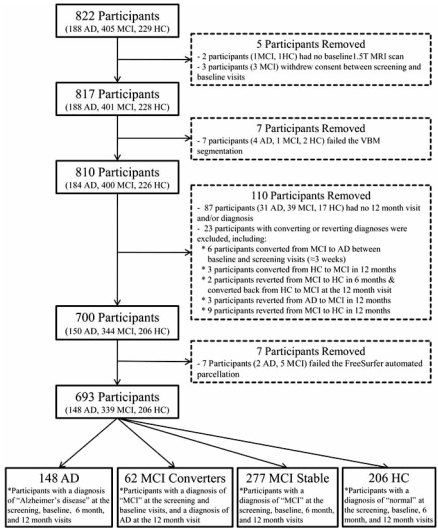
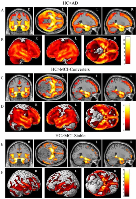
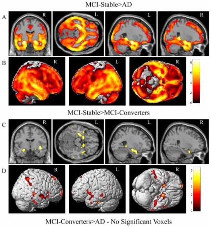
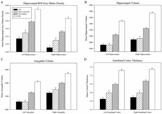
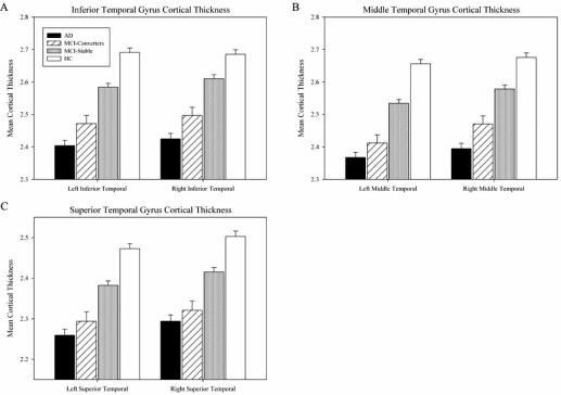
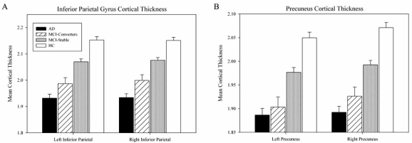
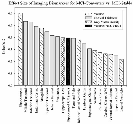
Verification
Claims the paper makes
The claims and statistics of this paper haven’t been checked yet.
Run the check — free, no account needed. It reads the open-access text and checks the sources it cites.
Claims other papers make about this one
Each is checked against this paper’s own text: does it say what the citing paper says it does?
In the citing paper
“they reliably predict cognitive decline [222]”
From Genetic architecture of Alzheimer's disease (2020) · cites this paper as [222]
In the citing paper
“a large number of novel loci and genes have been nominated by these analyses [222, 229–231]”
From Genetic architecture of Alzheimer's disease (2020) · cites this paper as [222]
Self-check
Percentages, sums and reported differences re-computed from the paper’s own numbers.
The self-check needs the paper’s full text.
Citations
Cites
Folstein, M. F., Folstein, S. E., & McHugh, P. R. (1975) · Journal of Psychiatric Research · doi:10.1016/0022-3956(75)90026-6 · Cited by 88,282
Cortical Surface-Based Analysis
Dale, A. M., Fischl, B., & Sereno, M. I. (1999) · NeuroImage · doi:10.1006/nimg.1998.0395 · Cited by 10,910
The Clinical Dementia Rating (CDR): Current version and scoring rules
Morris, J. C. (1993) · Neurology · doi:10.1212/wnl.43.11.2412-a · Cited by 9,882
Mild Cognitive Impairment: Clinical Characterization and Outcome
Petersen, R. C., Smith, G. E., Waring, S. C., Ivnik, R. J., Tangalos, E. G., & Kokmen, E. (1999) · Archives of Neurology · doi:10.1001/archneur.56.3.303 · Cited by 8,980
Voxel-Based Morphometry—The Methods
Ashburner, J., & Friston, K. J. (2000) · NeuroImage · doi:10.1006/nimg.2000.0582 · Cited by 8,676
Fischl, B., Salat, D. H., Busa, E., Albert, M., Dieterich, M., Haselgrove, C., van der Kouwe, A., Killiany, R., Kennedy, D., Klaveness, S., et al. (2002) · Neuron · doi:10.1016/s0896-6273(02)00569-x · Cited by 8,610
Mild cognitive impairment as a diagnostic entity
Petersen, R. C. (2004) · Journal of Internal Medicine · doi:10.1111/j.1365-2796.2004.01388.x · Cited by 7,864
Sheikh (1986) · doi:10.1300/j018v05n01_09 · Cited by 7,236
Cortical Surface-Based Analysis
Fischl, B., Sereno, M. I., & Dale, A. M. (1999) · NeuroImage · doi:10.1006/nimg.1998.0396 · Cited by 6,147
Measuring the thickness of the human cerebral cortex from magnetic resonance images
Fischl, B. R., & Dale, A. M. (2000) · Proceedings of the National Academy of Sciences · doi:10.1073/pnas.200033797 · Cited by 5,995
Research works printed but not linked: 1
- [35]Dickerson BC Goncharova I Sullivan MP Forchetti C Wilson RS Bennett DA MRI-derived entorhinal and hippocampal atrophy in incipient and very mild Alzheimer's disease Neurobiol Aging 2001 22 747 754 11705634
Cited by
Sorted by citations
Items 101–200 of 538
Khatri, U., & Kwon, G. (2022) · Frontiers in Aging Neuroscience · doi:10.3389/fnagi.2022.818871 · Cited by 84
Connectome-scale assessments of structural and functional connectivity in MCI
Zhu, D., Li, K., Terry, D. P., Puente, A. N., Wang, L., Shen, D. G., Miller, L. S., & Liu, T. (2013) · Human Brain Mapping · doi:10.1002/hbm.22373 · Cited by 84
Ruan, Q., D’Onofrio, G., Sancarlo, D., Bao, Z., Greco, A., & Yu, Z. (2016) · BMC Geriatrics · doi:10.1186/s12877-016-0281-7 · Cited by 83
Longitudinal Changes in Functional Brain Connectivity Predicts Conversion to Alzheimer’s Disease
Serra, L., Cercignani, M., Mastropasqua, C., Torso, M., Spanò, B., Makovac, E., Viola, V., Giulietti, G., Marra, C., Caltagirone, C., et al. (2016) · Journal of Alzheimer s Disease · doi:10.3233/jad-150961 · Cited by 80
Local Brain-Age: A U-Net Model
Popescu, S. G., Glocker, B., Sharp, D. J., & Cole, J. H. (2021) · Frontiers in Aging Neuroscience · doi:10.3389/fnagi.2021.761954 · Cited by 77
Initiative, A. D. N., Nho, K. T., Risacher, S. L., Crane, P. K., DeCarli, C. S., Glymour, M. M., Habeck, C. G., Kim, S., Lee, G. J., Mormino, E. C., et al. (2012) · Brain Imaging and Behavior · doi:10.1007/s11682-012-9203-2 · Cited by 83
Self-citationBrain MR Image Classification for Alzheimer’s Disease Diagnosis Based on Multifeature Fusion
Xiao, Z., Ding, Y., Lan, T., Zhang, C., Luo, C., & Qin, Z. (2017) · Computational and Mathematical Methods in Medicine · doi:10.1155/2017/1952373 · Cited by 77
Influence of age, disease onset and ApoE4 on visual medial temporal lobe atrophy cut‐offs
Pereira, J. B., Cavallin, L., Spulber, G., Aguilar, C., Mecocci, P., Vellas, B. J., Tsolaki, M., Kłoszewska, I., Soininen, H. S., Spenger, C., et al. (2013) · Journal of Internal Medicine · doi:10.1111/joim.12148 · Cited by 80
Mild Cognitive Impairment: Statistical Models of Transition Using Longitudinal Clinical Data
Abner, E. L., Kryscio, R. J., Cooper, G. E., Fardo, D. W., Jicha, G. A., Mendiondo, M. S., Nelson, P. T., Smith, C. D., Eldik, L. J. V., Wan, L., et al. (2012) · International Journal of Alzheimer s Disease · doi:10.1155/2012/291920 · Cited by 78
Prediction of Progressive Mild Cognitive Impairment by Multi-Modal Neuroimaging Biomarkers
Xu, L., Wu, X., Li, R., Chen, K., Long, Z., Zhang, J., Guo, X., Yao, L., & Initiative, T. A. D. N. (2016) · Journal of Alzheimer s Disease · doi:10.3233/jad-151010 · Cited by 79
Classifying Alzheimer's disease with brain imaging and genetic data using a neural network framework
Ning, K., Chen, B., Sun, F., Hobel, Z. B., Zhao, L., Matloff, W. J., & Toga, A. W. (2018) · Neurobiology of Aging · doi:10.1016/j.neurobiolaging.2018.04.009 · Cited by 79
Diep, C., Ftouni, S., Manousakis, J. E., Nicholas, C. L., Drummond, S. P. A., & Anderson, C. (2019) · SLEEP · doi:10.1093/sleep/zsz197 · Cited by 78
Xu, W., Initiative, A. D. N., Zhang, C. M., Li, J., Tan, C., Cao, X., Tan, L., & Yu, J. (2019) · Aging · doi:10.18632/aging.101971 · Cited by 74
Liang, P., Wang, Z., Yang, Y., & Li, K. (2012) · Journal of Alzheimer s Disease · doi:10.3233/jad-2012-111721 · Cited by 73
Biomarkers in Alzheimer's Disease: Past, Present and Future
Gustaw‐Rothenberg, K., Lerner, A. J., Bonda, D., Lee, H., Zhu, X., Perry, G. L., & Smith, M. A. (2010) · Biomarkers in Medicine · doi:10.2217/bmm.09.86 · Cited by 71
Preclinical AD Workgroup staging: Pathological correlates and potential challenges
Jicha, G. A., Abner, E. L., Schmitt, F. A., Kryscio, R. J., Riley, K. P., Cooper, G. E., Stiles, N., Mendiondo, M. S., Smith, C. D., Van Eldik, L. J., et al. (2012) · Neurobiology of Aging · doi:10.1016/j.neurobiolaging.2011.02.018 · Cited by 71
Virtual imaging laboratories for marker discovery in neurodegenerative diseases
Frisoni, G. B., Redolfi, A., Manset, D., Rousseau, M.-É., Toga, A. W., & Evans, A. C. (2011) · Nature Reviews Neurology · doi:10.1038/nrneurol.2011.99 · Cited by 71
Khanna, S., Domingo‐Fernándéz, D., Iyappan, A., Emon, M. A., Hofmann‐Apitius, M., & Fröhlich, H. (2018) · Scientific Reports · doi:10.1038/s41598-018-29433-3 · Cited by 76
Biochemical and Neuroimaging Studies in Subjective Cognitive Decline: Progress and Perspectives
Sun, Y., Yang, F., Lin, C., & Han, Y. (2015) · CNS Neuroscience & Therapeutics · doi:10.1111/cns.12395 · Cited by 71
Derivation of a New ADAS-cog Composite Using Tree-based Multivariate Analysis
Llano, D. A., Laforet, G. A., & Devanarayan, V. (2010) · Alzheimer Disease & Associated Disorders · doi:10.1097/wad.0b013e3181f5b8d8 · Cited by 70
Tábuas‐Pereira, M., Baldeiras, I., Duro, D., Santiago, B., Ribeiro, M. J., Leitão, M. J., Oliveira, C. R., & Santana, I. J. (2016) · Geriatrics · doi:10.3390/geriatrics1020011 · Cited by 72
Robust multi-label transfer feature learning for early diagnosis of Alzheimer’s disease
Initiative, A. D. N., Cheng, B., Liu, M., Zhang, D., & Shen, D. G. (2018) · Brain Imaging and Behavior · doi:10.1007/s11682-018-9846-8 · Cited by 69
Verfaillie, S. C. J., Tijms, B. M., Versteeg, A., Benedictus, M. R., Bouwman, F. H., Scheltens, P., Barkhof, F., Vrenken, H., & van der Flier, W. M. (2016) · Alzheimer s & Dementia Diagnosis Assessment & Disease Monitoring · doi:10.1016/j.dadm.2016.10.007 · Cited by 70
Biomarkers in translational research of Alzheimer’s Disease
Tarawneh, R., & Holtzman, D. M. (2010) · Neuropharmacology · doi:10.1016/j.neuropharm.2010.04.006 · Cited by 67
Liu, S., Ong, Y., Hilal, S., Loke, Y. M., Wong, T. Y., Chen, C., Cheung, C. Y.-L., & Zhou, J. H. (2016) · Journal of Alzheimer s Disease · doi:10.3233/jad-160067 · Cited by 65
Sendi, M. S. E., Zendehrouh, E., Miller, R. L., Fu, Z., Du, Y., Liu, J., Mormino, E. C., Salat, D. H., & Calhoun, V. D. (2021) · Frontiers in Neural Circuits · doi:10.3389/fncir.2020.593263 · Cited by 63
Ventricular maps in 804 ADNI subjects: correlations with CSF biomarkers and clinical decline
Chou, Y.-Y., Leporé, N., Saharan, P., Madsen, S. K., Hua, X., Jack, C. R., Shaw, L. M. J., Trojanowski, J. Q., Weiner, M. W., Toga, A. W., et al. (2010) · Neurobiology of Aging · doi:10.1016/j.neurobiolaging.2010.05.001 · Cited by 66
Alliance for aging research AD biomarkers work group: structural MRI
Jack, C. R. (2011) · Neurobiology of Aging · doi:10.1016/j.neurobiolaging.2011.09.011 · Cited by 64
Wan, J., Zhang, Z., Rao, B. D., Fang, S., Yan, J., Saykin, A. J., & Shen, L. (2014) · IEEE Transactions on Medical Imaging · doi:10.1109/tmi.2014.2314712 · Cited by 64
Self-citationLuk, C., Ishaque, A. H., Khan, M. H., Ta, D., Chenji, S., Yang, Y., Eurich, D. T., Kalra, S., & Initiative, T. A. D. N. (2018) · Alzheimer s & Dementia Diagnosis Assessment & Disease Monitoring · doi:10.1016/j.dadm.2018.09.002 · Cited by 67
Multicenter stability of resting state fMRI in the detection of Alzheimer’s disease and amnestic MCI
Teipel, S. J., Wohlert, A., Metzger, C., Grimmer, T., Sorg, C., Ewers, M., Meisenzahl, E., Klöppel, S., Borchardt, V., Grothe, M. J., et al. (2017) · NeuroImage: Clinical · doi:10.1016/j.nicl.2017.01.018 · Cited by 62
Tanpitukpongse, T. P., Mazurowski, M. A., Ikhena, J., & Petrella, J. R. (2017) · American Journal of Neuroradiology · doi:10.3174/ajnr.a5061 · Cited by 61
Franciotti, R., Falasca, N. W., Arnaldi, D., Famà, F., Babiloni, C., ONOFRJ, M. C., Nobili, F. M., & Bonanni, L. (2018) · Brain Topography · doi:10.1007/s10548-018-0674-3 · Cited by 66
Kim, J., Kim, Y., & Lee, J. (2012) · Brain Research · doi:10.1016/j.brainres.2012.12.011 · Cited by 61
Neuroimaging Measures as Endophenotypes in Alzheimer′s Disease
Braskie, M. N., Ringman, J. M., & Thompson, P. M. (2011) · International Journal of Alzheimer s Disease · doi:10.4061/2011/490140 · Cited by 62
Wang, S., Wang, H., Shen, Y., & Wang, X. (2018) · International Conference on Machine Learning and Applications · doi:10.1109/icmla.2018.00083 · Cited by 59
Spalletta, G., Piras, F., Piras, F., Sancesario, G., Iorio, M., Fratangeli, C., Cacciari, C., Caltagirone, C., & Orfei, M. D. (2014) · Cortex · doi:10.1016/j.cortex.2014.10.010 · Cited by 61
Zheng, W., Yao, Z., Xie, Y., Fan, J., & Hu, B. (2018) · Biological Psychiatry Cognitive Neuroscience and Neuroimaging · doi:10.1016/j.bpsc.2018.06.004 · Cited by 63
Balsters, J. H., O’Connell, R. G., Martin, M. P., Galli, A., Cassidy, S. M., Kilcullen, S. M., Delmonte, S., Brennan, S., Meaney, J. F., Fagan, A. J., et al. (2011) · PLoS ONE · doi:10.1371/journal.pone.0024126 · Cited by 56
Preti, M. G., Baglio, F., Laganà, M. M., Griffanti, L., Nemni, R., Clerici, M., Bozzali, M., & Baselli, G. (2012) · PLoS ONE · doi:10.1371/journal.pone.0035856 · Cited by 56
Lee, J. S., Kim, C., Shin, J., Cho, H., Shin, D.-S., Kim, N., Kim, H. J., Kim, Y., Lockhart, S. N., Na, D. L., et al. (2018) · Scientific Reports · doi:10.1038/s41598-018-22277-x · Cited by 58
Zhang, T., Zhao, Z., Zhang, C., Zhang, J., Jin, Z., & Li, L. (2019) · Frontiers in Psychiatry · doi:10.3389/fpsyt.2019.00572 · Cited by 57
Structural and Functional Hippocampal Changes in Subjective Cognitive Decline From the Community
Liang, L., Zhao, L., Wei, Y., Mai, W., Duan, G., Su, J., Nong, X., Yu, B., Li, C., Mo, X., et al. (2020) · Frontiers in Aging Neuroscience · doi:10.3389/fnagi.2020.00064 · Cited by 53
Domain Transfer Learning for MCI Conversion Prediction
Cheng, B., Zhang, D., & Shen, D. G. (2012) · Lecture notes in computer science · doi:10.1007/978-3-642-33415-3_11 · Cited by 55
Barnes, D. E., Cenzer, I. S., Yaffe, K. S., Ritchie, C. S., Lee, S. J., & Initiative, A. D. N. (2014) · Alzheimer s & Dementia · doi:10.1016/j.jalz.2013.12.014 · Cited by 57
Wei, R., Li, C., Fogelson, N., & Li, L. (2016) · Frontiers in Aging Neuroscience · doi:10.3389/fnagi.2016.00076 · Cited by 51
Kawagoe, T., Onoda, K., & Yamaguchi, S. (2019) · NeuroImage Clinical · doi:10.1016/j.nicl.2019.101675 · Cited by 55
Wu, Z., Peng, Y. F., Hong, M., & Zhang, Y. (2021) · Frontiers in Aging Neuroscience · doi:10.3389/fnagi.2021.593898 · Cited by 54
Association of cancer history with Alzheimer's disease onset and structural brain changes
Nudelman, K. N. H., Risacher, S. L., West, J. D., McDonald, B. C., Gao, S., Saykin, A. J., & Initiative, A. D. N. (2014) · Frontiers in Physiology · doi:10.3389/fphys.2014.00423 · Cited by 54
Self-citationLorenzi, M., Donohue, M., Paternicò, D., Scarpazza, C., Ostrowitzki, S., Blin, O., Irving, E. A., & Frisoni, G. B. (2010) · Neurobiology of Aging · doi:10.1016/j.neurobiolaging.2010.04.036 · Cited by 55
Swaminathan, S., Shen, L., Risacher, S. L., Yoder, K. K., West, J. D., Kim, S., Nho, K. T., Foroud, T., Inlow, M. H., Potkin, S. G., et al. (2011) · Brain Imaging and Behavior · doi:10.1007/s11682-011-9136-1 · Cited by 54
Self-citationPerry, B. L., Roth, A. R., Peng, S., Risacher, S. L., Saykin, A. J., & Apostolova, L. G. (2021) · The Journals of Gerontology Series B · doi:10.1093/geronb/gbab192 · Cited by 54
Self-citationLi, K., & Luo, S. (2017) · Statistical Methods in Medical Research · doi:10.1177/0962280217722177 · Cited by 53
A Focus on Structural Brain Imaging in the Alzheimer’s Disease Neuroimaging Initiative
Braskie, M. N., & Thompson, P. M. (2013) · Biological Psychiatry · doi:10.1016/j.biopsych.2013.11.020 · Cited by 56
Wang, C., Li, Y., Tsuboshita, Y., Sakurai, T., Goto, T., Yamaguchi, H., Yamashita, Y., Sekiguchi, A., Tachimori, H., Initiative, F. T. A. D. N., et al. (2022) · npj Digital Medicine · doi:10.1038/s41746-022-00577-x · Cited by 51
Neuroimaging in aging and neurologic diseases
Risacher, S. L., & Saykin, A. J. (2019) · Handbook of clinical neurology · doi:10.1016/b978-0-12-804766-8.00012-1 · Cited by 56
Self-citationA Survey of Deep Learning for Alzheimer’s Disease
Zhou, Q., Wang, J., Yu, X., Wang, S., & Zhang, Y. (2023) · Machine Learning and Knowledge Extraction · doi:10.3390/make5020035 · Cited by 58
The new Alzheimer’s criteria in a naturalistic series of patients with mild cognitive impairment
Galluzzi, S., Geroldi, C., Ghidoni, R., Paghera, B., Amicucci, G., Bonetti, M., Zanetti, O. O., Cotelli, M., Gennarelli, M., & Frisoni, G. B. (2010) · Journal of Neurology · doi:10.1007/s00415-010-5650-0 · Cited by 49
Agostini, S., Mancuso, R., Baglio, F., Cabinio, M., Hernis, A., Costa, A. S., Calabrese, E., Nemni, R., & Clerici, M. (2016) · Brain Behavior and Immunity · doi:10.1016/j.bbi.2016.07.153 · Cited by 51
Bagarinao, E., Watanabe, H., Maesawa, S., Mori, D., Hara, K., Kawabata, K., Yoneyama, N., Ohdake, R., Imai, K., Masuda, M., et al. (2017) · NeuroImage · doi:10.1016/j.neuroimage.2017.12.014 · Cited by 52
Gu, L., & Zhang, Z. (2019) · ACS Chemical Neuroscience · doi:10.1021/acschemneuro.9b00045 · Cited by 54
Xie, C., Li, W., Chen, G., Ward, B. D., Franczak, M. B., Jones, J. L., Antuono, P., Li, S., & Goveas, J. S. (2012) · Behavioural Brain Research · doi:10.1016/j.bbr.2012.08.007 · Cited by 52
Application of the National Institute on Aging-Alzheimer's Association AD criteria to ADNI
Lowe, V. J., Peller, P. J., Weigand, S. D., Quintero, C. M., Tosakulwong, N., Vemuri, P., Senjem, M. L., Jordan, L. G., Jack, C. R., Knopman, D. S., et al. (2013) · Neurology · doi:10.1212/wnl.0b013e318295d6cf · Cited by 50
Voxel-based morphometry: current perspectives
Scarpazza, C., & Simone, M. S. D. (2016) · Neuroscience and Neuroeconomics · doi:10.2147/nan.s66439 · Cited by 55
Beltrán, J. F., Wahba, B. M., Hose, N., Shasha, D. E., Kline, R., & Initiative, A. D. N. (2020) · PLoS ONE · doi:10.1371/journal.pone.0235663 · Cited by 50
Rapid olfactory decline during aging predicts dementia and GMV loss in AD brain regions
Pacyna, R. R., Han, S. D., Wroblewski, K., McClintock, M. K., & Pinto, J. M. (2022) · Alzheimer s & Dementia · doi:10.1002/alz.12717 · Cited by 46
A Novel Structure-Aware Sparse Learning Algorithm for Brain Imaging Genetics
Du, L., Yan, J., Kim, S., Risacher, S. L., Huang, H., Inlow, M. H., Moore, J. H., Saykin, A. J., & Shen, L. (2014) · Lecture notes in computer science · doi:10.1007/978-3-319-10443-0_42 · Cited by 49
Self-citationThe quantitative neuroradiology initiative framework: application to dementia
Goodkin, O., Pemberton, H., Vos, S. B., Prados, F., Sudre, C. H., Moggridge, J. C., Cardoso, M. J., Sebastien Ourselin, A., Bisdas, S., White, M. J., et al. (2019) · British Journal of Radiology · doi:10.1259/bjr.20190365 · Cited by 49
Neuroimaging in the early diagnosis of neurodegenerative disease
Stoessl, A. J. (2012) · Translational Neurodegeneration · doi:10.1186/2047-9158-1-5 · Cited by 52
Stage, E., Duran, T., Risacher, S. L., Goukasian, N., Do, T. M., West, J. D., Wilhalme, H. M., Nho, K. T., Phillips, M. L., Elashoff, D. A., et al. (2016) · Alzheimer s & Dementia Diagnosis Assessment & Disease Monitoring · doi:10.1016/j.dadm.2016.12.003 · Cited by 51
Self-citationMak, H. K., Zhang, Z., Yau, K. K. W., Zhang, L., Chan, Q., & Chu, L. (2011) · Journal of Alzheimer s Disease · doi:10.3233/jad-2010-101659 · Cited by 46
Luo, X., Li, M., Huang, L., Nho, K. T., Deng, M., Chen, Q., Weinberger, D. R., Vásquez, A. A., Rijpkema, M., Mattay, V. S., et al. (2012) · PLoS ONE · doi:10.1371/journal.pone.0050375 · Cited by 46
Self-citationLi, K., & Luo, S. (2017) · Statistics in Medicine · doi:10.1002/sim.7381 · Cited by 46
Nho, K. T., Nudelman, K. N. H., Allen, M., Hodges, A., Kim, S., Risacher, S. L., Apostolova, L. G., Lin, K., Lunnon, K., Wang, X., et al. (2020) · Alzheimer s & Dementia · doi:10.1002/alz.12092 · Cited by 48
Self-citationAssociation of plasma and cortical amyloid beta is modulated by APOE ε4 status
Swaminathan, S., Risacher, S. L., Yoder, K. K., West, J. D., Shen, L., Kim, S., Inlow, M. H., Foroud, T., Jagust, W., Koeppe, R. A., et al. (2013) · Alzheimer s & Dementia · doi:10.1016/j.jalz.2013.01.007 · Cited by 45
Self-citationGuo, Y., Zhang, Z., Zhou, B., Wang, P., Yao, H., Yuan, M., An, N., Dai, H., Wang, L., Zhang, X., et al. (2014) · Neuroscience Bulletin · doi:10.1007/s12264-013-1432-x · Cited by 47
Rygiel, K. (2016) · Journal of Postgraduate Medicine · doi:10.4103/0022-3859.188553 · Cited by 46
Zhou, Y., & Lui, Y. W. (2013) · ISRN Geriatrics · doi:10.1155/2013/542080 · Cited by 44
Neuroimaging Findings Related to Behavioral Disturbances in Alzheimer's Disease: A Systematic Review
Alves, G. S., Carvalho, A. F., de Carvalho, L. R. P., Sudo, F. K., Neto, J. I. S., Oertel‐Knöchel, V., Jurcoane, A., Knöchel, C., Boecker, H., Laks, J., et al. (2016) · Current Alzheimer Research · doi:10.2174/1567205013666160603010203 · Cited by 45
Li, K., & Luo, S. (2019) · Statistics in Medicine · doi:10.1002/sim.8334 · Cited by 44
Reynolds, G., Willment, K., & Gale, S. A. (2020) · The American Journal of Medicine · doi:10.1016/j.amjmed.2020.10.041 · Cited by 50
Dear reviewers: Responses to common reviewer critiques about infant neuroimaging studies
Korom, M., Camacho, M. C., Filippi, C. A., Licandro, R., Moore, L. A., Dufford, A. J., Zöllei, L., Graham, A. M., Spann, M. N., Howell, B., et al. (2021) · Developmental Cognitive Neuroscience · doi:10.1016/j.dcn.2021.101055 · Cited by 49
Quintana, M., Guàrdia‐Olmos, J., Sánchez‐Benavides, G., Aguilar, M., Molinuevo, J. L., Robles, A., Barquero, M. S., Antúnez, C., Martínez-Parra, C., Frank, A., et al. (2011) · Journal of Clinical and Experimental Neuropsychology · doi:10.1080/13803395.2011.630651 · Cited by 45
Multifold Bayesian Kernelization in Alzheimer’s Diagnosis
Liu, S., Song, Y., Cai, W., Pujol, S., Kikinis, R., Wang, X., & Feng, D. D. (2013) · Lecture notes in computer science · doi:10.1007/978-3-642-40763-5_38 · Cited by 43
Novel Cortical Thickness Pattern for Accurate Detection of Alzheimer’s Disease
Zheng, W., Yao, Z., Hu, B., Gao, X., Cai, H., Moore, P. T., & Initiative, A. F. T. A. D. N. (2015) · Journal of Alzheimer s Disease · doi:10.3233/jad-150311 · Cited by 42
Yoo, S.-H., Woo, S.-W., Shin, M., Yoon, J. A., Shin, Y., & Hong, K. (2021) · Current Alzheimer Research · doi:10.2174/1567205018666210212154941 · Cited by 43
High White Matter Lesion Load Is Associated with Hippocampal Atrophy in Mild Cognitive Impairment
Eckerström, C., Olsson, E., Klasson, N., Bjerke, M., Göthlin, M., Jonsson, M., Rolstad, S., Malmgren, H., Wallin, A., & Edman, Å. (2011) · Dementia and Geriatric Cognitive Disorders · doi:10.1159/000323014 · Cited by 43
Lee, E., Hongtu Zhu, B., Kong, D., Wang, Y., Giovanello, K. S., Ibrahim, J. G., & Initiative, A. D. N. (2015) · The Annals of Applied Statistics · doi:10.1214/15-aoas879 · Cited by 42
Genome‐wide mediation analysis of psychiatric and cognitive traits through imaging phenotypes
Bi, X., Yang, L., Li, T., Wang, B., Hongtu Zhu, B., & Zhang, H. (2017) · Human Brain Mapping · doi:10.1002/hbm.23650 · Cited by 41
Microstructural brain changes track cognitive decline in mild cognitive impairment
Reas, E. T., Hagler, D. J., White, N. S., Kuperman, J. M., Bartsch, H., Wierenga, C. E., Galasko, D. R., Brewer, J. B., Dale, A. M., & McEvoy, L. K. (2018) · NeuroImage Clinical · doi:10.1016/j.nicl.2018.09.027 · Cited by 39
Time-series visual explainability for Alzheimer’s disease progression detection for smart healthcare
Rahim, N., Abuhmed, T., Mirjalili, S., El–Sappagh, S., & Muhammad, K. (2023) · Alexandria Engineering Journal · doi:10.1016/j.aej.2023.09.050 · Cited by 42
Yeung, M. K.-C., Chau, A. K.-Y., Chiu, J. Y.-C., Shek, J. T.-L., Leung, J. P.-Y., & Wong, T. C.-H. (2022) · Ageing Research Reviews · doi:10.1016/j.arr.2022.101675 · Cited by 40
Initiative, A. D. N., Jefferson, A. L., Gifford, K. A., Damon, S. M., Chapman, G. W., Liu, D., Sparling, J. L., Dobromyslin, V. I., & Salat, D. H. (2014) · Brain Imaging and Behavior · doi:10.1007/s11682-014-9291-2 · Cited by 40
Hippocampal Subregions are Differentially Affected in the Progression to Alzheimer's Disease
Greene, S. J., Killiany, R., & Initiative, T. A. D. N. (2011) · The Anatomical Record · doi:10.1002/ar.21493 · Cited by 40
Xie, L., Das, S. R., Wisse, L. E. M., Ittyerah, R., de Florès, R., Shaw, L. M. J., Yushkevich, P. A., Wolk, D. A., & Initiative, A. D. N. (2023) · Alzheimer s Research & Therapy · doi:10.1186/s13195-023-01210-z · Cited by 50
Magnetic Resonance Imaging to Detect Early Molecular and Cellular Changes in Alzheimer's Disease
Knight, M. J., McCann, B., Kauppinen, R. A., & Coulthard, E. J. (2016) · Frontiers in Aging Neuroscience · doi:10.3389/fnagi.2016.00139 · Cited by 41
Wang, S., Wang, H., Cheung, A. C., Shen, Y., & Gan, M. (2020) · Advances in intelligent systems and computing · doi:10.1007/978-981-15-1816-4_4 · Cited by 40
Corpus Callosum Atrophy Rate in Mild Cognitive Impairment and Prodromal Alzheimer's Disease
Elahi, S., Initiative, A. D. N., Bachman, A. H., Lee, S. H., Sidtis, J. J., & Ardekani, B. A. (2015) · Journal of Alzheimer s Disease · doi:10.3233/jad-142631 · Cited by 40
Hall, A., Initiative, A. D. N., Muñoz‐Ruiz, M. Á., Mattila, J., Koikkalainen, J. R., Tsolaki, M., Mecocci, P., Kłoszewska, I., Vellas, B. J., Lovestone, S., et al. (2015) · Journal of Alzheimer s Disease · doi:10.3233/jad-140942 · Cited by 38
Defrancesco, M., Egger, K. E., Marksteiner, J., Esterhammer, R., Hinterhuber, H., Deisenhammer, E., & Schocke, M. (2014) · PLoS ONE · doi:10.1371/journal.pone.0106062 · Cited by 38
Metadata
- Authors
- Risacher, S., Saykin, A., Wes, J., Shen, L., Firpi, H., & McDonald, B.
- Affiliations
- Indiana University School of Medicine (US)
- Indiana University Bloomington (US)
- Corresponding author
- Saykin, Andrew
- Venue
- Current Alzheimer Research
- Publisher
- Bentham Science Publishers Ltd.
- Published
- 2009-08-01
- Volume
- 6
- Issue
- 4
- Pages
- 347-361
- ISSN
- 1567-2050 (ISSN-L)
- PMC
- PMC2764863
- OpenAlex
- W2128422359
- Cited by
- 538
- References
- 90
- Citations by year
- 2026: 18 · 2025: 11 · 2024: 21 · 2023: 23 · 2022: 20 · 2021: 29 · 2020: 39 · 2019: 36 · 2018: 44 · 2017: 38 · 2016: 41 · 2015: 29 · 2014: 31 · 2013: 46 · 2012: 33
- FWCI
- 22.68
- Type
- journal article
- Language
- en
- Open access
- bronze
- doi.org
- Locations
- Current Alzheimer Research (publishedVersion)
- PubMed Central (publishedVersion, cc-by)
- PubMed (publishedVersion)
- IUScholarWorks (Indiana University) (submittedVersion, cc-by)
- Funders
- Brigham Young University
- National Institutes of Health (5u01ag024904-10)
- School of Medicine, Indiana University
- National Institute on Aging (U01-AG-024904, P30AG010133, R01 AG19771, R01AG019771, U19AG010483, P30 AG-10133)
- National Institute of Biomedical Imaging and Bioengineering (R03EB008674)
- Topics
- Health Sciences › Medicine › Psychiatry and Mental health › Dementia and Cognitive Impairment Research
- Life Sciences › Neuroscience › Cellular and Molecular Neuroscience › Alzheimer's disease research and treatments
- Health Sciences › Medicine › Oncology › Cancer-related cognitive impairment studies
- Keywords
- Alzheimer's disease, amnestic mild cognitive impairment, neuroimaging biomarkers, hippocampal volume, gray matter density, amygdala, entorhinal cortex, cortical thickness, medial temporal lobe atrophy, structural MRI, voxel-based morphometry, MPRAGE, disease progression prediction
- MeSH terms
- Aged, Aged, 80 and over, Alzheimer Disease, Analysis of Variance, Atrophy, Brain Mapping, Cerebral Cortex, Cognition Disorders, Female, Hippocampus, Humans, Image Interpretation, Computer-Assisted, Image Processing, Computer-Assisted, Magnetic Resonance Imaging, Male, Middle Aged, Neuropsychological Tests, Organ Size, Predictive Value of Tests, Multivariate Analysis, Disease Progression
About these checks
Registry, retraction, Cites and statistics results are computed, not judged, with one exception: a printed reference that no index lists is read once by a model before it counts as a citation error. Claims are extracted by a model and kept only when their quote appears verbatim in the text; each cited source is then checked, with a model’s help, for whether it says what the claim relies on. Those verdicts can be wrong — read the claim’s quote and the cited source before relying on one.
Statistics and arithmetic are recomputed, not judged.
Confidence bands are shown only for fields calibrated on our annotated test set. None is calibrated yet, so no band is shown.
What counts as a claim ›How we measure confidence ›How we check this ›
Are you an author, or do you see a problem with this page? Request a review
