Cited by 579Neural dynamics and brain functionNeuroscience and Neuropharmacology ResearchDeep Brain Stimulation and Movement Disorders

The DOI matches its Crossref record; 234 of the 234 references it cites were found in a registry or an index.

Brain rhythms and neural syntax: implications for efficient coding of cognitive content and neuropsychiatric disease.

Open access
Buzsáki, György, Watson, Brendon O.·2012·Dialogues in Clinical Neuroscience

Abstract

The perpetual activity of the cerebral cortex is largely supported by the variety of oscillations the brain generates, spanning a number of frequencies and anatomical locations, as well as behavioral correlates. First, we review findings from animal studies showing that most forms of brain rhythms are inhibition-based, producing rhythmic volleys of inhibitory inputs to principal cell populations, thereby providing alternating temporal windows of relatively reduced and enhanced excitability in neuronal networks. These inhibition-based mechanisms offer natural temporal frames to group or "chunk" neuronal activity into cell assemblies and sequences of assemblies, with more complex multi-oscillation interactions creating syntactical rules for the effective exchange of information among cortical networks. We then review recent studies in human psychiatric patients demonstrating a variety alterations in neural oscillations across all major psychiatric diseases, and suggest possible future research directions and treatment approaches based on the fundamental properties of brain rhythms.

Overview

The claims and statistics of this paper haven’t been checked yet.

Run the check — free, no account needed. It reads the open-access text and checks the sources it cites.

A few free checks a day · papers already verified open instantly · results are public

Integrity signals

Registry

Crossref · 10.31887/dcns.2012.14.4/gbuzsaki

Cites

234 of 234 references verified0 citation errors · 0 not checkable

Statistics

Figures

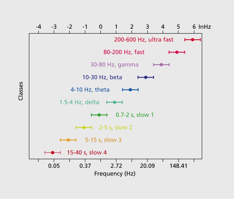
Figure 1. A system of interacting brain oscillations. Oscillatory classes in the cortex. Note the linear progression of the frequency classes (written next to commonly used name for each rhythm), on the natural log scale. This geometrical order is despite the fact that these frequency families were defined based on phenomenological correlates. Reproduced from ref 7: Buzsaki G, Draguhn A. Neuronal oscillations in cortical networks. Science. 2004;304:1926-1929. Copyright © American Association for the Advancement of Science 2004
Figure 1. A system of interacting brain oscillations. Oscillatory classes in the cortex. Note the linear progression of the frequency classes (written next to commonly used name for each rhythm), on the natural log scale. This geometrical order is despite the fact that these frequency families were defined based on phenomenological correlates. Reproduced from ref 7: Buzsaki G, Draguhn A. Neuronal oscillations in cortical networks. Science. 2004;304:1926-1929. Copyright © American Association for the Advancement of Science 2004CC BY-NC-ND · PubMed Central
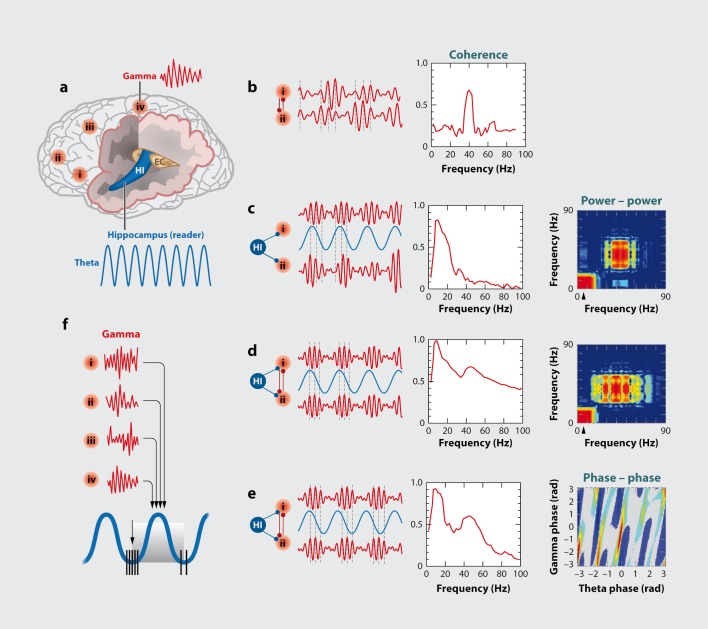
Figure 2. Oscillations can route information by multiple mechanisms, (a) View of the brain showing location of computation as revealed by transient γ oscillations (i-iv) and θ oscillation in the hippocampus (HI) entorhinal cortex (EC). Brain rhythms of the same and different kind (eg, θ, γ) can influence each other within and across structures by modulating the phase and/or the amplitude of the oscillations, (b) Phasephase coupling of “synthetic” γ oscillations between two structures for illustration purposes. Phase is shared between the waves — see dashed gray lines and coherence plot regardless of amplitude. Coherence spectrum between the two rhythms can reveal the strength of phase coupling, (c) Cross-frequency phase-amplitude coupling. Here the phase relationship between the respective γ oscillations can be random but the envelope of γ waves at both sites is modulated by the common θ rhythm and can be revealed by the power-power correlation (comodugram; right), (d) In addition to phase-amplitude coupling (θ-γ), the two sites may display γ coherence as well; a sign of strong inter-site interaction, (e) Cross-frequency phase-phase coupling. See also Figure 4. (f) A slow rhythm (eg, hippocampal θ oscillation) can modulate γ power at multiple neocortical areas so that the results of the local computations are returned to the hippocampus when the hippocampal network is in “readiness” phase of the slow oscillation. Reproduced from ref 13: Buzsáki G, WangX-J. Mechanisms of γ oscillations. Ann Rev Neurosci. 2012;35:203-225. Copyright © Annual Reviews 2012
Figure 2. Oscillations can route information by multiple mechanisms, (a) View of the brain showing location of computation as revealed by transient γ oscillations (i-iv) and θ oscillation in the hippocampus (HI) entorhinal cortex (EC). Brain rhythms of the same and different kind (eg, θ, γ) can influence each other within and across structures by modulating the phase and/or the amplitude of the oscillations, (b) Phasephase coupling of “synthetic” γ oscillations between two structures for illustration purposes. Phase is shared between the waves — see dashed gray lines and coherence plot regardless of amplitude. Coherence spectrum between the two rhythms can reveal the strength of phase coupling, (c) Cross-frequency phase-amplitude coupling. Here the phase relationship between the respective γ oscillations can be random but the envelope of γ waves at both sites is modulated by the common θ rhythm and can be revealed by the power-power correlation (comodugram; right), (d) In addition to phase-amplitude coupling (θ-γ), the two sites may display γ coherence as well; a sign of strong inter-site interaction, (e) Cross-frequency phase-phase coupling. See also Figure 4. (f) A slow rhythm (eg, hippocampal θ oscillation) can modulate γ power at multiple neocortical areas so that the results of the local computations are returned to the hippocampus when the hippocampal network is in “readiness” phase of the slow oscillation. Reproduced from ref 13: Buzsáki G, WangX-J. Mechanisms of γ oscillations. Ann Rev Neurosci. 2012;35:203-225. Copyright © Annual Reviews 2012CC BY-NC-ND · PubMed Central
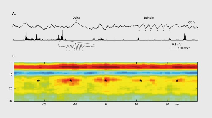
Figure 3. Cross-frequency coupling contributes to the hierarchy of brain rhythms, (a) Local field potential trace from layer 5 of the rat neocortex (1 Hz - 3 kHz) and a filtered (140-240 Hz) and rectified derivative of a trace from the hippocampal CA1 pyramidal layer, illustrating the emergence of “ripples.” One ripple event is shown at an expanded time scale. Peak of a δ wave and troughs of a sleep spindle are marked by asterisks. (b) Hippocampal ripple-triggered power spectrogram of neocortical activity centered on hippocampal ripples. Note that ripple activity is modulated by the sleep spindles (as revealed by the power in the 10-1 8 Hz band), and both events are modulated by the slow oscillation (strong red band 0-3 Hz), and all three oscillations are biased by the phase of the ultraslow rhythm (approximately 0.1 Hz, asterisks). CX, V, Cortex, Layer 5 Reproduced from ref 21: Sirota A, Csicsvari J, Buhl D, Buzsaki G. Communication between neocortex and hippocampus during sleep in rodents. Proc Natl AcadSciUSA. 2003;100:2065-2069. Copyright © National Academy of Science 2003
Figure 3. Cross-frequency coupling contributes to the hierarchy of brain rhythms, (a) Local field potential trace from layer 5 of the rat neocortex (1 Hz - 3 kHz) and a filtered (140-240 Hz) and rectified derivative of a trace from the hippocampal CA1 pyramidal layer, illustrating the emergence of “ripples.” One ripple event is shown at an expanded time scale. Peak of a δ wave and troughs of a sleep spindle are marked by asterisks. (b) Hippocampal ripple-triggered power spectrogram of neocortical activity centered on hippocampal ripples. Note that ripple activity is modulated by the sleep spindles (as revealed by the power in the 10-1 8 Hz band), and both events are modulated by the slow oscillation (strong red band 0-3 Hz), and all three oscillations are biased by the phase of the ultraslow rhythm (approximately 0.1 Hz, asterisks). CX, V, Cortex, Layer 5 Reproduced from ref 21: Sirota A, Csicsvari J, Buhl D, Buzsaki G. Communication between neocortex and hippocampus during sleep in rodents. Proc Natl AcadSciUSA. 2003;100:2065-2069. Copyright © National Academy of Science 2003CC BY-NC-ND · PubMed Central
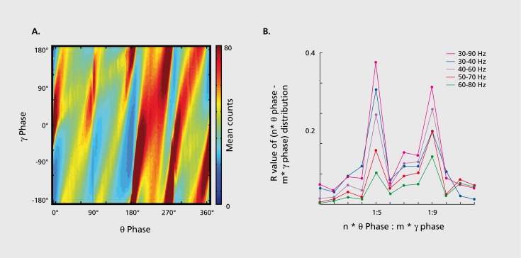
Figure 4. Phase-phase coupling between oscillations, (a) Plot illustrates phase-phase relationship between hippocampal θ and γ oscillations. Note that faster and slower γ oscillations (different diagonal bands) are simultaneously present, (b) Mean radial distance values (R value) from the distribution of the difference between θ and γ phases calculated for different n:m (θ:γ) relationships (1:1, 1:2....1:12) for several filtered γ frequency bands. Note large peaks (also known as Arnold's tongues) at 5 (mainly for band 30-40 Hz) and 9 (mainly for 50-80 Hz). Reproduced from ref 1 14: Belluscio MA, Mizuseki K, Schmidt R, Kempter R, Buzsaki G. Cross-frequency phase-phase coupling between θ and γ oscillations in the hippocampus. J Neurosci. 2012:32:423-435. Copyright © Society for Neuroscience 2012
Figure 4. Phase-phase coupling between oscillations, (a) Plot illustrates phase-phase relationship between hippocampal θ and γ oscillations. Note that faster and slower γ oscillations (different diagonal bands) are simultaneously present, (b) Mean radial distance values (R value) from the distribution of the difference between θ and γ phases calculated for different n:m (θ:γ) relationships (1:1, 1:2....1:12) for several filtered γ frequency bands. Note large peaks (also known as Arnold's tongues) at 5 (mainly for band 30-40 Hz) and 9 (mainly for 50-80 Hz). Reproduced from ref 1 14: Belluscio MA, Mizuseki K, Schmidt R, Kempter R, Buzsaki G. Cross-frequency phase-phase coupling between θ and γ oscillations in the hippocampus. J Neurosci. 2012:32:423-435. Copyright © Society for Neuroscience 2012CC BY-NC-ND · PubMed Central
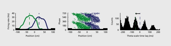
Figure 5. Multiple temporal representations in the hippocampus. Two CA1 pyramidal cells (green and blue) with overlapping place field representations on a linear track (black). It takes several seconds for the animal to pass from the peak (dashed line) of place field 1 (green) to place field 2 (blue). Middle panel, plots of the spikes as a function of the rat's position on the track and their θ phase. Each dot is an action potential. Ordinate, phase of θ shown twice for better visualization. Note that as the animal moves closer to the center of the place field for each cell, say running from left to right, the action potentials move closer to the 180-degree phase of the oscillation and as it moves away, the action potentials move further away. Right panel, time offset between the spikes of the reference (blue) and the green neurons at the θ time scale (arrow), as shown in a cross-correlogram. The time offsets of place cell spikes within the θ cycle and their distance (or travel time) representation are correlated. Reproduced from ref 128: Diba K, Buzsaki G. Hippocampal network dynamics constrain the time lag between pyramidal cells across modified environments. J Neurosci. 2008:28:1 3448-1 3456. Copyright © Society for Neuroscience 2008
Figure 5. Multiple temporal representations in the hippocampus. Two CA1 pyramidal cells (green and blue) with overlapping place field representations on a linear track (black). It takes several seconds for the animal to pass from the peak (dashed line) of place field 1 (green) to place field 2 (blue). Middle panel, plots of the spikes as a function of the rat's position on the track and their θ phase. Each dot is an action potential. Ordinate, phase of θ shown twice for better visualization. Note that as the animal moves closer to the center of the place field for each cell, say running from left to right, the action potentials move closer to the 180-degree phase of the oscillation and as it moves away, the action potentials move further away. Right panel, time offset between the spikes of the reference (blue) and the green neurons at the θ time scale (arrow), as shown in a cross-correlogram. The time offsets of place cell spikes within the θ cycle and their distance (or travel time) representation are correlated. Reproduced from ref 128: Diba K, Buzsaki G. Hippocampal network dynamics constrain the time lag between pyramidal cells across modified environments. J Neurosci. 2008:28:1 3448-1 3456. Copyright © Society for Neuroscience 2008CC BY-NC-ND · PubMed Central
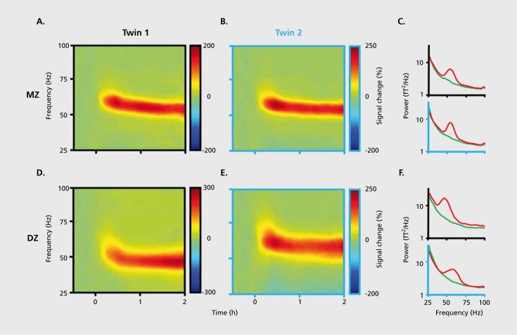
Figure 6. Brain rhythms are gene-regulated and unique, (a, b) Time-frequency display of visually induced γ band activity in a monozygotic (MZ) twin pair. C, Average spectra! power of magnetoencephalogram activity during control (green) and visual stimulation (red) epochs. D-F, same as A-C in a dizygotic (DZ) twin pair. Note stronger similarity of frequency and temporal dynamic changes of the MEG in the MZ pair relative to the DZ pair. Reproduced from ref 198: van Pelt S, Boomsma Dl, Fries P. Magnetoencephalography in twins reveals a strong genetic determination of the peak frequency of visually induced y-band synchronization. J Neurosci. 201 2:32:3388-3392. Copyright © Society for Neuroscience 2012
Figure 6. Brain rhythms are gene-regulated and unique, (a, b) Time-frequency display of visually induced γ band activity in a monozygotic (MZ) twin pair. C, Average spectra! power of magnetoencephalogram activity during control (green) and visual stimulation (red) epochs. D-F, same as A-C in a dizygotic (DZ) twin pair. Note stronger similarity of frequency and temporal dynamic changes of the MEG in the MZ pair relative to the DZ pair. Reproduced from ref 198: van Pelt S, Boomsma Dl, Fries P. Magnetoencephalography in twins reveals a strong genetic determination of the peak frequency of visually induced y-band synchronization. J Neurosci. 201 2:32:3388-3392. Copyright © Society for Neuroscience 2012CC BY-NC-ND · PubMed Central
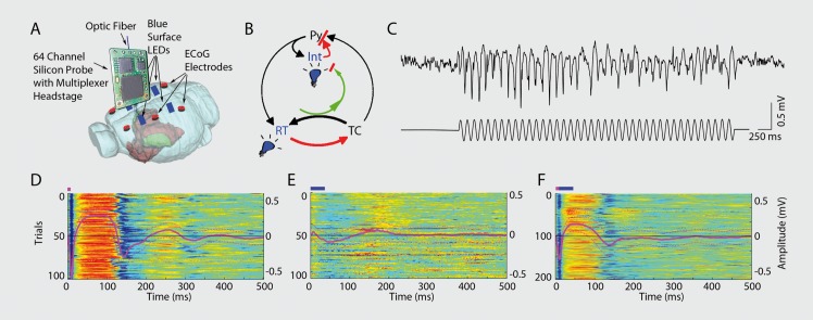
Figure 7. Closed-loop interaction in the thalamocortical loop, (a) Experimental setup. Optic fiber is placed into the reticular nucleus of thalamus in a FValb-IRES-Cre:Ai32 double transgenic mouse to induce spike-wave seizure-like pattern; shown in (c). Blue LEDs (squares) placed epidurally at two positions in each hemisphere, (b). Schematic of the reverberation in the thalamocortical loop. Neurons of the thalamus: reticular nucleus cells (RT), thalamocortical projection neurons TC). Neurons of the cortex: pyramidal cells (Py) and inhibitory interneurons (int). (d) Light stimulation of the pan/albumin RT neurons alone induces spike-waves, whereas light stimulation of cortical pan/albumin interneurons alone induces rebound excitation in cortical pyramidal cells (Py). Combined and phase shifted stimulation of RT and cortex attenuates the induced spike-wave activity. Reproduced from ref 252: Berenyi A, Belluscio M, Mao D, Buzsaki G. Closed-loop control of epilepsy by transcranial electrical stimulation. Science. 2012;337:735-737. Copyright ©American Association for the Advancement of Science 2012
Figure 7. Closed-loop interaction in the thalamocortical loop, (a) Experimental setup. Optic fiber is placed into the reticular nucleus of thalamus in a FValb-IRES-Cre:Ai32 double transgenic mouse to induce spike-wave seizure-like pattern; shown in (c). Blue LEDs (squares) placed epidurally at two positions in each hemisphere, (b). Schematic of the reverberation in the thalamocortical loop. Neurons of the thalamus: reticular nucleus cells (RT), thalamocortical projection neurons TC). Neurons of the cortex: pyramidal cells (Py) and inhibitory interneurons (int). (d) Light stimulation of the pan/albumin RT neurons alone induces spike-waves, whereas light stimulation of cortical pan/albumin interneurons alone induces rebound excitation in cortical pyramidal cells (Py). Combined and phase shifted stimulation of RT and cortex attenuates the induced spike-wave activity. Reproduced from ref 252: Berenyi A, Belluscio M, Mao D, Buzsaki G. Closed-loop control of epilepsy by transcranial electrical stimulation. Science. 2012;337:735-737. Copyright ©American Association for the Advancement of Science 2012CC BY-NC-ND · PubMed Central

Verification

Claims the paper makes

The claims and statistics of this paper haven’t been checked yet.

Run the check — free, no account needed. It reads the open-access text and checks the sources it cites.

A few free checks a day · papers already verified open instantly · results are public

Claims other papers make about this one

Each is checked against this paper’s own text: does it say what the citing paper says it does?

Self-check

Percentages, sums and reported differences re-computed from the paper’s own numbers.

The self-check needs the paper’s full text.

Citations

Cites

Show all 233 references

Research works printed but not linked: 24

  1. Sherrington CS., 1941, Man On His Nature.New York, NY; Cambridge, UK: The Macmillan Company; The University Press.
  2. Nunez PL., 2005, Electric Fields of the Brain: the Neurophysics of EEG. 2nd ed. New York, NY: Oxford University Press.
  3. Jerison HJ., 1973, Evolution of the Brain and Intelligence. London, UK: Academic Press.
  4. Hebb DO., 1949, The Organization of Behavior: a Neuropsychological Theory New York, NY: Wiley
  5. Johnston D., 1994, Foundations of Cellular Neurophysiology Cambridge, MA: MIT Press, Bradford Books
  6. Lachaux J-P., 2012, Prog Neurobiol In press
  7. O'Keefe J., 1978, The Hippocampus as a Cognitive Map New York, NY: Clarendon Press
  8. Royer S., 2012, Neurosci., 15, 769
  9. O'Neill J., 2008, Neurosci., 11, 209
  10. Girardeau G., 2009, Neurosci., 12, 1222
  11. Buzsaki G., 2012, Prog Neurobiol In press
  12. Szatmáry B., PLoS Comput Biol In press
  13. Nestler E., 2008, Molecular Neuropharmacology: A Foundation for Clinical Neuroscience, Second Edition New York, NY: McGrawHill Professional
  14. Kandel E., 2005, Psychiatry, Psychoanalysis, And The New Biology Of Mind Arlington, VA: American Psychiatric Publishing
  15. Vogel F., 1970, Human Genetics., 10, 91
  16. Matousek M., Electroencephalogr Clin Neurophysiol., 3, 603
  17. van Beijsterveldt CE., 1996, Am J Hum Genet., 58, 562
  18. Marescaux C., 1992, J Neural Trans Suppl., 35, 37
  19. Noebels JL., 1999, Adv Neurol., 79, 227
  20. Sadock BJ., 2000, Kaplan & Sadock Comprehensive Textbook of Psychiatry Philadelphia, PA: Lippincott Williams & Wilkins, 48
  21. Mathers C., 2008, The Global Burden of Disease: 2004 Update Geneva, Switzerland: World Health Organization
  22. rivedi MH., 2006, Psychiatry., 163, 28
  23. Weinberg RA., 2006, The Biology of Cancer New York, NY; London, UK: Garland Science
  24. Casals JB., 2011, Compar Med., 61, 305

Cited by

Sorted by citations

Items 101–101 of 101

Metadata

Authors
Buzsáki, G., & Watson, B. O.
Affiliations
  • New York University (US)
  • Weill Cornell Medicine (US)
Corresponding author
Buzsáki, György
Venue
Dialogues in Clinical Neuroscience
Published
2012-12-31
Volume
14
Issue
4
Pages
345-367
ISSN
  • 1294-8322 (ISSN-L)
  • 1958-5969
OpenAlex
W4214953342
Cited by
579
References
233
Citations by year
2026: 37 · 2025: 47 · 2024: 40 · 2023: 36 · 2022: 64 · 2021: 61 · 2020: 50 · 2019: 48 · 2018: 41 · 2017: 33 · 2016: 47 · 2015: 34 · 2014: 24 · 2013: 9
FWCI
10.98
Type
journal article
Language
en
Open access
Locations
  • Dialogues in Clinical Neuroscience (publishedVersion, cc-by-nc-nd)
  • Europe PMC (PubMed Central) (publishedVersion, cc-by-nc-nd)
  • PubMed Central (publishedVersion, cc-by-nc-nd)
  • PubMed (publishedVersion)
  • PubMed
Funders
National Institute of Mental Health (R01MH054671)
Topics
  • Life Sciences › Neuroscience › Cognitive Neuroscience › Neural dynamics and brain function
  • Life Sciences › Neuroscience › Cellular and Molecular Neuroscience › Neuroscience and Neuropharmacology Research
  • Health Sciences › Medicine › Clinical Neurology › Deep Brain Stimulation and Movement Disorders
Keywords
neural oscillations, efficient coding, neuronal excitability, cell assemblies, cortical networks, mental illness, neuropsychiatric disorders, neuromodulation
MeSH terms
Animals, Mental Disorders, Brain, Cognition, Cortical Synchronization, Humans, Periodicity

About these checks

Registry, retraction, Cites and statistics results are computed, not judged, with one exception: a printed reference that no index lists is read once by a model before it counts as a citation error. Claims are extracted by a model and kept only when their quote appears verbatim in the text; each cited source is then checked, with a model’s help, for whether it says what the claim relies on. Those verdicts can be wrong — read the claim’s quote and the cited source before relying on one.

Statistics and arithmetic are recomputed, not judged.

Confidence bands are shown only for fields calibrated on our annotated test set. None is calibrated yet, so no band is shown.

What counts as a claim ›How we measure confidence ›How we check this ›

Are you an author, or do you see a problem with this page? Request a review