

The DOI matches its Crossref record; 234 of the 237 references it cites were found in a registry or an index.
Oxidative Stress, Synaptic Dysfunction, and Alzheimer’s Disease
Open accessAbstract
Alzheimer's disease (AD) is a devastating neurodegenerative disorder without a cure. Most AD cases are sporadic where age represents the greatest risk factor. Lack of understanding of the disease mechanism hinders the development of efficacious therapeutic approaches. The loss of synapses in the affected brain regions correlates best with cognitive impairment in AD patients and has been considered as the early mechanism that precedes neuronal loss. Oxidative stress has been recognized as a contributing factor in aging and in the progression of multiple neurodegenerative diseases including AD. Increased production of reactive oxygen species (ROS) associated with age- and disease-dependent loss of mitochondrial function, altered metal homeostasis, and reduced antioxidant defense directly affect synaptic activity and neurotransmission in neurons leading to cognitive dysfunction. In addition, molecular targets affected by ROS include nuclear and mitochondrial DNA, lipids, proteins, calcium homeostasis, mitochondrial dynamics and function, cellular architecture, receptor trafficking and endocytosis, and energy homeostasis. Abnormal cellular metabolism in turn could affect the production and accumulation of amyloid-β (Aβ) and hyperphosphorylated Tau protein, which independently could exacerbate mitochondrial dysfunction and ROS production, thereby contributing to a vicious cycle. While mounting evidence implicates ROS in the AD etiology, clinical trials with antioxidant therapies have not produced consistent results. In this review, we will discuss the role of oxidative stress in synaptic dysfunction in AD, innovative therapeutic strategies evolved based on a better understanding of the complexity of molecular mechanisms of AD, and the dual role ROS play in health and disease.
Overview
The claims and statistics of this paper haven’t been checked yet.
Run the check — free, no account needed. It reads the open-access text and checks the sources it cites.
Integrity signals
Registry
Cites
Statistics
Figures
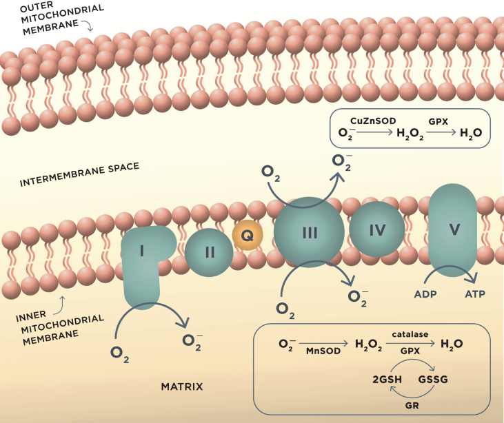
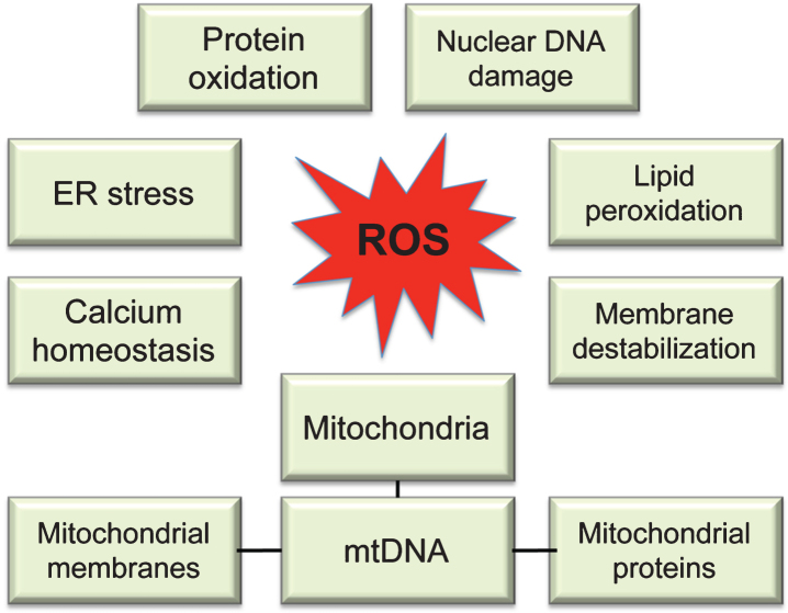
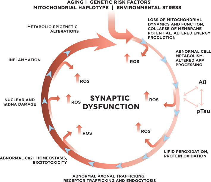
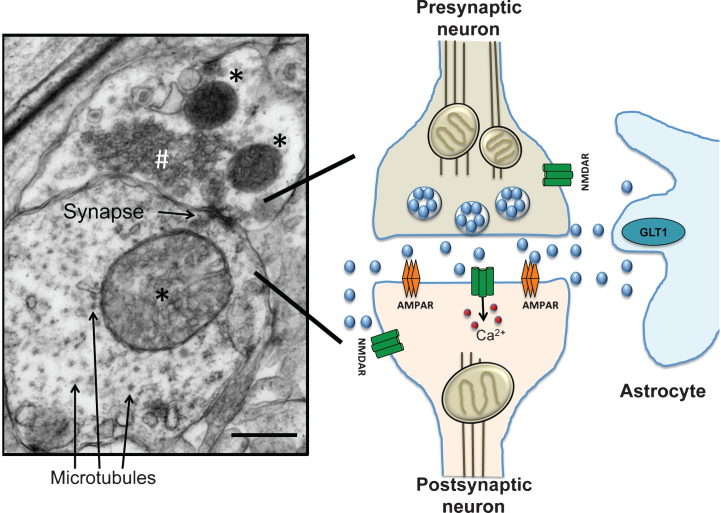
Verification
Claims the paper makes
The claims and statistics of this paper haven’t been checked yet.
Run the check — free, no account needed. It reads the open-access text and checks the sources it cites.
Claims other papers make about this one
Each is checked against this paper’s own text: does it say what the citing paper says it does?
The source backs part of the claim, not all of it — or backs it for a different population, condition or comparison.
In the citing paper
“Oxidative stress is associated with a number of inflammatory and degenerative pathologies and is a significant biomarker common to the pathophysiology of both AD and PD. 55 , 58”
From Therapeutic Properties and Pharmacological Activities of Asiaticoside and Madecassoside: A Review (2023) · cites this paper as [58]
Experimental — accuracy not yet measuredIn the citing paper
“since there is a strong correlation between immune activation and oxidative damage in neurodegenerative disease [42,43,44]”
From ROS Generation in Microglia: Understanding Oxidative Stress and Inflammation in Neurodegenerative Disease (2020) · cites this paper as [43]
Self-check
Percentages, sums and reported differences re-computed from the paper’s own numbers.
The self-check needs the paper’s full text.
Citations
Cites
Free Radicals in the Physiological Control of Cell Function
Dröge, W. (2002) · Physiological Reviews · doi:10.1152/physrev.00018.2001 · Cited by 9,855
Oxyl radicals, redox-sensitive signalling cascades and antioxidants
Genestra, M. (2007) · Cellular Signalling · doi:10.1016/j.cellsig.2007.04.009 · Cited by 4,930
Hypothetical model of dynamic biomarkers of the Alzheimer's pathological cascade
Jack, C. R., Knopman, D. S., Jagust, W. J., Shaw, L. M., Aisen, P. S., Weiner, M. W., Petersen, R. C., & Trojanowski, J. Q. (2010) · The Lancet Neurology · doi:10.1016/s1474-4422(09)70299-6 · Cited by 4,806
Terry, R. D., Masliah, E., Salmon, D. P., Butters, N., DeTeresa, R., Hill, R., Hansen, L. A., & Katzman, R. (1991) · Annals of Neurology · doi:10.1002/ana.410300410 · Cited by 4,397
Reactive oxygen species (ROS) homeostasis and redox regulation in cellular signaling
Ray, P. D., Huang, B.-W., & Tsuji, Y. (2012) · Cellular Signalling · doi:10.1016/j.cellsig.2012.01.008 · Cited by 4,373
Oxidative mechanisms in the toxicity of metal ions
Stohs, S. J., & Bagchi, D. (1995) · Free Radical Biology and Medicine · doi:10.1016/0891-5849(94)00159-h · Cited by 4,219
Triple-Transgenic Model of Alzheimer’s Disease with Plaques and Tangles
Oddo, S., Caccamo, A., Shepherd, J. D., Murphy, M. P., Golde, T. E., Kayed, R., Metherate, R., Mattson, M. P., Akbari, Y., & LaFerla, F. M. (2003) · Neuron · doi:10.1016/s0896-6273(03)00434-3 · Cited by 4,126
Malenka, R. C., & Bear, M. F. (2004) · Neuron · doi:10.1016/j.neuron.2004.09.012 · Cited by 3,968
Clinical and biomarker changes in dominantly inherited Alzheimer's disease
Bateman, R. J., Xiong, C., Benzinger, T. L. S., Fagan, A. M., Goate, A., Fox, N. C., Marcus, D. S., Cairns, N. J., Xie, X., Blazey, T. M., et al. (2012) · New England Journal of Medicine · doi:10.1056/nejmoa1202753 · Cited by 3,814
Cell Survival Responses to Environmental Stresses Via the Keap1-Nrf2-ARE Pathway
Kensler, T. W., Wakabayashi, N., & Biswal, S. (2007) · Annual Review of Pharmacology and Toxicology · doi:10.1146/annurev.pharmtox.46.120604.141046 · Cited by 3,630
Research works printed but not linked: 1
- 10.1007/s12603-013-0004-0 · DOI
Cited by
Sorted by citations
Oxidative stress, aging, and diseases
Liguori, I., Russo, G., Curcio, F., Bulli, G., Aran, L., Della-Morte, D., Gargiulo, G., Testa, G., Cacciatore, F., Bonaduce, D., et al. (2018) · Clinical Interventions in Aging · doi:10.2147/cia.s158513 · Cited by 3,979
Simpson, D. S. A., & Oliver, P. L. (2020) · Antioxidants · doi:10.3390/antiox9080743 · Cited by 1,044
Molecular and cellular mechanisms underlying the pathogenesis of Alzheimer’s disease
Guo, T., Zhang, D., Zeng, Y., Huang, T. Y., Xu, H., & Zhao, Y. (2020) · Molecular Neurodegeneration · doi:10.1186/s13024-020-00391-7 · Cited by 994
Role of oxidative stress in depression
Bhatt, S., Nagappa, A. N., & Patil, C. R. (2020) · Drug Discovery Today · doi:10.1016/j.drudis.2020.05.001 · Cited by 854
The plastic brain: neurotoxicity of micro- and nanoplastics
Prüst, M., Meijer, J., & Westerink, R. H. S. (2020) · Particle and Fibre Toxicology · doi:10.1186/s12989-020-00358-y · Cited by 811
Metadata
- Authors
- Tönnies, E., & Trushina, E.
- Affiliations
- Mayo Clinic (US)
- Corresponding author
- Trushina, Eugenia
- ORCID
- Trushina, Eugenia
- Venue
- Journal of Alzheimer s Disease
- Publisher
- SAGE Publications
- Published
- 2017-04-19
- Volume
- 57
- Issue
- 4
- Pages
- 1105-1121
- ISSN
- 1387-2877 (ISSN-L)
- 1875-8908
- PMC
- PMC5409043
- OpenAlex
- W2568450381
- Cited by
- 1,750
- References
- 237
- Citations by year
- 2026: 135 · 2025: 220 · 2024: 259 · 2023: 258 · 2022: 253 · 2021: 246 · 2020: 193 · 2019: 101 · 2018: 54 · 2017: 30
- FWCI
- 12.72
- Type
- journal article
- Language
- en
- Open access
- bronze
- doi.org
- Locations
- Journal of Alzheimer s Disease (publishedVersion)
- PubMed Central (publishedVersion, cc-by-nc)
- PubMed (publishedVersion)
- Funders
- National Institutes of Health
- National Institute on Aging (RF1AG055549)
- National Institute of Environmental Health Sciences (R01ES020715)
- Topics
- Life Sciences › Neuroscience › Cellular and Molecular Neuroscience › Alzheimer's disease research and treatments
- Life Sciences › Biochemistry, Genetics and Molecular Biology › Molecular Biology › Mitochondrial Function and Pathology
- Health Sciences › Medicine › Psychiatry and Mental health › Dementia and Cognitive Impairment Research
- Keywords
- Alzheimer's disease, oxidative stress
- MeSH terms
- Alzheimer Disease, Animals, Humans, Synapses, Oxidative Stress
About these checks
Registry, retraction, Cites and statistics results are computed, not judged, with one exception: a printed reference that no index lists is read once by a model before it counts as a citation error. Claims are extracted by a model and kept only when their quote appears verbatim in the text; each cited source is then checked, with a model’s help, for whether it says what the claim relies on. Those verdicts can be wrong — read the claim’s quote and the cited source before relying on one.
Statistics and arithmetic are recomputed, not judged.
Confidence bands are shown only for fields calibrated on our annotated test set. None is calibrated yet, so no band is shown.
What counts as a claim ›How we measure confidence ›How we check this ›
Are you an author, or do you see a problem with this page? Request a review
