

The DOI matches its Crossref record; 234 of the 237 references it cites were found in a registry or an index.
Oxidative Stress, Synaptic Dysfunction, and Alzheimer’s Disease
Open accessAbstract
Alzheimer's disease (AD) is a devastating neurodegenerative disorder without a cure. Most AD cases are sporadic where age represents the greatest risk factor. Lack of understanding of the disease mechanism hinders the development of efficacious therapeutic approaches. The loss of synapses in the affected brain regions correlates best with cognitive impairment in AD patients and has been considered as the early mechanism that precedes neuronal loss. Oxidative stress has been recognized as a contributing factor in aging and in the progression of multiple neurodegenerative diseases including AD. Increased production of reactive oxygen species (ROS) associated with age- and disease-dependent loss of mitochondrial function, altered metal homeostasis, and reduced antioxidant defense directly affect synaptic activity and neurotransmission in neurons leading to cognitive dysfunction. In addition, molecular targets affected by ROS include nuclear and mitochondrial DNA, lipids, proteins, calcium homeostasis, mitochondrial dynamics and function, cellular architecture, receptor trafficking and endocytosis, and energy homeostasis. Abnormal cellular metabolism in turn could affect the production and accumulation of amyloid-β (Aβ) and hyperphosphorylated Tau protein, which independently could exacerbate mitochondrial dysfunction and ROS production, thereby contributing to a vicious cycle. While mounting evidence implicates ROS in the AD etiology, clinical trials with antioxidant therapies have not produced consistent results. In this review, we will discuss the role of oxidative stress in synaptic dysfunction in AD, innovative therapeutic strategies evolved based on a better understanding of the complexity of molecular mechanisms of AD, and the dual role ROS play in health and disease.
Overview
The claims and statistics of this paper haven’t been checked yet.
Run the check — free, no account needed. It reads the open-access text and checks the sources it cites.
Integrity signals
Registry
Cites
Statistics
Figures
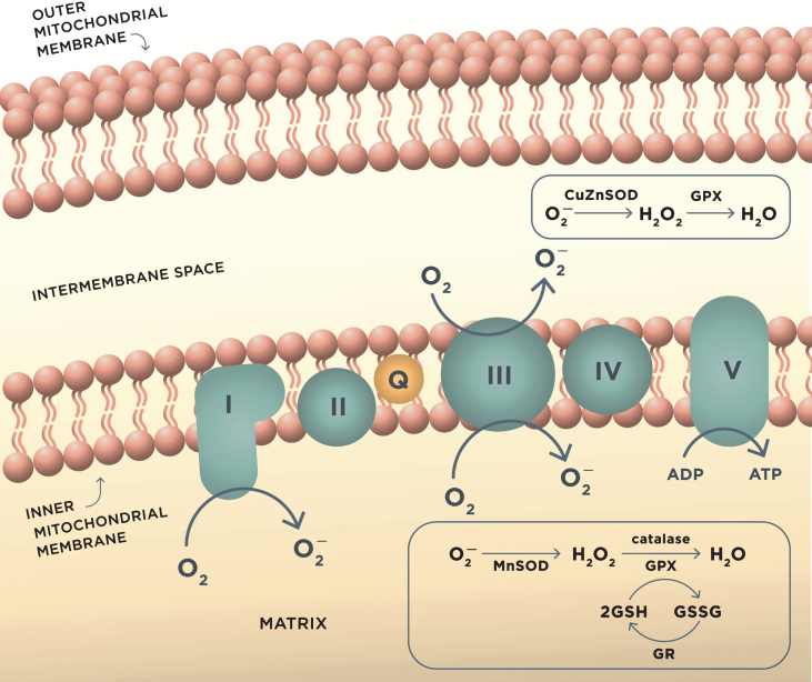
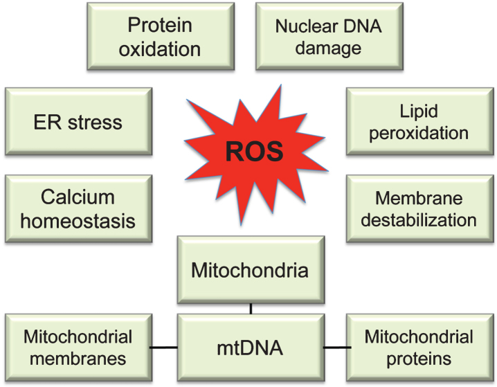
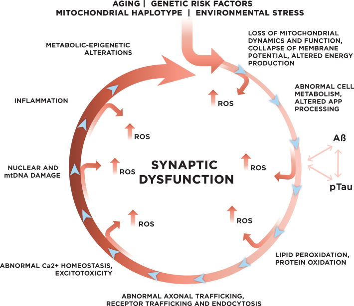
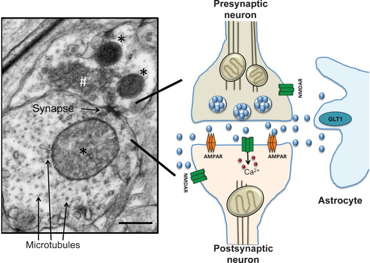
Verification
Claims the paper makes
The claims and statistics of this paper haven’t been checked yet.
Run the check — free, no account needed. It reads the open-access text and checks the sources it cites.
Claims other papers make about this one
Each is checked against this paper’s own text: does it say what the citing paper says it does?
The source backs part of the claim, not all of it — or backs it for a different population, condition or comparison.
In the citing paper
“Oxidative stress is associated with a number of inflammatory and degenerative pathologies and is a significant biomarker common to the pathophysiology of both AD and PD. 55 , 58”
From Therapeutic Properties and Pharmacological Activities of Asiaticoside and Madecassoside: A Review (2023) · cites this paper as [58]
Experimental — accuracy not yet measuredIn the citing paper
“since there is a strong correlation between immune activation and oxidative damage in neurodegenerative disease [42,43,44]”
From ROS Generation in Microglia: Understanding Oxidative Stress and Inflammation in Neurodegenerative Disease (2020) · cites this paper as [43]
Self-check
Percentages, sums and reported differences re-computed from the paper’s own numbers.
The self-check needs the paper’s full text.
Citations
Cites
Free Radicals in the Physiological Control of Cell Function
Dröge, W. (2002) · Physiological Reviews · doi:10.1152/physrev.00018.2001 · Cited by 9,855
Oxyl radicals, redox-sensitive signalling cascades and antioxidants
Genestra, M. (2007) · Cellular Signalling · doi:10.1016/j.cellsig.2007.04.009 · Cited by 4,930
Hypothetical model of dynamic biomarkers of the Alzheimer's pathological cascade
Jack, C. R., Knopman, D. S., Jagust, W. J., Shaw, L. M., Aisen, P. S., Weiner, M. W., Petersen, R. C., & Trojanowski, J. Q. (2010) · The Lancet Neurology · doi:10.1016/s1474-4422(09)70299-6 · Cited by 4,806
Terry, R. D., Masliah, E., Salmon, D. P., Butters, N., DeTeresa, R., Hill, R., Hansen, L. A., & Katzman, R. (1991) · Annals of Neurology · doi:10.1002/ana.410300410 · Cited by 4,397
Reactive oxygen species (ROS) homeostasis and redox regulation in cellular signaling
Ray, P. D., Huang, B.-W., & Tsuji, Y. (2012) · Cellular Signalling · doi:10.1016/j.cellsig.2012.01.008 · Cited by 4,373
Oxidative mechanisms in the toxicity of metal ions
Stohs, S. J., & Bagchi, D. (1995) · Free Radical Biology and Medicine · doi:10.1016/0891-5849(94)00159-h · Cited by 4,219
Triple-Transgenic Model of Alzheimer’s Disease with Plaques and Tangles
Oddo, S., Caccamo, A., Shepherd, J. D., Murphy, M. P., Golde, T. E., Kayed, R., Metherate, R., Mattson, M. P., Akbari, Y., & LaFerla, F. M. (2003) · Neuron · doi:10.1016/s0896-6273(03)00434-3 · Cited by 4,126
Malenka, R. C., & Bear, M. F. (2004) · Neuron · doi:10.1016/j.neuron.2004.09.012 · Cited by 3,968
Clinical and biomarker changes in dominantly inherited Alzheimer's disease
Bateman, R. J., Xiong, C., Benzinger, T. L. S., Fagan, A. M., Goate, A., Fox, N. C., Marcus, D. S., Cairns, N. J., Xie, X., Blazey, T. M., et al. (2012) · New England Journal of Medicine · doi:10.1056/nejmoa1202753 · Cited by 3,814
Cell Survival Responses to Environmental Stresses Via the Keap1-Nrf2-ARE Pathway
Kensler, T. W., Wakabayashi, N., & Biswal, S. (2007) · Annual Review of Pharmacology and Toxicology · doi:10.1146/annurev.pharmtox.46.120604.141046 · Cited by 3,630
Research works printed but not linked: 1
- 10.1007/s12603-013-0004-0 · DOI
Cited by
Sorted by citations
Oxidative stress, aging, and diseases
Liguori, I., Russo, G., Curcio, F., Bulli, G., Aran, L., Della-Morte, D., Gargiulo, G., Testa, G., Cacciatore, F., Bonaduce, D., et al. (2018) · Clinical Interventions in Aging · doi:10.2147/cia.s158513 · Cited by 3,979
Simpson, D. S. A., & Oliver, P. L. (2020) · Antioxidants · doi:10.3390/antiox9080743 · Cited by 1,044
Molecular and cellular mechanisms underlying the pathogenesis of Alzheimer’s disease
Guo, T., Zhang, D., Zeng, Y., Huang, T. Y., Xu, H., & Zhao, Y. (2020) · Molecular Neurodegeneration · doi:10.1186/s13024-020-00391-7 · Cited by 994
Role of oxidative stress in depression
Bhatt, S., Nagappa, A. N., & Patil, C. R. (2020) · Drug Discovery Today · doi:10.1016/j.drudis.2020.05.001 · Cited by 854
The plastic brain: neurotoxicity of micro- and nanoplastics
Prüst, M., Meijer, J., & Westerink, R. H. S. (2020) · Particle and Fibre Toxicology · doi:10.1186/s12989-020-00358-y · Cited by 811
Emerging roles of oxidative stress in brain aging and Alzheimer's disease
Ionescu-Tucker, A., & Cotman, C. W. (2021) · Neurobiology of Aging · doi:10.1016/j.neurobiolaging.2021.07.014 · Cited by 737
Dash, U. C., Bhol, N. K., Swain, S. K., Samal, R. R., Nayak, P. K., Raina, V., Panda, S. K., Kerry, R. G., Duttaroy, A. K., & Jena, A. B. (2024) · Acta Pharmaceutica Sinica B · doi:10.1016/j.apsb.2024.10.004 · Cited by 806
Lipids and Alzheimer’s Disease
Kao, Y.-C., Ho, P.-C., Tu, Y.-K., Jou, I.-M., & Tsai, K.-J. (2020) · International Journal of Molecular Sciences · doi:10.3390/ijms21041505 · Cited by 485
The initiation of oxidative stress and therapeutic strategies in wound healing
Wang, G., Yang, F., Zhou, W., Xiao, N., Luo, M., & Tang, Z. (2022) · Biomedicine & Pharmacotherapy · doi:10.1016/j.biopha.2022.114004 · Cited by 451
Mitochondrial dysfunction in neurodegenerative disorders
Klemmensen, M. M., Borrowman, S. H., Pearce, C., Pyles, B., & Chandra, B. (2024) · Neurotherapeutics · doi:10.1016/j.neurot.2023.10.002 · Cited by 396
Therapeutic application of quercetin in aging-related diseases: SIRT1 as a potential mechanism
Cui, Z., Zhao, X., Amevor, F. K., Du, X., Wang, Y., Li, D., Shu, G., Tian, Y., & Zhao, X. (2022) · Frontiers in Immunology · doi:10.3389/fimmu.2022.943321 · Cited by 413
Gadhave, D. G., Sugandhi, V. V., Jha, S. K., Nangare, S., Gupta, G., Singh, S. K., Dua, K., Cho, H., Hansbro, P. M., & Paudel, K. R. (2024) · Ageing Research Reviews · doi:10.1016/j.arr.2024.102357 · Cited by 420
Therapeutic Potential of Centella asiatica and Its Triterpenes: A Review
Sun, B., Wu, L., Wu, Y., Zhang, C., Qin, L., Hayashi, M., Kudo, M., Gao, M., & Liu, T. (2020) · Frontiers in Pharmacology · doi:10.3389/fphar.2020.568032 · Cited by 353
The Role of Oxidative Stress in Parkinson’s Disease
Chang, K., & Chen, C. (2020) · Antioxidants · doi:10.3390/antiox9070597 · Cited by 369
Exercise benefits on Alzheimer’s disease: State-of-the-science
Valenzuela, P. L., Castillo-García, A., Morales, J. S., de la Villa, P., Hampel, H., Emanuele, E., Lista, S., & Lucia, A. (2020) · Ageing Research Reviews · doi:10.1016/j.arr.2020.101108 · Cited by 337
García-Sánchez, A., Miranda‐Díaz, A. G., & Cardona-Muñóz, E. G. (2020) · Oxidative Medicine and Cellular Longevity · doi:10.1155/2020/2082145 · Cited by 369
Neuroinflammation in Alzheimer’s Disease
Onyango, I. G., Jauregui, G. V., Čarná, M., Bennett, J. P., & Stokin, G. B. (2021) · Biomedicines · doi:10.3390/biomedicines9050524 · Cited by 330
Shandilya, S., Kumar, S., Jha, N. K., Kesari, K. K., & Ruokolainen, J. (2021) · Journal of Advanced Research · doi:10.1016/j.jare.2021.09.005 · Cited by 310
Oxidative stress in alzheimer’s disease: A review on emergent natural polyphenolic therapeutics
Cassidy, L., Fernandez, F., Johnson, J. B., Naiker, M., Owoola, A. G., & Broszczak, D. A. (2019) · Complementary Therapies in Medicine · doi:10.1016/j.ctim.2019.102294 · Cited by 288
Application of Metabolomics in Alzheimer’s Disease
Wilkins, J. M., & Trushina, E. (2018) · Frontiers in Neurology · doi:10.3389/fneur.2017.00719 · Cited by 268
Self-citationThe Association of Tau With Mitochondrial Dysfunction in Alzheimer’s Disease
Cheng, Y., & Bai, F. (2018) · Frontiers in Neuroscience · doi:10.3389/fnins.2018.00163 · Cited by 239
Jamwal, S., Blackburn, J. K., & Elsworth, J. D. (2021) · Pharmacology & Therapeutics · doi:10.1016/j.pharmthera.2020.107705 · Cited by 236
Dewanjee, S., Chakraborty, P., Bhattacharya, H., Chacko, L., Singh, B., Chaudhary, A., Javvaji, K., Pradhan, S. R., Vallamkondu, J., Dey, A., et al. (2022) · Free Radical Biology and Medicine · doi:10.1016/j.freeradbiomed.2022.09.032 · Cited by 243
Neuroprotective Natural Products for Alzheimer’s Disease
Chen, X., Drew, J., Berney, W., & Lei, W. (2021) · Cells · doi:10.3390/cells10061309 · Cited by 236
The PI3K/Akt signaling axis in Alzheimer’s disease: a valuable target to stimulate or suppress?
Razani, E., Pourbagheri‐Sigaroodi, A., Safaroghli‐Azar, A., Zoghi, A., Bavarsad, M. S., & Bashash, D. (2021) · Cell Stress and Chaperones · doi:10.1007/s12192-021-01231-3 · Cited by 232
Alzheimer’s Disease: Key Insights from Two Decades of Clinical Trial Failures
Kim, C. K., Lee, Y. R., Ong, L., Gold, M., Kalali, A., & Sarkar, J. (2022) · Journal of Alzheimer s Disease · doi:10.3233/jad-215699 · Cited by 241
Ochalek, A., Mihalik, B., Avci, H. X., Chandrasekaran, A., Téglási, A., Bock, I., Giudice, M. L., Táncos, Z., Molnár, K., László, L., et al. (2017) · Alzheimer's Research & Therapy · doi:10.1186/s13195-017-0317-z · Cited by 225
A review on advances of treatment modalities for Alzheimer's disease
Thoe, E. S., Fauzi, A., Tang, Y.-Q., Chamyuang, S., & Chia, A. Y. Y. (2021) · Life Sciences · doi:10.1016/j.lfs.2021.119129 · Cited by 234
Therapeutic Properties and Pharmacological Activities of Asiaticoside and Madecassoside: A Review
Bandopadhyay, S., Mandal, S., Ghorai, M., Jha, N. K., Kumar, M., Radha, Ghosh, A., Proćków, J., Pérez de la Lastra, J. M., & Dey, A. (2023) · Journal of Cellular and Molecular Medicine · doi:10.1111/jcmm.17635 · Cited by 220
Mitochondrial Dysfunction: a Potential Therapeutic Target to Treat Alzheimer’s Disease
Nand, S., Singh, C., Singh, A., Singh, M. P., & Singh, S. P. (2020) · Molecular Neurobiology · doi:10.1007/s12035-020-01945-y · Cited by 215
Oxidative Stress and the Microbiota‐Gut‐Brain Axis
Dumitrescu, L., Mitrea, I., Cozma, L., Tulbă, D., Hinescu, M. E., Ceafalan, L. C., Gherghiceanu, M., & Popescu, B. O. (2018) · Oxidative Medicine and Cellular Longevity · doi:10.1155/2018/2406594 · Cited by 222
Hannan, M. A., Dash, R., Sohag, A. A. M., Haque, M. N., & Moon, I. S. (2020) · Frontiers in Molecular Neuroscience · doi:10.3389/fnmol.2020.00116 · Cited by 215
A Review for the Pharmacological Effect of Lycopene in Central Nervous System Disorders
Chen, D., Huang, C., & Chen, Z. (2019) · Biomedicine & Pharmacotherapy · doi:10.1016/j.biopha.2018.12.151 · Cited by 206
Griñán-Ferré, C., Bellver-Sanchis, A., Izquierdo, V., Corpas, R., Roig-Soriano, J., Chillón, M., Andres-Lacueva, C., Somogyvári, M., Sőti, C., Sanfeliu, C., et al. (2021) · Ageing Research Reviews · doi:10.1016/j.arr.2021.101271 · Cited by 208
Role of Nrf2 in aging, Alzheimer’s and other neurodegenerative diseases
George, M., Tharakan, M., Culberson, J. W., Reddy, A. P., & Reddy, P. H. (2022) · Ageing Research Reviews · doi:10.1016/j.arr.2022.101756 · Cited by 216
Role of reactive oxygen species in the progression of Alzheimer’s disease
Bhatt, S., Puli, L., & Patil, C. R. (2020) · Drug Discovery Today · doi:10.1016/j.drudis.2020.12.004 · Cited by 217
Updates in Alzheimer's disease: from basic research to diagnosis and therapies
Liu, E., Zhang, Y., & Wang, J. (2024) · Translational Neurodegeneration · doi:10.1186/s40035-024-00432-x · Cited by 241
Mitochondrial dynamics and transport in Alzheimer's disease
Flannery, P. J., & Trushina, E. V. (2019) · Molecular and Cellular Neuroscience · doi:10.1016/j.mcn.2019.06.009 · Cited by 203
Self-citationNovel target for treating Alzheimer’s Diseases: Crosstalk between the Nrf2 pathway and autophagy
Zhang, W., Feng, C., & Jiang, H. (2020) · Ageing Research Reviews · doi:10.1016/j.arr.2020.101207 · Cited by 207
Interplay between MicroRNAs and Oxidative Stress in Neurodegenerative Diseases
Konovalova, J., Gerasymchuk, D., Parkkinen, I., Chmielarz, P., & Domanskyi, A. (2019) · International Journal of Molecular Sciences · doi:10.3390/ijms20236055 · Cited by 204
The duality of amyloid-β: its role in normal and Alzheimer’s disease states
Azargoonjahromi, A. (2024) · Molecular Brain · doi:10.1186/s13041-024-01118-1 · Cited by 214
Moulton, M. J., Barish, S., Ralhan, I., Chang, J., Goodman, L. D., Harland, J. G., Marcogliese, P. C., Johansson, J. O., Ioannou, M. S., & Bellen, H. J. (2021) · Proceedings of the National Academy of Sciences · doi:10.1073/pnas.2112095118 · Cited by 180
HOOK, V. Y. H., Yoon, M. C., Mosier, C., Ito, G., Podvin, S., Head, B. P., Rissman, R. A., O’Donoghue, A. J., & Hook, G. (2020) · Biochimica et Biophysica Acta (BBA) - Proteins and Proteomics · doi:10.1016/j.bbapap.2020.140428 · Cited by 177
Zierold, K. M., & Odoh, C. (2020) · Reviews on Environmental Health · doi:10.1515/reveh-2019-0039 · Cited by 183
Lau, V. Z., Ramer, L. M., & Tremblay, M. (2023) · Nature Communications · doi:10.1038/s41467-023-37304-3 · Cited by 175
Lee, H.-J., Lee, K.-E., Kim, J.-K., & Kim, D.-H. (2019) · Scientific Reports · doi:10.1038/s41598-019-48342-7 · Cited by 164
Docosahexaenoic and Arachidonic Acids as Neuroprotective Nutrients throughout the Life Cycle
Sambra, V., Echeverría, F., Valenzuela, A., Chouinard‐Watkins, R., & Valenzuela, R. (2021) · Nutrients · doi:10.3390/nu13030986 · Cited by 166
An Overview of Cognitive Impairment
Morley, J. E. (2018) · Clinics in Geriatric Medicine · doi:10.1016/j.cger.2018.06.003 · Cited by 172
The role of mitophagy in the regulation of mitochondrial energetic status in neurons
Han, S., Zhang, M., Jeong, Y. Y., Margolis, D. J., & Cai, Q. (2021) · Autophagy · doi:10.1080/15548627.2021.1907167 · Cited by 151
Resveratrol Derivatives as Potential Treatments for Alzheimer’s and Parkinson’s Disease
Arbo, B. D., André‐Miral, C., Nasre‐Nasser, R. G., Schimith, L. E., dos Santos, M. G., da Costa-Silva, D. G., Muccillo-Baisch, A. L., & Hort, M. A. (2020) · Frontiers in Aging Neuroscience · doi:10.3389/fnagi.2020.00103 · Cited by 159
Mediterranean Diet, Alzheimer Disease Biomarkers, and Brain Atrophy in Old Age
Ballarini, T., Melo van Lent, D., Brunner, J., Schröder, A., Wolfsgruber, S., Altenstein, S., Brosseron, F., Buerger, K., Dechent, P., Dobisch, L., et al. (2021) · Neurology · doi:10.1212/wnl.0000000000012067 · Cited by 153
Molecular Mechanisms for Herpes Simplex Virus Type 1 Pathogenesis in Alzheimer’s Disease
Harris, S. A., & Harris, E. A. (2018) · Frontiers in Aging Neuroscience · doi:10.3389/fnagi.2018.00048 · Cited by 154
Lee, Y. M., He, W., & Liou, Y. (2021) · Cell Death and Disease · doi:10.1038/s41419-020-03355-3 · Cited by 157
Shi, L., Zhu, Q., Wang, Y., Hao, H., Zhang, H., Schwartz, J., Amini, H., van Donkelaar, A., Martin, R. V., Steenland, K., et al. (2023) · Proceedings of the National Academy of Sciences · doi:10.1073/pnas.2211282119 · Cited by 145
Gold Nanoparticles Treatment Reverses Brain Damage in Alzheimer’s Disease Model
Tramontin, N. D. S., da Silva, S., Arruda, R., Simon, K. U., Canteiro, P. B., Silveira, G. D. B., Mendes, C., Silveira, P. C. L., & Muller, A. P. (2019) · Molecular Neurobiology · doi:10.1007/s12035-019-01780-w · Cited by 150
Chen, C., & Nakagawa, S. (2023) · Ageing Research Reviews · doi:10.1016/j.arr.2023.101868 · Cited by 178
Liu, Y., Chu, J. M. T., Yan, T., Zhang, Y., Chen, Y., Chang, R. C., & Wong, G. T. C. (2020) · Journal of Neuroinflammation · doi:10.1186/s12974-019-1653-7 · Cited by 156
Air Pollution and Alzheimer’s Disease: A Systematic Review and Meta-Analysis
Fu, P., & Yung, K. K. L. (2020) · Journal of Alzheimer's Disease · doi:10.3233/jad-200483 · Cited by 146
MIND diet and cognitive performance in older adults: a systematic review
Kheirouri, S., & Alizadeh, M. (2021) · Critical Reviews in Food Science and Nutrition · doi:10.1080/10408398.2021.1925220 · Cited by 148
Omics sciences for systems biology in Alzheimer’s disease: State-of-the-art of the evidence
Hampel, H. J., Nisticò, R., Seyfried, N. T., Levey, A. I., Modeste, E. S., Lemercier, P., Baldacci, F., Toschi, N., Garaci, F., Perry, G. L., et al. (2021) · Ageing Research Reviews · doi:10.1016/j.arr.2021.101346 · Cited by 147
Signal pathways in the treatment of Alzheimer’s disease with traditional Chinese medicine
Ding, M.-R., Qu, Y.-J., Hu, B., & An, H. (2022) · Biomedicine & Pharmacotherapy · doi:10.1016/j.biopha.2022.113208 · Cited by 145
Role of natural products for the treatment of Alzheimer’s disease
Noori, T., Dehpour, A. R., Sureda, A., Sobarzo-Sanchez, E., & Shirooie, S. (2021) · European Journal of Pharmacology · doi:10.1016/j.ejphar.2021.173974 · Cited by 141
Glutamatergic synaptic plasticity and dysfunction in Alzheimer disease: Emerging mechanisms
Benarroch, E. E. (2018) · Neurology · doi:10.1212/wnl.0000000000005807 · Cited by 115
Lower brain and blood nutrient status in Alzheimer's disease: Results from meta‐analyses
de Wilde, M. C., Vellas, B. J., Girault, E. M., Cetinyurek‐Yavuz, A., & Sijben, J. (2017) · Alzheimer s & Dementia Translational Research & Clinical Interventions · doi:10.1016/j.trci.2017.06.002 · Cited by 144
Antioxidant Therapies for Neuroprotection—A Review
Teleanu, R. I., Chircov, C., Grumezescu, A. M., Volceanov, A., & Teleanu, D. M. (2019) · Journal of Clinical Medicine · doi:10.3390/jcm8101659 · Cited by 146
Villavicencio-Tejo, F., & Quintanilla, R. A. (2021) · Antioxidants · doi:10.3390/antiox10071069 · Cited by 143
Mitochondria‐targeted nanoparticles in treatment of neurodegenerative diseases
Zhang, Y., Yang, H. N., Wei, D., Zhang, X., Wang, J., Wu, X., & Chang, J. K. (2021) · Exploration · doi:10.1002/exp.20210115 · Cited by 136
Cheignon, C., Jones, M., Atrián-Blasco, E., Kieffer, I., Faller, P., Collin, F., & Hureau, C. (2017) · Chemical Science · doi:10.1039/c7sc00809k · Cited by 132
Mrakic‐Sposta, S., Santo, S. G. D., Franchini, F., Arlati, S., Zangiacomi, A., Greci, L., Moretti, S., Jesuthasan, N., Marzorati, M., Rizzo, G., et al. (2018) · Frontiers in Aging Neuroscience · doi:10.3389/fnagi.2018.00282 · Cited by 134
From birth to death: A role for reactive oxygen species in neuronal development
Wilson, C., Muñoz-Palma, E., & González-Billault, C. E. (2017) · Seminars in Cell and Developmental Biology · doi:10.1016/j.semcdb.2017.09.012 · Cited by 131
Khan, M. S., Ali, T., Kim, M. W., Jo, M. H., Chung, J. I., & Kim, M. O. (2018) · Molecular Neurobiology · doi:10.1007/s12035-018-1101-1 · Cited by 129
RetractedMitochondrial complex I as a therapeutic target for Alzheimer's disease
Trushina, E. V., Trushin, S., & Hasan, M. F. (2021) · Acta Pharmaceutica Sinica B · doi:10.1016/j.apsb.2021.11.003 · Cited by 131
Self-citationWang, C., Cai, X., Hu, W., Li, Z., Kong, F., Chen, X., & Wang, D. (2018) · International Journal of Molecular Medicine · doi:10.3892/ijmm.2018.4032 · Cited by 129
Nanozymes for Regenerative Medicine
Mou, X., Wu, Q., Zhang, Z., Liu, Y., Zhang, J., Zhang, C., Chen, X., Fan, K., & Liu, H. (2022) · Small Methods · doi:10.1002/smtd.202200997 · Cited by 127
Alzheimer's disease: Updated multi-targets therapeutics are in clinical and in progress
Sang, Z., Wang, K., Dong, J., & Tang, L. (2022) · European Journal of Medicinal Chemistry · doi:10.1016/j.ejmech.2022.114464 · Cited by 127
The Role of Vitamins in Neurodegenerative Disease: An Update
Nand, S., Singh, P., Steinbusch, H. W. M., Vamanu, E., Ashraf, G. M., & Singh, M. P. (2021) · Biomedicines · doi:10.3390/biomedicines9101284 · Cited by 126
Alzheimer’s Disease: Novel Targets and Investigational Drugs for Disease Modification
Cummings, J. L., Osse, A. M. L., & Kinney, J. W. (2023) · Drugs · doi:10.1007/s40265-023-01938-w · Cited by 125
Zinc promotes liquid–liquid phase separation of tau protein
Singh, V. P., Xu, L., Boyko, S., Surewicz, K. A., & Surewicz, W. K. (2020) · Journal of Biological Chemistry · doi:10.1074/jbc.ac120.013166 · Cited by 122
Discovery of Soluble Epoxide Hydrolase Inhibitors from Chemical Synthesis and Natural Products
Sun, C., Zhang, X., Morisseau, C. H. P., Hwang, S. H., Zhang, Z., Hammock, B. D., & Ma, X. (2020) · Journal of Medicinal Chemistry · doi:10.1021/acs.jmedchem.0c01507 · Cited by 123
Teixeira, J. P., de Castro, A. A., Soares, F. V., da Cunha, E. F. F., & Ramalho, T. C. (2019) · Molecules · doi:10.3390/molecules24234410 · Cited by 124
Newsholme, E. A., Newsholme, P., & Curi, R. (1987) · PubMed · Cited by 121
Alzheimer’s Disease, Inflammation, and the Role of Antioxidants
Sinyor, B., Mineo, J., & Ochner, C. (2020) · Journal of Alzheimer s Disease Reports · doi:10.3233/adr-200171 · Cited by 120
The potential of blood neurofilament light as a marker of neurodegeneration for Alzheimer's disease
Jung, Y., & Damoiseaux, J. S. (2023) · Brain · doi:10.1093/brain/awad267 · Cited by 123
Advance of sporadic Alzheimer's disease animal models
Zhang, L., Chen, C., Mak, M. S., Lu, J., Wu, Z., Chen, Q., Han, Y., Li, Y., & Pi, R. (2020) · Medicinal Research Reviews · doi:10.1002/med.21624 · Cited by 116
Nand, S., Mishra, D., Singh, P., Vamanu, E., & Singh, M. P. (2021) · Biomedicine & Pharmacotherapy · doi:10.1016/j.biopha.2021.111377 · Cited by 118
Wu, J., Yu, Y., Wan, Y., Xia, J., Xu, J., Zhang, L., Liu, D., Chen, L., Tang, F., Ao, H., et al. (2022) · Biomedicine & Pharmacotherapy · doi:10.1016/j.biopha.2022.113207 · Cited by 119
The neuroprotective potential of carotenoids in vitro and in vivo
Manochkumar, J., Doss, C. G. P., El-Seedi, H. R., Efferth, T., & Ramamoorthy, S. (2021) · Phytomedicine · doi:10.1016/j.phymed.2021.153676 · Cited by 117
The Role of Astaxanthin as a Nutraceutical in Health and Age-Related Conditions
Bjørklund, G., Gasmi, A., Lenchyk, L. V., Shanaida, M., Zafar, S., Mujawdiya, P. K., Lysiuk, R., Antonyak, H., Noor, S., Akram, M., et al. (2022) · Molecules · doi:10.3390/molecules27217167 · Cited by 123
Olugbemide, A. S., Ben‐Azu, B., Bakre, A. G., Ajayi, A. M., Femi-Akinlosotu, O. M., & Umukoro, S. (2020) · Brain Research Bulletin · doi:10.1016/j.brainresbull.2020.12.003 · Cited by 110
The Role of Mitochondrial Calcium Homeostasis in Alzheimer’s and Related Diseases
Ryan, K. C., Ashkavand, Z., & Norman, K. R. (2020) · International Journal of Molecular Sciences · doi:10.3390/ijms21239153 · Cited by 106
Oxidative Stress, Endoplasmic Reticulum Stress and Apoptosis in the Pathology of Alzheimer’s Disease
Ekundayo, B. E., Obafemi, T. O., Adewale, O. B., Obafemi, B. A., Oyinloye, B. E., & Ekundayo, S. K. (2024) · Cell Biochemistry and Biophysics · doi:10.1007/s12013-024-01248-2 · Cited by 110
Guo, Q., Xu, S., Yang, P., Wang, P., Lü, S., Sheng, D., Qian, K., Cao, J., Lü, W., & Zhang, Q. (2020) · Journal of Controlled Release · doi:10.1016/j.jconrel.2020.01.039 · Cited by 107
Venkataraman, A. V., Mansur, A., Rizzo, G., Bishop, C., Lewis, Y., Kocagoncu, E., Lingford-Hughes, A., Huiban, M., Passchier, J., Rowe, J. B., et al. (2022) · Science Translational Medicine · doi:10.1126/scitranslmed.abk1051 · Cited by 101
Liuzzi, G. M., Petraglia, T., Latronico, T., Crescenzi, A., & Rossano, R. (2023) · Nutrients · doi:10.3390/nu15081913 · Cited by 111
Vinceti, M., Chiari, A., Eichmüller, M., Rothman, K. J., Filippini, T., Malagoli, C., Weuve, J., Tondelli, M., Zamboni, G., Nichelli, P., et al. (2017) · Alzheimer s Research & Therapy · doi:10.1186/s13195-017-0323-1 · Cited by 112
Dietary antioxidants remodel DNA methylation patterns in chronic disease
Beetch, M., Harandi‐Zadeh, S., Shen, K., Lubecka, K., Kitts, D. D., O’Hagan, H. M., & Stefañska, B. (2019) · British Journal of Pharmacology · doi:10.1111/bph.14888 · Cited by 105
Mitochondrial Dysfunction: A Key Player in Brain Aging and Diseases
Bartman, S., Coppotelli, G., & Ross, J. M. (2024) · Current Issues in Molecular Biology · doi:10.3390/cimb46030130 · Cited by 110
Inada, K. O. P., Leite, I. B., Martins, A. B. N., Fialho, E., Tomás-Barberán, F. A., Perrone, D., & Monteiro, M. C. (2021) · Food Research International · doi:10.1016/j.foodres.2021.110518 · Cited by 114
Esmieu, C., Guettas, D., Conte‐Daban, A., Sabater, L., Faller, P., & Hureau, C. (2019) · Inorganic Chemistry · doi:10.1021/acs.inorgchem.9b00995 · Cited by 103
Mitophagy regulates integrity of mitochondria at synapses and is critical for synaptic maintenance
Han, S., Jeong, Y. Y., Sheshadri, P., Su, X. Y., & Cai, Q. (2020) · EMBO Reports · doi:10.15252/embr.201949801 · Cited by 96
Metadata
- Authors
- Tönnies, E., & Trushina, E.
- Affiliations
- Mayo Clinic (US)
- Corresponding author
- Trushina, Eugenia
- ORCID
- Trushina, Eugenia
- Venue
- Journal of Alzheimer s Disease
- Publisher
- SAGE Publications
- Published
- 2017-04-19
- Volume
- 57
- Issue
- 4
- Pages
- 1105-1121
- ISSN
- 1387-2877 (ISSN-L)
- 1875-8908
- PMC
- PMC5409043
- OpenAlex
- W2568450381
- Cited by
- 1,750
- References
- 237
- Citations by year
- 2026: 135 · 2025: 220 · 2024: 259 · 2023: 258 · 2022: 253 · 2021: 246 · 2020: 193 · 2019: 101 · 2018: 54 · 2017: 30
- FWCI
- 12.72
- Type
- journal article
- Language
- en
- Open access
- bronze
- doi.org
- Locations
- Journal of Alzheimer s Disease (publishedVersion)
- PubMed Central (publishedVersion, cc-by-nc)
- PubMed (publishedVersion)
- Funders
- National Institutes of Health
- National Institute on Aging (RF1AG055549)
- National Institute of Environmental Health Sciences (R01ES020715)
- Topics
- Life Sciences › Neuroscience › Cellular and Molecular Neuroscience › Alzheimer's disease research and treatments
- Life Sciences › Biochemistry, Genetics and Molecular Biology › Molecular Biology › Mitochondrial Function and Pathology
- Health Sciences › Medicine › Psychiatry and Mental health › Dementia and Cognitive Impairment Research
- Keywords
- Alzheimer's disease, oxidative stress
- MeSH terms
- Alzheimer Disease, Animals, Humans, Synapses, Oxidative Stress
About these checks
Registry, retraction, Cites and statistics results are computed, not judged, with one exception: a printed reference that no index lists is read once by a model before it counts as a citation error. Claims are extracted by a model and kept only when their quote appears verbatim in the text; each cited source is then checked, with a model’s help, for whether it says what the claim relies on. Those verdicts can be wrong — read the claim’s quote and the cited source before relying on one.
Statistics and arithmetic are recomputed, not judged.
Confidence bands are shown only for fields calibrated on our annotated test set. None is calibrated yet, so no band is shown.
What counts as a claim ›How we measure confidence ›How we check this ›
Are you an author, or do you see a problem with this page? Request a review
