

The DOI matches its Crossref record; 5 of the 5 references it cites were found in a registry or an index.
Imaging biofilms using fluorescence in situ hybridization: seeing is believing
Abstract
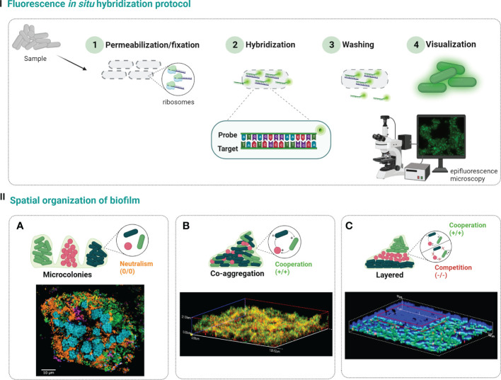
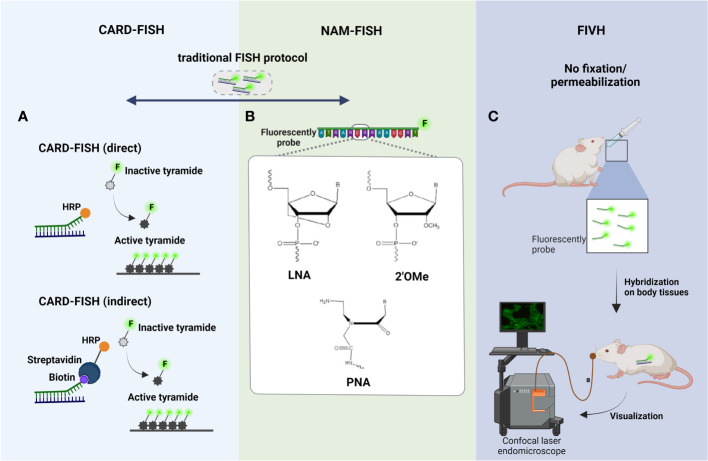
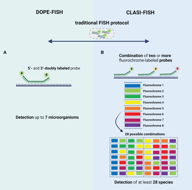
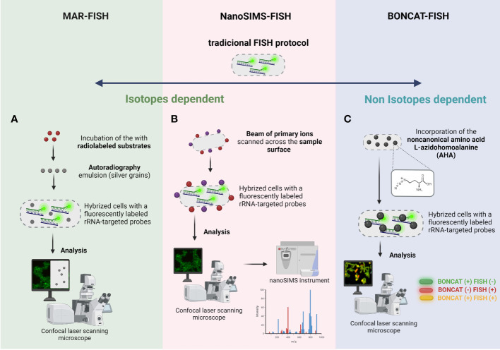
Biofilms are complex structures with an intricate relationship between the resident microorganisms, the extracellular matrix, and the surrounding environment. Interest in biofilms is growing exponentially given its ubiquity in so diverse fields such as healthcare, environmental and industry. Molecular techniques (e.g., next-generation sequencing, RNA-seq) have been used to study biofilm properties. However, these techniques disrupt the spatial structure of biofilms; therefore, they do not allow to observe the location/position of biofilm components (e.g., cells, genes, metabolites), which is particularly relevant to explore and study the interactions and functions of microorganisms. Fluorescence in situ hybridization (FISH) has been arguably the most widely used method for an in situ analysis of spatial distribution of biofilms. In this review, an overview on different FISH variants already applied on biofilm studies (e.g., CLASI-FISH, BONCAT-FISH, HiPR-FISH, seq-FISH) will be explored. In combination with confocal laser scanning microscopy, these variants emerged as a powerful approach to visualize, quantify and locate microorganisms, genes, and metabolites inside biofilms. Finally, we discuss new possible research directions for the development of robust and accurate FISH-based approaches that will allow to dig deeper into the biofilm structure and function.
Overview
The claims and statistics of this paper haven’t been checked yet.
Run the check — free, no account needed. It reads the open-access text and checks the sources it cites.
Integrity signals
Registry
Cites
Statistics
Figures
Verification
Claims the paper makes
The claims and statistics of this paper haven’t been checked yet.
Run the check — free, no account needed. It reads the open-access text and checks the sources it cites.
Claims other papers make about this one
Each is checked against this paper’s own text: does it say what the citing paper says it does?
In the citing paper
“Advanced methods such as peptide nucleic acid fluorescence in situ hybridization (PNA-FISH)44 provide insights into the microbial composition and distribution but do not offer details about the structural and topographic interactions between biofilms and tissues that SEM provides.”
From SEMTWIST Quantification of Biofilm Infection in Human Chronic Wound Using Scanning Electron Microscopy and Machine Learning (2025) · cites this paper as [44]
In the citing paper
“PNA-FISH specifically identifies bacterial species and displays aggregation characteristics thus helping identify and characterize BFI.44”
From SEMTWIST Quantification of Biofilm Infection in Human Chronic Wound Using Scanning Electron Microscopy and Machine Learning (2025) · cites this paper as [44]
Self-check
Percentages, sums and reported differences re-computed from the paper’s own numbers.
The self-check needs the paper’s full text.
Citations
Cites
Spatially resolved, highly multiplexed RNA profiling in single cells
Chen, K. H., Boettiger, A. N., Moffitt, J. R., Wang, S., & Zhuang, X. (2015) · Science · doi:10.1126/science.aaa6090 · Cited by 2,969
Imaging individual mRNA molecules using multiple singly labeled probes
Raj, A., van den Bogaard, P., Rifkin, S. A., van Oudenaarden, A., & Tyagi, S. (2008) · Nature Methods · doi:10.1038/nmeth.1253 · Cited by 2,028
Strategies for combating bacterial biofilm infections
Wu, H., Moser, C., Wang, H.-Z., Høiby, N., & Song, Z.-J. (2015) · International Journal of Oral Science · doi:10.1038/ijos.2014.65 · Cited by 925
Swidsinski, A., Weber, J., Loening-Baucke, V., Hale, L. P., & Lochs, H. (2005) · Journal of Clinical Microbiology · doi:10.1128/jcm.43.7.3380-3389.2005 · Cited by 894 · cited 1×
“As mentioned in previous sections, several FISH variants provide important phylogenetic information in complex and heterogeneous biofilms (Swidsinski et al., 2005).”
BackgroundShaping the growth behaviour of biofilms initiated from bacterial aggregates
Melaugh, G., Hutchison, J., Kragh, K. N., Irie, Y., Roberts, A., Bjarnsholt, T., Diggle, S. P., Gordon, V. D., & Allen, R. J. (2016) · PLoS ONE · doi:10.1371/journal.pone.0149683 · Cited by 93
Research works printed but not linked: 163
- Allkja, 2022, Interactions of microorganisms within a urinary catheter polymicrobial biofilm model, Biotechnol. Bioengineering, 120, 239, 10.1002/bit.28241 · DOI
- Almeida, 2021, An introduction to fluorescence in situ hybridization in microorganisms, Methods Mol. Biol., 2246, 1, 10.1007/978-1-0716-1115-9_1 · DOI
- Almeida, 2011, Discriminating multi-species populations in biofilms with peptide nucleic acid fluorescence in situ hybridization (PNA FISH), PLoS One, 6, 10.1371/journal.pone.0014786 · DOI
- Amann, 2008, Single-cell identification in microbial communities by improved fluorescence in situ hybridization techniques, Nat. Rev. Microbiol., 6, 339, 10.1038/nrmicro1888 · DOI
- Aquino de Muro, 2008, Probe design, production, and applications
- Artini, 2022, Essential oils biofilm modulation activity and machine learning analysis on pseudomonas aeruginosa isolates from cystic fibrosis patients, Microorganisms, 10, 10.3390/microorganisms10050887 · DOI
- Azeredo, 2017, Critical review on biofilm methods, Crit. Rev. Microbiol., 43, 313, 10.1080/1040841X.2016.1208146 · DOI
- Azevedo, 2021, Biofilms vs. cities and humans vs. aliens - a tale of reproducibility in biofilms, Trends Microbiol., 29, 1062, 10.1016/j.tim.2021.05.003 · DOI
- Azevedo, 2015, Detection and discrimination of biofilm populations using locked nucleic acid/2′-O-methyl-RNA fluorescence in situ hybridization (LNA/2′OMe-FISH), Biochem. Eng. J., 104, 64, 10.1016/j.bej.2015.04.024 · DOI
- Azevedo, 2016, Impact of delftia tsuruhatensis and achromobacter xylosoxidans on escherichia coli dual-species biofilms treated with antibiotic agents, Biofouling, 32, 227, 10.1080/08927014.2015.1124096 · DOI
- Azevedo, 2022, Spectral imaging and nucleic acid mimics fluorescence in situ hybridization (SI-NAM-FISH) for multiplex detection of clinical pathogens, Front. Microbiol., 13, 10.3389/fmicb.2022.976639 · DOI
- Azevedo, 2020, Increased intraspecies diversity in escherichia coli biofilms promotes cellular growth at the expense of matrix production, Antibiotics, 9, 818, 10.3390/antibiotics9110818 · DOI
- Batani, 2019, Fluorescence in situ hybridization (FISH) and cell sorting of living bacteria, Sci. Rep., 9, 18618, 10.1038/s41598-019-55049-2 · DOI
- Behnam, 2012, A straightforward DOPE (Double labeling of oligonucleotide probes)-FISH (Fluorescence In situ hybridization) method for simultaneous multicolor detection of six microbial populations, Appl. Environ. Microbiol., 78, 5138, 10.1128/AEM.00977-12 · DOI
- Behrens, 2008, Linking microbial phylogeny to metabolic activity at the single-cell level by using enhanced element labeling-catalyzed reporter deposition fluorescence in situ hybridization (EL-FISH) and NanoSIMS, Appl. Environ. Microbiol., 74, 3143, 10.1128/AEM.00191-08 · DOI
- Berk, 2012, Molecular architecture and assembly principles of vibrio cholerae biofilms, Science, 337, 236, 10.1126/science.1222981 · DOI
- Booth, 2020, Influence of interspecies interactions on the spatial organization of dual species bacterial communities, Biofilm, 2, 100035, 10.1016/j.bioflm.2020.100035 · DOI
- Braasch, 2001, Locked nucleic acid (LNA): fine-tuning the recognition of DNA and RNA, Chem. Biol., 8, 1, 10.1016/S1074-5521(00)00058-2 · DOI
- Brileya, 2014, 3D-fluorescence in situ hybridization of intact, anaerobic biofilm, 10.1007/978-1-4939-0554-6_13 · DOI
- Burmølle, 2014, Interactions in multispecies biofilms: do they actually matter, Trends Microbiol., 22, 84, 10.1016/j.tim.2013.12.004 · DOI
- Cerqueira, 2008, DNA Mimics for the rapid identification of microorganisms by fluorescence in situ hybridization (FISH), Int. J. Mol. Sci., 9, 1944, 10.3390/ijms9101944 · DOI
- Cerqueira, 2013, Biofilm formation with mixed cultures of pseudomonas aeruginosa/Escherichia coli on silicone using artificial urine to mimic urinary catheters, Biofouling, 29, 829, 10.1080/08927014.2013.807913 · DOI
- Costa, 2017, It is all about location: how to pinpoint microorganisms and their functions in multispecies biofilms, Future Microbiol., 12, 987, 10.2217/fmb-2017-0053 · DOI
- Cruz, 2021, The two weapons against bacterial biofilms: detection and treatment, Antibiotics (Basel)., 10, 10.3390/antibiotics10121482 · DOI
- Dai, 2022, Advances and trends in omics technology development, Front. Med. (Lausanne)., 9, 10.3389/fmed.2022.911861 · DOI
- Dar, 2021, Spatial transcriptomics of planktonic and sessile bacterial populations at single-cell resolution, Science, 373, 10.3389/fmed.2022.911861 · DOI
- Di Gregorio, 2018, Biofilm diversity, structure and matrix seasonality in a full-scale cooling tower, Biofouling, 34, 1093, 10.1080/08927014.2018.1541454 · DOI
- Di Martino, 2018, Extracellular polymeric substances, a key element in understanding biofilm phenotype, AIMS Microbiol., 4, 274, 10.3934/microbiol.2018.2.274 · DOI
- Dimauro, 2020, A novel approach for biofilm detection based on a convolutional neural network, Electronics, 9, 881, 10.3390/electronics9060881 · DOI
- Donlan, 2002, Biofilms: survival mechanisms of clinically relevant microorganisms, Clin. Microbiol. Rev., 15, 167, 10.1128/CMR.15.2.167-193.2002 · DOI · Paper page
- Dufour, 2010, Bacterial biofilm: structure, function, and antimicrobial resistance, Endodontic Topics, 22, 2, 10.1111/j.1601-1546.2012.00277.x · DOI
- Dutta, 2021, Analysis of biofilm matrix by multiplex fluorescence in situ hybridization (M-FISH) and confocal laser scanning microscopy (CLSM) during nosocomial infections, 10.1007/978-1-0716-1378-8_8 · DOI
- Ebersole, 2019, Biofilm-induced profiles of immune response gene expression by oral epithelial cells, Mol. Oral. Microbiol., 34, 10.1111/omi.12251 · DOI · Paper page
- Elayadi, 2002, Implications of high-affinity hybridization by locked nucleic acid oligomers for inhibition of human telomerase, Biochemistry, 41, 9973, 10.1021/bi025907j · DOI
- Elias, 2012, Multi-species biofilms: living with friendly neighbors, FEMS Microbiol. Rev., 36, 990, 10.1111/j.1574-6976.2012.00325.x · DOI
- Eriksson, 2017, Saliva and tooth biofilm bacterial microbiota in adolescents in a low caries community, Sci. Rep., 7, 5861, 10.1038/s41598-017-06221-z · DOI
- Escudero, 2018, Active microbial biofilms in deep poor porous continental subsurface rocks, Sci. Rep., 8, 1538, 10.1038/s41598-018-19903-z · DOI
- Fernandez, 2008, Microbial community dynamics in a chemolithotrophic denitrification reactor inoculated with methanogenic granular sludge, Chemosphere, 70, 462, 10.1016/j.chemosphere.2007.06.062 · DOI
- Ferrari, 2006, Catalyzed reporter deposition-fluorescence in situ hybridization allows for enrichment-independent detection of microcolony-forming soil bacteria, Appl. Environ. Microbiol., 72, 918, 10.1128/AEM.72.1.918-922.2006 · DOI
- Fike, 2008, Micron-scale mapping of sulfur cycling across the oxycline of a cyanobacterial mat: a paired nanoSIMS and CARD-FISH approach, ISME J., 2, 749, 10.1038/ismej.2008.39 · DOI
- Flemming, 2010, The biofilm matrix, Nat. Rev. Microbiol., 8, 623, 10.1038/nrmicro2415 · DOI · Paper page
- Flemming, 2016, Biofilms: an emergent form of bacterial life, Nat. Rev. Microbiol., 14, 563, 10.1038/nrmicro.2016.94 · DOI · Paper page
- Folsom, 2010, Physiology of pseudomonas aeruginosa in biofilms as revealed by transcriptome analysis, BMC Microbiol., 10, 1, 10.1186/1471-2180-10-294 · DOI
- Fontenete, 2013, Hybridization-based detection of helicobacter pylori at human body temperature using advanced locked nucleic acid (LNA) probes, PloS One, 8, 10.1371/journal.pone.0081230 · DOI
- Fontenete, 2016, Fluorescence In vivo hybridization (FIVH) for detection of helicobacter pylori infection in a C57BL/6 mouse model, PLoS One, 11, 10.1371/journal.pone.0148353 · DOI
- Frank, 2007, Poly-n-acetylglucosamine is not a major component of the extracellular matrix in biofilms formed by icaADBC-positive staphylococcus lugdunensis isolates, Infect. Immun., 75, 4728, 10.1128/IAI.00640-07 · DOI
- Franklin, 2015, New technologies for studying biofilms, Microbiol Spectr., 3, 1, 10.1128/microbiolspec.MB-0016-2014 · DOI
- Funari, 2022, Detection and characterization of bacterial biofilms and biofilm-based sensors, ACS Sensors., 7, 347, 10.1021/acssensors.1c02722 · DOI
- Gao, 2016, A critical review of NanoSIMS in analysis of microbial metabolic activities at single-cell level, Crit. Rev. Biotechnol., 36, 884, 10.3109/07388551.2015.1057550 · DOI
- Garini, 2006, Spectral imaging: principles and applications, Cytometry A., 69, 735, 10.1002/cyto.a.20311 · DOI
- Gu, 2022, Application of fluorescence In situ hybridization (FISH) in oral microbial detection, Pathogens, 11, 1450, 10.3390/pathogens11121450 · DOI
- Guzmán-Soto, 2021, Mimicking biofilm formation and development: recent progress in in vitro and in vivo biofilm models, iScience, 24, 102443, 10.1016/j.isci.2021.102443 · DOI
- Habimana, 2010, Enhanced surface colonization by escherichia coli O157:H7 in biofilms formed by an acinetobacter calcoaceticus isolate from meat-processing environments, Appl. Environ. Microbiol., 76, 4557, 10.1128/AEM.02707-09 · DOI
- Hall-Stoodley, 2004, Bacterial biofilms: from the natural environment to infectious diseases, Nat. Rev. Microbiol., 2, 95, 10.1038/nrmicro821 · DOI
- Handelsman, 2004, Metagenomics: application of genomics to uncultured microorganisms, Microbiol. Mol. Biol. Rev., 68, 669, 10.1128/MMBR.68.4.669-685.2004 · DOI
- Hasan, 2014, Microbial community profiling of human saliva using shotgun metagenomic sequencing, PLoS One, 9, 10.1371/journal.pone.0097699 · DOI
- Hatzenpichler, 2016, Visualizing in situ translational activity for identifying and sorting slow-growing archaeal−bacterial consortia, Proc. Natl. Acad. Sci., 113, E4069, 10.1073/pnas.1603757113 · DOI
- Hatzenpichler, 2014, In situ visualization of newly synthesized proteins in environmental microbes using amino acid tagging and click chemistry, Environ. Microbiol., 16, 2568, 10.1111/1462-2920.12436 · DOI
- Heim, 2017, Frutexites-like structures formed by iron oxidizing biofilms in the continental subsurface (Äspö hard rock laboratory, Sweden), PLoS One, 12, 10.1371/journal.pone.0177542 · DOI
- Hesselsoe, 2005, Isotope labeling and microautoradiography of active heterotrophic bacteria on the basis of assimilation of 14CO(2), Appl. Environ. Microbiol., 71, 646, 10.1128/AEM.71.2.646-655.2005 · DOI
- Hibbing, 2010, Bacterial competition: surviving and thriving in the microbial jungle, Nat. Rev. Microbiol., 8, 15, 10.1038/nrmicro2259 · DOI
- Hoehler, 2013, Microbial life under extreme energy limitation, Nat. Rev. Microbiol., 11, 83, 10.1038/nrmicro2939 · DOI
- Hoshino, 2008, Quantification of target molecules needed to detect microorganisms by fluorescence in situ hybridization (FISH) and catalyzed reporter deposition-FISH, Appl. Environ. Microbiol., 74, 5068, 10.1128/AEM.00208-08 · DOI
- Huber, 2018, Fluorescence in situ hybridization (FISH): history, limitations and what to expect from micro-scale FISH, Micro Nano Engineering., 1, 15, 10.1016/j.mne.2018.10.006 · DOI
- Karkare, 2006, Promising nucleic acid analogs and mimics: characteristic features and applications of PNA, LNA, and morpholino, Appl. Microbiol. Biotechnol., 71, 575, 10.1007/s00253-006-0434-2 · DOI
- Khatoon, 2018, Bacterial biofilm formation on implantable devices and approaches to its treatment and prevention, Heliyon, 4, 10.1016/j.heliyon.2018.e01067 · DOI
- Kierzek, 2005, The influence of locked nucleic acid residues on the thermodynamic properties of 2′-o-methyl RNA/RNA heteroduplexes, Nucleic Acids Res., 33, 5082, 10.1093/nar/gki789 · DOI
- Kinaret, 2020, Transcriptomics in toxicogenomics, part I: experimental design, technologies, publicly available data, and regulatory aspects, Nanomaterials (Basel)., 10, 10.3390/nano10040750 · DOI
- Kindaichi, 2004, Ecophysiological interaction between nitrifying bacteria and heterotrophic bacteria in autotrophic nitrifying biofilms as determined by microautoradiography-fluorescence in situ hybridization, Appl. Environ. Microbiol., 70, 1641, 10.1128/AEM.70.3.1641-1650.2004 · DOI
- Kitzinger, 2021, Assigning function to phylogeny: FISH-nanoSIMS, Methods Mol. Biol., 2246, 207, 10.1007/978-1-0716-1115-9_13 · DOI
- Kostakioti, 2013, Bacterial biofilms: development, dispersal, and therapeutic strategies in the dawn of the postantibiotic era, Cold Spring Harbor Perspect. Med., 3, a010306, 10.1101/cshperspect.a010306 · DOI
- Kubota, 2013, CARD-FISH for environmental microorganisms: technical advancement and future applications, Microbes Environ., 28, 3, 10.1264/jsme2.ME12107 · DOI
- Lee, 1999, Combination of fluorescent in situ hybridization and microautoradiography–a new tool for structure-function analyses in microbial ecology, Appl. Environ. Microbiol., 65, 1289, 10.1128/AEM.65.3.1289-1297.1999 · DOI
- Levenson, 2008, Multiplexing with multispectral imaging: from mice to microscopy, Ilar J., 49, 78, 10.1093/ilar.49.1.78 · DOI
- Li, 2008, Simultaneous analysis of microbial identity and function using NanoSIMS, Environ. Microbiol., 10, 580, 10.1111/j.1462-2920.2007.01478.x · DOI
- Lopes, 2017, Discriminating typical and atypical cystic fibrosis-related bacteria by multiplex PNA-FISH, Biotechnol. Bioengineering., 114, 355, 10.1002/bit.26085 · DOI
- Lowe, 2017, Transcriptomics technologies, PloS Comput. Biol., 13, 10.1371/journal.pcbi.1005457 · DOI
- Lubeck, 2014, Single-cell in situ RNA profiling by sequential hybridization, Nat. Methods, 11, 360, 10.1038/nmeth.2892 · DOI · Paper page
- Lukumbuzya, 2019, A multicolor fluorescence in situ hybridization approach using an extended set of fluorophores to visualize microorganisms, Front. Microbiol., 10, 10.3389/fmicb.2019.01383 · DOI
- Lupini, 2011, CARD-FISH and confocal laser scanner microscopy to assess successional changes of the bacterial community in freshwater biofilms, J. Microbiol. Methods, 86, 248, 10.1016/j.mimet.2011.05.011 · DOI
- Magalhaes, 2019, RNA-Based qPCR as a tool to quantify and to characterize dual-species biofilms, Sci. Rep., 9, 13639, 10.1038/s41598-019-50094-3 · DOI
- Malic, 2009, Detection and identification of specific bacteria in wound biofilms using peptide nucleic acid fluorescent in situ hybridization (PNA FISH), Microbiology, 155, 2603, 10.1099/mic.0.028712-0 · DOI · Paper page
- Mansfield, 2005, Autofluorescence removal, multiplexing, and automated analysis methods for in-vivo fluorescence imaging, J Biomed Opt., 10, 41207, 10.1117/1.2032458 · DOI · Paper page
- Mark Welch, 2016, Biogeography of a human oral microbiome at the micron scale, Proc. Natl. Acad. Sci. USA, 113, E791, 10.1073/pnas.1522149113 · DOI · Paper page
- Matturro, 2021, CAtalyzed reporter deposition fluorescence in situ hybridization (CARD-FISH) for complex environmental samples, Methods Mol. Biol., 2246, 129, 10.1007/978-1-0716-1115-9_9 · DOI
- Miller, 2011, In vivo fluorescence-based endoscopic detection of colon dysplasia in the mouse using a novel peptide probe, PloS One, 6, 10.1371/journal.pone.0017384 · DOI
- Miller, 2018, Transcriptome analysis of porphyromonas gingivalis and acinetobacter baumannii in polymicrobial communities, Mol. Oral. Microbiol., 33, 364, 10.1111/omi.12238 · DOI · Paper page
- Monticolo, 2020, The role of DNA in the extracellular environment: a focus on NETs, RETs and biofilms, Front. Plant Sci., 11, 10.3389/fpls.2020.589837 · DOI
- Moreau, 2007, Extracellular proteins limit the dispersal of biogenic nanoparticles, Science, 316, 1600, 10.1126/science.1141064 · DOI
- Moreira, 2022, Liposome delivery of nucleic acids in bacteria: toward In vivo labeling of human microbiota, ACS Infect. Diseases., 8, 1218, 10.1021/acsinfecdis.1c00601 · DOI
- Moter, 2000, Fluorescence in situ hybridization (FISH) for direct visualization of microorganisms, J. Microbiol. Methods, 41, 85, 10.1016/S0167-7012(00)00152-4 · DOI
- Musat, 2012, Detecting metabolic activities in single cells, with emphasis on nanoSIMS, FEMS Microbiol. Rev., 36, 486, 10.1111/j.1574-6976.2011.00303.x · DOI
- Nácher-Vázquez, 2022, The role of nucleic acid mimics (NAMs) on FISH-based techniques and applications for microbial detection, Microbiological Res., 262, 127086, 10.1016/j.micres.2022.127086 · DOI
- Nadell, 2016, Spatial structure, cooperation and competition in biofilms, Nat. Rev. Microbiol., 14, 589, 10.1038/nrmicro.2016.84 · DOI
- Naidoo, 2013, Treated wastewater effluent as a source of microbial pollution of surface water resources, Int. J. Environ. Res. Public Health, 11, 249, 10.3390/ijerph110100249 · DOI
- Nguyen, 2014, Probing of exopolysaccharides with green fluorescence protein-labeled carbohydrate-binding module in escherichia coli biofilms and flocs induced by bcsB overexpression, J. Biosci. Bioeng., 118, 400, 10.1016/j.jbiosc.2014.03.005 · DOI
- Nielsen, 2021, Assigning function to phylogeny: MAR-FISH, 10.1007/978-1-0716-1115-9_14 · DOI
- Nielsen, 2002, Quantification of cell-specific substrate uptake by probe-defined bacteria under in situ conditions by microautoradiography and fluorescence in situ hybridization, Environ. Microbiol., 5, 202, 10.1046/j.1462-2920.2003.00402.x · DOI
- Nielsen, 2002, Abundance and phylogenetic affiliation of iron reducers in activated sludge as assessed by fluorescence in situ hybridization and microautoradiography, Appl. Environ. Microbiol., 68, 4629, 10.1128/AEM.68.9.4629-4636.2002 · DOI
- Nielsen, 2010, Combined microautoradiography and fluorescence in situ hybridization (MAR-FISH) for the identification of metabolically active microorganisms, 10.1007/978-3-540-77587-4_319 · DOI
- Nielsen, 2000, Role of commensal relationships on the spatial structure of a surface-attached microbial consortium, Environ. Microbiol., 2, 59, 10.1046/j.1462-2920.2000.00084.x · DOI
- Nistico, 2014, Imaging bacteria and biofilms on hardware and periprosthetic tissue in orthopedic infections, Methods Mol. Biol., 1147, 105, 10.1007/978-1-4939-0467-9_8 · DOI
- Nunez, 2017, NanoSIMS for biological applications: current practices and analyses, Biointerphases, 13, 03B301, 10.1116/1.4993628 · DOI
- Okabe, 2004, MAR-FISH–an ecophysiological approach to link phylogenetic affiliation and in situ metabolic activity of microorganisms at a single-cell resolution, Microbes Environ, 19, 83, 10.1264/jsme2.19.83 · DOI
- Okabe, 2011, Chapter seven - a polyphasic approach to study ecophysiology of complex multispecies nitrifying biofilms
- Partoazar, 2019, Antibiofilm activity of natural zeolite supported NanoZnO: inhibition of esp gene expression of enterococcus faecalis, Nanomedicine, 14, 675, 10.2217/nnm-2018-0173 · DOI
- Patwardhan, 2014, Molecular markers in phylogenetic studies-a review, J. Phylogenet. Evolutionary Biol., 2014, 10.4172/2329-9002.1000131 · DOI
- Pereira, 2018, Pseudomonas-specific NGS assay provides insight into abundance and dynamics of pseudomonas species including p. aeruginosa in a cooling tower, Front. Microbiol., 9, 10.3389/fmicb.2018.01958 · DOI
- Petersen, 2003, LNA: a versatile tool for therapeutics and genomics, Trends Biotechnol., 21, 74, 10.1016/S0167-7799(02)00038-0 · DOI
- Peterson, 2014, Functional expression of dental plaque microbiota, Front. Cell Infect. Microbiol., 4, 10.3389/fcimb.2014.00108 · DOI
- Peyrusson, 2020, Intracellular staphylococcus aureus persisters upon antibiotic exposure, Nat. Commun., 11, 2200, 10.1038/s41467-020-15966-7 · DOI · Paper page
- Polglase, 2005, A fluorescence confocal endomicroscope for in vivo microscopy of the upper- and the lower-GI tract, Gastrointest Endosc., 62, 686, 10.1016/j.gie.2005.05.021 · DOI
- Rajput, 2022, Machine learning from pseudomonas aeruginosa transcriptomes identifies independently modulated sets of genes associated with known transcriptional regulators, Nucleic Acids Res., 50, 3658, 10.1093/nar/gkac187 · DOI
- Ramírez-Puebla, 2022, Spatial organization of the kelp microbiome at micron scales, Microbiome, 10, 52, 10.1186/s40168-022-01235-w · DOI
- Rehman, 2020, Metagenomic analysis of sludge and early-stage biofilm communities of a submerged membrane bioreactor, Sci. Total Environment., 701, 134682, 10.1016/j.scitotenv.2019.134682 · DOI
- Resch, 2005, Differential gene expression profiling of staphylococcus aureus cultivated under biofilm and planktonic conditions, Appl. Environ. Microbiol., 71, 2663, 10.1128/AEM.71.5.2663-2676.2005 · DOI
- Reuter, 2015, High-throughput sequencing technologies, Mol. Cell., 58, 586, 10.1016/j.molcel.2015.05.004 · DOI
- Robertson, 2009, LNA flow–FISH: a flow cytometry–fluorescence in situ hybridization method to detect messenger RNA using locked nucleic acid probes, Analytical Biochem., 390, 109, 10.1016/j.ab.2009.04.026 · DOI
- Rocha, 2019, Development and application of peptide nucleic acid fluorescence in situ hybridization for the specific detection of listeria monocytogenes, Food Microbiol., 80, 1, 10.1016/j.fm.2018.12.009 · DOI
- Roder, 2020, Unravelling interspecies interactions across heterogeneities in complex biofilm communities, Environ. Microbiol., 22, 5, 10.1111/1462-2920.14834 · DOI
- Røder, 2021, Biofilms can act as plasmid reserves in the absence of plasmid specific selection, NPJ Biofilms Microbiomes., 7, 78, 10.1038/s41522-021-00249-w · DOI
- Römling, 2012, Biofilm infections, their resilience to therapy and innovative treatment strategies, J. Internal Med., 272, 541, 10.1111/joim.12004 · DOI
- Rosenberg, 2019, Propidium iodide staining underestimates viability of adherent bacterial cells, Sci. Rep., 9, 6483, 10.1038/s41598-019-42906-3 · DOI · Paper page
- Saarbach, 2019, Peptide nucleic acid (PNA) and its applications in chemical biology, diagnostics, and therapeutics, Curr. Opin. Chem. Biol., 52, 112, 10.1016/j.cbpa.2019.06.006 · DOI
- Sauer, 2022, The biofilm life cycle: expanding the conceptual model of biofilm formation, Nat. Rev. Microbiol., 20, 608, 10.1038/s41579-022-00767-0 · DOI
- Schaible, 2022, Correlative SIP-FISH-Raman-SEM-NanoSIMS links identity, morphology, biochemistry, and physiology of environmental microbes, ISME Commun., 2, 52, 10.1038/s43705-022-00134-3 · DOI
- Schimak, 2016, MiL-FISH: multilabeled oligonucleotides for fluorescence in situ hybridization improve visualization of bacterial cells, Appl. Environ. Microbiol., 82, 62, 10.1128/AEM.02776-15 · DOI
- Schlafer, 2017, Confocal microscopy imaging of the biofilm matrix, J. Microbiological Methods, 138, 50, 10.1016/j.mimet.2016.03.002 · DOI
- Schlundt, 2020, Spatial structure in the "Plastisphere": molecular resources for imaging microscopic communities on plastic marine debris, Mol. Ecol. Resour., 20, 620, 10.1111/1755-0998.13119 · DOI
- Seneviratne, 2020, Multi-omics tools for studying microbial biofilms: current perspectives and future directions, Crit. Rev. Microbiol., 46, 759, 10.1080/1040841X.2020.1828817 · DOI
- Shemesh, 2007, Differential gene expression profiling of streptococcus mutans cultured under biofilm and planktonic conditions, Microbiol. (Reading)., 153, 1307, 10.1099/mic.0.2006/002030-0 · DOI
- Shi, 2020, Highly multiplexed spatial mapping of microbial communities, Nature, 588, 676, 10.1038/s41586-020-2983-4 · DOI
- Silahtaroglu, 2004, LNA-modified oligonucleotides are highly efficient as FISH probes, Cytogenet. Genome Res., 107, 32, 10.1159/000079569 · DOI
- Sime-Ngando, 2022, Technique for identifying and counting infective chytrid sporangia using the chitinaceous fluorochrome calcofluor white, 10.1007/978-3-030-83749-5_4 · DOI
- Slaveykova, 2009, Dynamic NanoSIMS ion imaging of unicellular freshwater algae exposed to copper, Anal. Bioanal Chem., 393, 583, 10.1007/s00216-008-2486-x · DOI
- So, 2011, General properties of transcriptional time series in escherichia coli, Nat. Genet., 43, 554, 10.1038/ng.821 · DOI
- Sousa, 2021, A new PNA-FISH probe targeting fannyhessea vaginae, Front. Cell Infect. Microbiol., 11, 10.3389/fcimb.2021.779376 · DOI
- Srinivasan, 2021, Bacterial biofilm inhibition: a focused review on recent therapeutic strategies for combating the biofilm mediated infections, Front. Microbiol., 12, 10.3389/fmicb.2021.676458 · DOI
- Stacy, 2016, The biogeography of polymicrobial infection, Nat. Rev. Microbiol., 14, 93, 10.1038/nrmicro.2015.8 · DOI
- Stark, 2019, RNA Sequencing: the teenage years, Nat. Rev. Genet., 20, 631, 10.1038/s41576-019-0150-2 · DOI
- Stender, 2002, PNA for rapid microbiology, J. Microbiological Methods, 48, 1, 10.1016/S0167-7012(01)00340-2 · DOI
- Stewart, 2008, Physiological heterogeneity in biofilms, Nat. Rev. Microbiol., 6, 199, 10.1038/nrmicro1838 · DOI · Paper page
- Stoecker, 2010, Double labeling of oligonucleotide probes for fluorescence in situ hybridization (DOPE-FISH) improves signal intensity and increases rRNA accessibility, J. Appl. Environ. Microbiol., 76, 922, 10.1128/AEM.02456-09 · DOI
- Stoodley, 2002, Biofilms as complex differentiated communities, Annu. Rev. Microbiol., 56, 187, 10.1146/annurev.micro.56.012302.160705 · DOI
- Stuart, 2016, Light regimes shape utilization of extracellular organic c and n in a cyanobacterial biofilm, mBio, 7, 10.1128/mBio.00650-16 · DOI
- Subhadra, 2018, Control of biofilm formation in healthcare: recent advances exploiting quorum-sensing interference strategies and multidrug efflux pump inhibitors, Materials, 11, 1676, 10.3390/ma11091676 · DOI
- Teixeira, 2021, Bioinformatic tools and guidelines for the design of fluorescence in situ hybridization probes, 10.1007/978-1-0716-1115-9_3 · DOI
- Thomsen, 2005, Dramatically improved RNA in situ hybridization signals using LNA-modified probes, RNA, 11, 1745, 10.1261/rna.2139705 · DOI
- Tolker-Nielsen, 2015, Biofilm development, Microbiol. spectrum., 3, 3.2.21, 10.1128/9781555817466.ch3 · DOI
- Valm, 2012, CLASI-FISH: principles of combinatorial labeling and spectral imaging, Syst. Appl. Microbiol., 35, 496, 10.1016/j.syapm.2012.03.004 · DOI
- Valm, 2011, Systems-level analysis of microbial community organization through combinatorial labeling and spectral imaging, Proc. Natl. Acad. Sci. U.S.A., 108, 4152, 10.1073/pnas.1101134108 · DOI
- Valm, 2016, Multiplexed spectral imaging of 120 different fluorescent labels, PLoS One, 11, 10.1371/journal.pone.0158495 · DOI
- van Dijk, 2018, The third revolution in sequencing technology, Trends Genet., 34, 666, 10.1016/j.tig.2018.05.008 · DOI · Paper page
- Veedu, 2009, Locked nucleic acid as a novel class of therapeutic agents, RNA Biol., 6, 321, 10.4161/rna.6.3.8807 · DOI
- Vilas Boas, 2016, Discrimination of bacteriophage infected cells using locked nucleic acid fluorescent in situ hybridization (LNA-FISH), Biofouling, 32, 179, 10.1080/08927014.2015.1131821 · DOI
- Wu, 2019, The pathogenicity and transcriptome analysis of methicillin-resistant staphylococcus aureus in response to water extract of galla chinensis, Evid Based Complement Alternat Med., 2019, 3276156, 10.1155/2019/3276156 · DOI
- Xu, 2020, Electrochemical impedance spectroscopic detection of e.coli with machine learning, J. Electrochemical Society., 167, 047508, 10.1149/1945-7111/ab732f · DOI
- Yamashita, 2014, Nitrogen and phosphorus removal from wastewater treatment plant effluent via bacterial sulfate reduction in an anoxic bioreactor packed with wood and iron, Int. J. Environ. Res. Public Health, 11, 9835, 10.3390/ijerph110909835 · DOI
- Yan, 2019, Surviving as a community: antibiotic tolerance and persistence in bacterial biofilms, Cell Host Microbe, 26, 15, 10.1016/j.chom.2019.06.002 · DOI
- Yilmaz, 2004, Mechanistic approach to the problem of hybridization efficiency in fluorescent in situ hybridization, Appl. Environ. Microbiol., 70, 7126, 10.1128/AEM.70.12.7126-7139.2004 · DOI
- Yilmaz, 2006, Making all parts of the 16S rRNA of escherichia coli accessible in situ to single DNA oligonucleotides, Appl. Environ. Microbiol., 72, 733, 10.1128/AEM.72.1.733-744.2006 · DOI
- Zemanick, 2010, Reliability of quantitative real-time PCR for bacterial detection in cystic fibrosis airway specimens, PLoS One, 5, 10.1371/journal.pone.0015101 · DOI
- Zimmermann, 2014, Clearing up the signal: spectral imaging and linear unmixing in fluorescence microscopy, 10.1007/978-1-60761-847-8_5 · DOI · Paper page
Cited by
Sorted by citations
Singh, S., Muniz De Oliveira, F., Wang, C., Kumar, M., Xuan, Y., DeMazumder, D., Sen, C. K., & Roy, S. (2025) · Advances in Wound Care · doi:10.1089/wound.2024.0291 · Cited by 6
Metadata
- Authors
- Barbosa, A., Miranda, S., Azevedo, N. F., Cerqueira, L., & Azevedo, A. S.
- Venue
- Frontiers in Cellular and Infection Microbiology
- Publisher
- Frontiers Media SA
- Published
- 2023-05-22
- Volume
- 13
- ISSN
- 2235-2988
- PMC
- PMC10239779
- Cited by
- 63
- References
- 5
About these checks
Registry, retraction, Cites and statistics results are computed, not judged, with one exception: a printed reference that no index lists is read once by a model before it counts as a citation error. Claims are extracted by a model and kept only when their quote appears verbatim in the text; each cited source is then checked, with a model’s help, for whether it says what the claim relies on. Those verdicts can be wrong — read the claim’s quote and the cited source before relying on one.
Statistics and arithmetic are recomputed, not judged.
Confidence bands are shown only for fields calibrated on our annotated test set. None is calibrated yet, so no band is shown.
What counts as a claim ›How we measure confidence ›How we check this ›
Are you an author, or do you see a problem with this page? Request a review
How to cite
Cite CoChat for the checks on this page, and the paper for its own findings.
- This page
- CoChat. (2026). Citation check: Imaging biofilms using fluorescence in situ hybridization: seeing is believing. https://app.cochat.ai/papers/10.3389/fcimb.2023.1195803
- The paper
- Barbosa, A., Miranda, S., Azevedo, N. F., Cerqueira, L., & Azevedo, A. S. (2023). Imaging biofilms using fluorescence in situ hybridization: seeing is believing. Frontiers in Cellular and Infection Microbiology. https://doi.org/10.3389/fcimb.2023.1195803
